Scalable Production of MSC-Derived Exosomes: Optimizing Culture Conditions for Clinical Translation
This article provides a comprehensive guide for researchers and drug development professionals on scaling up mesenchymal stem cell (MSC) culture to produce therapeutic exosomes.
Scalable Production of MSC-Derived Exosomes: Optimizing Culture Conditions for Clinical Translation
Abstract
This article provides a comprehensive guide for researchers and drug development professionals on scaling up mesenchymal stem cell (MSC) culture to produce therapeutic exosomes. It covers foundational principles of MSC-exosome biology, explores advanced 3D bioreactor systems and purification methodologies, and offers practical strategies for troubleshooting batch variability and optimizing critical quality attributes. The content synthesizes the latest 2025 research to present a validated framework for achieving high-yield, consistent, and potent exosome production, directly addressing the key challenges in translating cell-free therapies from the laboratory to the clinic.
MSC-Exosome Biology and Preconditioning for Enhanced Potency
Frequently Asked Questions (FAQs): Core Concepts
Q1: What are the primary safety advantages of using MSC-derived exosomes over whole MSC therapy?
MSC-derived exosomes offer a cell-free therapeutic profile that circumvents key risks associated with whole cell transplantation. Major safety advantages include:
- Lower Immunogenicity: Exosomes lack major histocompatibility complex (MHC) molecules, significantly reducing the risk of immune rejection [1] [2].
- Reduced Tumorigenic Risk: Unlike live cells, exosomes are non-replicating, eliminating the risk of unwanted differentiation or tumor formation [1] [2].
- Avoidance of Vascular Complications: Their nanoscale size prevents capillary blockage (embolism), a potential risk with larger cell infusions [3].
- Higher Stability and Ease of Storage: Exosomes are easier to preserve and store compared to live cells, simplifying logistics for clinical applications [4].
Q2: Through what key mechanisms do MSC-derived exosomes exert their therapeutic effects?
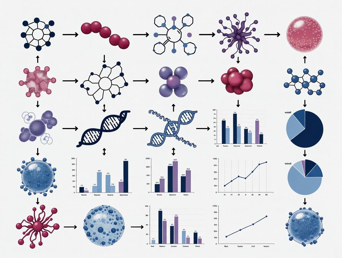
MSC-derived exosomes function primarily through intercellular communication, mediating their effects via several core mechanisms summarized in the diagram below:
The primary mechanisms are:
- Bioactive Cargo Delivery: Exosomes transfer functional miRNAs, proteins, and lipids to recipient cells, modulating key signaling pathways (e.g., Wnt/β-catenin for hair regeneration, AKT/ERK for renal repair) [1] [3].
- Immune Cell Modulation: They directly influence immune cells by suppressing T and B cell proliferation, promoting anti-inflammatory M2 macrophage polarization, and reducing natural killer cell cytotoxicity [1] [4].
- Promotion of Tissue Repair: Exosomes enhance cell proliferation and survival, reduce apoptosis and oxidative stress, and stimulate angiogenesis, facilitating tissue regeneration [3] [5].
Troubleshooting Guides: Common Experimental Challenges
Challenge 1: Low Yield and Scalability in Exosome Production
Low exosome yield is a major bottleneck for preclinical and clinical studies [6].
Potential Causes and Solutions:
- Cause: Suboptimal Cell Culture Conditions.
- Solution: Transition from 2D static culture to 3D bioreactor systems (e.g., Hollow Fiber bioreactors). These systems significantly increase cell density and prolong productive culture duration, enhancing yield [7]. Optimization of basal culture media (e.g., α-MEM may support higher yields than DMEM) and the use of xeno-free supplements like human platelet lysate (hPL) can also improve cell growth and exosome secretion [5].
- Cause: Inefficient Isolation Method.
- Solution: Replace traditional ultracentrifugation (UC) with more efficient, scalable methods like Tangential Flow Filtration (TFF). TFF allows for processing large volumes of conditioned media with higher recovery rates and better preservation of exosome integrity [5].
Recommended Protocol: Scalable Production using a Hollow Fiber Bioreactor [7]
- Cell Expansion: Seed human umbilical cord-derived MSCs (hUC-MSCs) into a Hollow Fiber 3D bioreactor.
- Conditioned Media Collection: Use a defined exosome-production medium (e.g., RoosterBio exosome-harvesting system) and harvest conditioned media periodically over a 28-day production cycle.
- Primary Concentration: Use TFF to concentrate the conditioned media.
- Exosome Isolation & Purification: Further purify the concentrated solution using size-exclusion chromatography (SEC) or density gradient centrifugation to remove contaminating proteins.
- Characterization: Analyze the final product using Nanoparticle Tracking Analysis (NTA), transmission electron microscopy (TEM), and western blot (for CD63, CD81, TSG101) to confirm yield, size, and marker expression.
Challenge 2: Heterogeneity and Lack of Functional Consistency
Exosome preparations are often heterogeneous, leading to variable experimental outcomes [7] [8].
Potential Causes and Solutions:
- Cause: Inherent Biological Variability.
- Solution: Standardize the cell source and passage number. Implement rigorous quality control for the parent MSCs, including surface marker profiling and differentiation potential assays. Establish a consistent "collection window" (e.g., specific passages) for harvesting exosomes to ensure subpopulation stability [7].
- Cause: Presence of Non-Exosomal Contaminants.
Recommended Protocol: Functional Consistency Testing in a Disease Model [7]
To ensure functional consistency, especially when scaling up, validate batches in a relevant disease model.
- In Vivo Model: Utilize a silica-induced mouse model of silicosis.
- Administration: Test the therapeutic efficacy via respiratory delivery (nebulization).
- Assessment: Evaluate outcomes through histology (e.g., lung tissue fibrosis scoring) and analysis of inflammatory biomarkers (e.g., cytokine levels in bronchoalveolar lavage fluid).
- Benchmarking: Compare the effects of new exosome batches against a well-characterized reference batch to ensure consistent biological activity.
Challenge 3: Inefficient Delivery and Biodistribution
The route of administration critically determines exosome delivery and therapeutic efficacy [7] [8].
Potential Causes and Solutions:
- Cause: Unsuitable Administration Route.
- Cause: Lack of Biodistribution Data.
- Solution: Track exosomes in vivo using isotopic labeling (e.g., with Zirconium-89, ⁸⁹Zr) to study their tissue tropism and pharmacokinetics, which informs optimal dosing and route selection [7].
Recommended Protocol: Evaluating Administration Routes [7] [8]
- Labeling: Label purified exosomes with a radioactive isotope (e.g., ⁸⁹Zr) or a fluorescent dye (e.g., DiR).
- Administration: Administer the labeled exosomes to animal models via two different routes (e.g., intravenous vs. nebulization).
- Tracking: Use imaging techniques (e.g., PET/CT for radioactive labels, IVIS for fluorescent labels) at multiple time points to monitor real-time biodistribution.
- Correlation with Efficacy: Sacrifice the animals and correlate the biodistribution data with therapeutic outcomes in the target tissue.
The Scientist's Toolkit: Essential Reagents and Materials
The following table details key reagents and tools critical for optimizing MSC culture and exosome production.
| Research Reagent / Material | Function / Application | Key Consideration |
|---|---|---|
| Hollow Fiber Bioreactor | 3D culture system for high-density cell expansion and continuous exosome harvest [7]. | Enables long-term (e.g., 28-day) production, improving yield and scalability over 2D flasks. |
| Tangential Flow Filtration (TFF) | Scalable isolation and concentration of exosomes from large volumes of conditioned media [5]. | Superior to ultracentrifugation for yield, processing time, and preserving exosome integrity. |
| RoosterBio Exosome System | A commercially available, integrated system including culture media and harvest supplements [7]. | Designed to enhance exosome yield and provide a standardized, xeno-free platform. |
| Human Platelet Lysate (hPL) | Xeno-free supplement for MSC culture media, replacing fetal bovine serum (FBS) [5]. | Avoids introduction of non-human vesicles and aligns with clinical translation requirements. |
| Size-Exclusion Chromatography (SEC) | High-purity purification of exosomes after initial concentration [2]. | Effectively separates exosomes from soluble proteins and other contaminants. |
| Nanoparticle Tracking Analysis (NTA) | Characterizes exosome particle size distribution and concentration [5]. | Essential for quality control and dose standardization (particles/mL). |
To aid in experimental design, key quantitative findings from recent studies are summarized below.
Table 1: Quantitative Data on Production and Dosing
| Parameter | Findings / Value Range | Context / Source |
|---|---|---|
| Production Yield | TFF isolation yielded statistically higher particle counts than Ultracentrifugation (UC) [5]. | Critical for selecting isolation methodology. |
| Production Duration | A 28-day biomanufacturing workflow in a Hollow Fiber bioreactor demonstrated stable subpopulation harvesting [7]. | Informs long-term production planning. |
| Effective Nebulized Dose | Therapeutic effects in lung disease models observed at doses around 10⁸ particles [8]. | Significantly lower than IV doses, highlighting route efficiency. |
| Intravenous Biodistribution | Predominant accumulation in the liver following IV injection [7]. | Crucial for planning systemic administration studies. |
Frequently Asked Questions (FAQs)
Q1: What are the primary therapeutic effects associated with miR-21, miR-146, and miR-181a in MSC-derived exosomes? A1: These miRNAs are key mediators of the immunomodulatory and regenerative effects of MSC-exosomes. miR-21 promotes cell survival and reduces apoptosis, miR-146a is a potent anti-inflammatory agent that suppresses the NF-κB pathway, and miR-181a regulates immune cell differentiation and function, particularly T-cell responses.
Q2: How can I efficiently isolate and quantify these specific miRNAs from my MSC-exosome preparations? A2: The recommended protocol involves total RNA isolation using kits optimized for small RNAs (e.g., miRNeasy Micro Kit), followed by reverse transcription with stem-loop primers specific to each miRNA. Quantification is best performed via RT-qPCR using TaqMan or SYBR Green assays designed for mature miRNA sequences.
Q3: My MSC culture conditions seem to alter the miRNA cargo profile. How can I standardize this?
A3: miRNA cargo is highly sensitive to culture conditions. To standardize production, control for passage number (use low passage MSCs,
Q4: What is the best method to confirm the functional delivery of these miRNAs to target cells? A4: Transfect MSC-exosomes with Cy3 or Cy5 fluorescently labeled mimics of your miRNA of interest and image uptake in target cells. For functional confirmation, transfect MSCs with a miRNA inhibitor (antagomir) prior to exosome collection, then demonstrate loss of the expected therapeutic effect in the target assay.
Troubleshooting Guides
Problem: Low yield of total RNA from isolated exosomes.
- Potential Cause 1: Inefficient exosome lysis.
- Solution: Add a membrane disruption reagent like 2% β-mercaptoethanol directly to the lysis buffer. Vortex vigorously for 15 seconds after adding the lysis buffer.
- Potential Cause 2: RNA loss during precipitation or column binding.
- Solution: Include a carrier (e.g., glycogen) during precipitation steps. Ensure ethanol concentrations are correct for column-based kits. Perform a second elution with pre-heated nuclease-free water to maximize yield.
Problem: High Ct values or non-detectable levels of target miRNAs in RT-qPCR.
- Potential Cause 1: Inefficient reverse transcription of mature miRNAs.
- Solution: Use stem-loop RT primers specifically designed for the mature miRNA sequence, not the pre-miRNA. Confirm the primer sequences are correct for the species (human, mouse, rat).
- Potential Cause 2: Purity of exosome preparation.
- Solution: Contaminating proteins from FBS or cell debris can inhibit RT and qPCR. Re-pellet exosomes and wash with PBS. Use a more stringent isolation method like density gradient centrifugation.
Problem: Inconsistent therapeutic effects between MSC-exosome batches.
- Potential Cause 1: Variability in MSC source and culture.
- Solution: Use standardized, characterized, low-passage MSCs from a reliable source. Maintain meticulous records of culture confluence at harvest and media batch numbers. Implement a serum-free media protocol.
- Potential Cause 2: Fluctuations in key miRNA cargo levels.
- Solution: Perform QC on each exosome batch by quantifying a panel of key miRNAs (miR-21, -146a, -181a) via RT-qPCR and normalize to particle number (e.g., by NTA). Only use batches that meet a pre-defined miRNA/potency threshold.
Experimental Protocols
Protocol 1: Quantifying miRNA Cargo from MSC-Exosomes
Title: miRNA Extraction & Quantification from Exosomes
Methodology:
- Exosome Isolation: Isolve exosomes from 10 mL of conditioned MSC media via ultracentrifugation (100,000 × g, 70 min) or size-exclusion chromatography. Resuspend the pellet in 200-500 µL of PBS.
- RNA Extraction: Use the miRNeasy Micro Kit (Qiagen) or equivalent.
- Add 700 µL Qiazol Lysis Reagent to 200 µL of exosome suspension. Vortex for 1 min.
- Incubate for 5 min at room temperature.
- Add 140 µL chloroform, shake vigorously for 15 sec, incubate 3 min.
- Centrifuge at 12,000 × g for 15 min at 4°C.
- Transfer the upper aqueous phase to a new tube.
- Add 1.5 volumes of 100% ethanol. Mix.
- Transfer mixture to an RNeasy MinElute spin column. Centrifuge.
- Wash with RWT and RPE buffers per kit instructions.
- Elute RNA in 14 µL nuclease-free water.
- Reverse Transcription: Use the TaqMan MicroRNA Reverse Transcription Kit with gene-specific stem-loop primers.
- Prepare RT master mix per kit instructions.
- Use 5 µL of extracted RNA.
- Run in a thermal cycler: 16°C for 30 min, 42°C for 30 min, 85°C for 5 min.
- qPCR Amplification:
- Dilute RT product 1:5.
- Prepare qPCR mix with TaqMan Universal Master Mix II and the specific TaqMan miRNA assay.
- Run in a real-time PCR system using standard cycling conditions.
- Use snoRNA234 or U6 snRNA as an endogenous control for data normalization (ΔΔCt method).
Protocol 2: Modifying MSC miRNA Cargo via Pre-conditioning
Title: Pre-conditioning MSCs to Modulate Exosomal miRNA
Methodology:
- Select Pre-conditioning Stimulus:
- Inflammatory priming: Treat MSCs at 70-80% confluence with 20 ng/mL IFN-γ and 15 ng/mL TNF-α for 24-48 hours.
- Hypoxic conditioning: Culture MSCs in a hypoxia chamber (1-3% O₂) for 48 hours.
- Harvest Conditioned Media: Collect media after preconditioning. Centrifuge at 2,000 × g for 10 min to remove cells and debris.
- Isolate Exosomes: Proceed with standard exosome isolation (e.g., ultracentrifugation, SEC, or TFF).
- Validate Cargo Changes: Extract RNA from the isolated exosomes and perform RT-qPCR for miR-146a, miR-21, and miR-181a. Compare fold-changes to exosomes from untreated, normoxic MSCs.
Table 1: miRNA Cargo Changes in MSC-Exosomes Under Different Culture Conditions
| miRNA | Condition (vs. Standard 2D) | Fold Change | Measured Effect | Reference Model |
|---|---|---|---|---|
| miR-21 | 3D Spheroid Culture | 3.5 - 5.0 ↑ | Enhanced cardiomyocyte survival | In vitro (H9C2 cells) |
| miR-146a | IFN-γ/TNF-α Priming | 8.0 - 12.0 ↑ | Suppressed macrophage TNF-α secretion | In vitro (LPS-stimulated macrophages) |
| miR-181a | Hypoxia (1% O₂) | 2.0 - 4.0 ↑ | Reduced T-cell proliferation | In vitro (PBMC assay) |
| miR-21 | High Passage (P10 vs P3) | 0.4 - 0.6 ↓ | Reduced anti-apoptotic effect | In vitro (HK-2 cells) |
Table 2: Functional Outcomes of MSC-Exosome miRNA Modulation
| Target miRNA | Modulation Method | In Vivo Model | Key Quantitative Outcome |
|---|---|---|---|
| miR-21 | Overexpression in MSCs | Mouse MI Model | 40% reduction in infarct size; 2.1-fold increase in capillary density vs. control exosomes. |
| miR-146a | Knockdown in MSCs | Mouse Colitis Model | Abolished protective effect: Disease Activity Index increased from 3.2 to 7.8 (control exo vs. KO exo). |
| miR-181a | Hypoxic Pre-conditioning | Mouse GvHD Model | 60% increase in survival rate at day 60; 50% reduction in pathological score for liver and skin. |
Pathway Diagrams
Title: MSC-Exosome miRNA Signaling Pathways
Title: miRNA Cargo Analysis Workflow
The Scientist's Toolkit: Research Reagent Solutions
| Reagent / Material | Function / Application |
|---|---|
| miRNeasy Micro Kit (Qiagen) | Isolation of high-quality total RNA, including small RNAs < 200 nt, from low-yield samples like exosomes. |
| TaqMan MicroRNA Assays (Thermo Fisher) | Sequence-specific primers and probes for highly sensitive and specific detection of mature miRNAs via RT-qPCR. |
| CD63/CD81/CD9 Antibodies | Antibodies for western blot analysis to confirm the presence of exosomal tetraspanin markers during characterization. |
| Exosome-depleted FBS | Fetal bovine serum processed to remove bovine exosomes, preventing contamination in MSC culture media. |
| SYBR Green PCR Master Mix | A fluorescent dye for qPCR that binds double-stranded DNA, used as an alternative to TaqMan probes for miRNA quantification. |
| Stem-loop RT Primers | Specialized reverse transcription primers that create a longer cDNA template from the short mature miRNA sequence for qPCR. |
| PBS (pH 7.4) | Phosphate-buffered saline for washing cell pellets, resuspending exosomes, and as a diluent for various reagents. |
| RNase Zap / RNase-free reagents | Critical for preventing degradation of low-abundance miRNA molecules during all steps of RNA work. |
Frequently Asked Questions (FAQs)
Q1: What is the primary goal of preconditioning MSCs for exosome production? A1: The primary goal is to modulate the MSC secretome, enhancing the yield, altering the cargo, and boosting the therapeutic efficacy (e.g., anti-inflammatory, pro-angiogenic) of the harvested exosomes for downstream applications.
Q2: What is the recommended duration for hypoxia preconditioning? A2: Most protocols use a duration between 24 and 72 hours. The optimal time can vary based on MSC source and desired exosome profile. Shorter times (24-48h) are common for inducing pro-angiogenic factors, while longer times may increase stress-related markers.
Q3: What are the critical quality control checkpoints after preconditioning? A3:
- Cell Viability: Confirm >90% viability via Trypan Blue exclusion or similar.
- Phenotypic Stability: Verify retention of MSC surface markers (CD73, CD90, CD105) and absence of hematopoietic markers via flow cytometry.
- Exosome Characterization: Validate exosome identity using NTA (for size/concentration), TEM (for morphology), and Western Blot (for markers like CD63, CD81, TSG101).
Q4: How do I choose between LPS and cytokine (TNF-α/IL-1β) preconditioning? A4: The choice depends on the intended therapeutic outcome.
- LPS: Mimics a bacterial infection, strongly polarizing MSCs towards an anti-inflammatory, immunomodulatory phenotype. Ideal for applications in sepsis or combating excessive inflammation.
- Cytokines (TNF-α/IL-1β): Mimic a sterile inflammatory environment (e.g., arthritis). This often leads to a more complex secretome with a mix of immunomodulatory and tissue-reparative factors.
Q5: Can preconditioning strategies be combined? A5: Yes, sequential combination is an active area of research. For example, priming with a cytokine followed by hypoxia can have a synergistic effect, potentially further enhancing exosome potency. However, this requires extensive optimization to avoid inducing senescence or apoptosis.
Troubleshooting Guide
Problem: Low Exosome Yield After Preconditioning
- Cause 1: Preconditioning stress-induced MSC senescence or apoptosis.
- Solution: Reduce the preconditioning stimulus concentration or duration. Perform a dose-response and time-course experiment to find the sub-toxic "sweet spot."
- Cause 2: Inefficient exosome isolation method.
- Solution: Compare ultracentrifugation with commercial polymer-based precipitation kits or size-exclusion chromatography. Ensure the protocol is optimized for your conditioned media volume.
Problem: High Levels of Contaminating Proteins in Exosome Prep
- Cause: Co-isolation of non-exosomal proteins and aggregates, common with precipitation kits.
- Solution: Incorporate a purification step, such as size-exclusion chromatography, after the initial isolation. Always include a PBS wash step during ultracentrifugation.
Problem: Inconsistent Results Between Batches
- Cause 1: Variability in MSC population (donor-to-donor, passage number).
- Solution: Use low-passage MSCs (P4-P8), fully characterize master cell banks, and use cells from a consistent donor source when possible.
- Cause 2: Inconsistent preconditioning environment.
- Solution: For hypoxia, use a dedicated, calibrated tri-gas incubator. For reagents, use aliquots from the same batch, and confirm LPS/cytokine activity with a reference assay (e.g., NF-κB reporter assay).
Problem: Preconditioning Induces Unwanted MSC Differentiation
- Cause: Specific stimuli (e.g., prolonged inflammatory signaling) may inadvertently trigger lineage commitment.
- Solution: After preconditioning and before exosome harvest, assess differentiation potential by staining for key lineage markers (e.g., Oil Red O for adipogenesis, Alizarin Red for osteogenesis).
Table 1: Impact of Preconditioning on MSC Exosome Characteristics
| Preconditioning Stimulus | Typical Concentration / Level | Key Cargo Changes (Example Proteins/miRNAs) | Reported Fold-Change in Yield | Primary Functional Outcome |
|---|---|---|---|---|
| Hypoxia (1-3% O₂) | 24 - 72 hours | ↑ miR-21, miR-31, miR-125b; ↑ VEGF, HIF-1α | 1.5 - 3.0x | Enhanced Angiogenesis, Cell Survival |
| LPS | 100 ng/mL - 1 µg/mL (24h) | ↑ miR-146a, let-7b; ↑ PGE2, IDO, TSG-6 | 1.2 - 2.0x | Potent Anti-inflammatory, Immunomodulation |
| TNF-α | 10 - 50 ng/mL (24-48h) | ↑ miR-146a, miR-155; ↑ IL-10, GRO-α | 1.5 - 2.5x | Enhanced Immunomodulation, Tissue Repair |
| IL-1β | 10 - 20 ng/mL (24-48h) | ↑ miR-146a, miR-21; ↑ IL-6, IL-8 | 1.3 - 2.0x | Enhanced Anti-inflammatory, Matrix Remodeling |
Experimental Protocols
Protocol 1: Hypoxia Preconditioning of MSCs for Exosome Production
- Cell Preparation: Seed MSCs at 70-80% confluence in complete growth medium.
- Adherence: Allow cells to adhere for 24 hours in a standard incubator (37°C, 5% CO₂, 21% O₂).
- Preconditioning:
- Replace medium with fresh, exosome-depleted (via ultracentrifugation or commercial kits) growth medium.
- Transfer culture flasks/plates to a pre-equilibrated hypoxia chamber or tri-gas incubator set to 37°C, 5% CO₂, and 1% O₂.
- Incubate for 48 hours.
- Conditioned Media Collection:
- Post-incubation, collect the conditioned media.
- Centrifuge at 300 × g for 10 min to remove cells.
- Centrifuge the supernatant at 2,000 × g for 20 min to remove dead cells and debris.
- Centrifuge again at 10,000 × g for 30 min to remove larger vesicles.
- The resulting supernatant is ready for exosome isolation (e.g., ultracentrifugation at 100,000 × g for 70 min).
Protocol 2: Inflammatory Preconditioning with TNF-α and IL-1β
- Cell Preparation: Seed MSCs as in Protocol 1.
- Stimulus Preparation: Reconstitute recombinant human TNF-α and IL-1β as per manufacturer's instructions. Prepare a working stock in PBS with 0.1% BSA.
- Preconditioning:
- Replace medium with fresh, exosome-depleted growth medium.
- Add TNF-α to a final concentration of 20 ng/mL and IL-1β to a final concentration of 10 ng/mL.
- Return cells to the standard incubator (37°C, 5% CO₂, 21% O₂) for 24 hours.
- Conditioned Media Collection: Follow the same centrifugation steps as in Protocol 1, Step 4.
Signaling Pathways and Workflows
Hypoxia Signaling in MSCs
Inflammatory Preconditioning Pathway
MSC Exosome Production Workflow
The Scientist's Toolkit
Table 2: Essential Research Reagents and Materials
| Item | Function / Application |
|---|---|
| Tri-Gas Incubator | Precise control of O₂, CO₂, and temperature for reliable hypoxia studies. |
| Recombinant Human TNF-α & IL-1β | High-purity, bioactive cytokines for inflammatory preconditioning. |
| Ultra-Low Attachment Flasks/Plates | To culture MSCs in suspension (e.g., as spheroids) which can synergize with preconditioning. |
| Exosome-Depleted FBS | Fetal Bovine Serum processed to remove bovine exosomes, preventing contamination of MSC-exosome preps. |
| Differential Ultracentrifuge | The gold-standard method for isolating exosomes via high-speed pelleting. |
| Nanoparticle Tracking Analysis (NTA) | Instrumentation to determine exosome particle size and concentration. |
| CD63/CD81/TSG101 Antibodies | Antibodies for Western Blot validation of exosome markers. |
| Annexin V / Propidium Iodide | Reagents for flow cytometry-based assessment of apoptosis/necrosis post-preconditioning. |
Impact of Preconditioning on Exosomal miRNA Profiles and Subsequent Immunomodulatory and Regenerative Outcomes
Troubleshooting Guide: Common Experimental Challenges
1. Problem: Low exosome yield after MSC preconditioning.
- Potential Causes: Suboptimal preconditioning agent concentration; insufficient stimulation duration; decline in MSC health.
- Solutions:
- Conduct a dose-response experiment for the preconditioning agent (e.g., test LPS at 0.1, 0.5, and 1.0 μg/mL) [9].
- Ensure MSCs are at 70-80% confluence at the start of preconditioning and are in a healthy, proliferative state [10].
- Standardize the duration of preconditioning; a common effective timeframe is 48 hours [10].
2. Problem: Inconsistent miRNA profiles in exosomes between batches.
- Potential Causes: Variability in MSC sources/passages; differences in exosome isolation methods; contamination of isolates.
- Solutions:
- Use MSCs at low, consistent passages (e.g., 3rd to 5th) and characterize them regularly for surface markers and viability [10] [11].
- Strictly adhere to a single, validated exosome isolation protocol (e.g., differential ultracentrifugation) across all experiments [10] [12].
- Include a size-exclusion chromatography (SEC) step post-isolation to remove contaminating proteins [12].
3. Problem: Isolated exosomes lack functional effects in recipient cells.
- Potential Causes: Exosome degradation due to improper storage; inefficient uptake by recipient cells; inaccurate exosome quantification.
- Solutions:
- Store exosomes in PBS with 0.1% BSA at -80°C and avoid repeated freeze-thaw cycles [13].
- Verify exosome uptake by labeling them with a lipophilic dye (e.g., DiI) and visualizing with confocal microscopy [10].
- Quantify exosomes using nanoparticle tracking analysis (NTA) rather than relying solely on protein concentration, which can be inaccurate [10] [13].
4. Problem: Poor targeting of exosomes to specific cell types.
- Potential Causes: Native exosomes lack tissue-specific tropism.
- Solutions:
Frequently Asked Questions (FAQs)
Q1: What are the most effective preconditioning strategies to enhance immunomodulatory miRNA content? Preconditioning MSCs with inflammatory cytokines is highly effective. Key strategies and their resulting miRNAs include:
- IFN-γ: Upregulates miR-21, which improves cardiac function post-infarction via the STAT1/miR-21/BTG2 axis [15].
- IL-1β: Increases miR-146a and miR-21 levels, promoting anti-inflammatory M2 macrophage polarization [15] [9].
- TNF-α: Upregulates miR-146a in a dose-dependent manner, enhancing immunomodulatory potential [9].
- LPS: At 1 μg/mL, significantly increases miR-150-5p, which modulates macrophage plasticity via the PI3K/Akt/mTOR pathway [10].
Q2: How does hypoxic preconditioning alter the exosomal miRNA profile for regenerative outcomes? Hypoxic preconditioning (typically 1-5% O₂) mimics the physiological niche and robustly enhances pro-angiogenic and pro-regenerative miRNA content [15]:
- It upregulates miR-612 and miR-486-5p, stimulating HIF-1α-VEGF signaling and promoting angiogenesis [15].
- It enriches miR-125a-5p, which protects vascular endothelial cells and blood-brain barrier integrity by targeting RTEF-1 under hypoxic stress [16].
Q3: What is the best method for isolating exosomes for miRNA profiling? No single method is perfect, and the choice can impact miRNA profiles [12]. The most common and reliable method is differential ultracentrifugation [10] [12]. However, it can co-isolate contaminants. For higher purity, especially from complex biofluids like plasma, following ultracentrifugation with size-exclusion chromatography (SEC) is recommended to remove soluble proteins and improve the accuracy of downstream miRNA analysis [12] [13].
Q4: Are there specific markers to confirm the successful isolation of MSC-derived exosomes? No single universal marker exists. The International Society for Extracellular Vesicles (ISEV) recommends a combination of positive and negative markers [13] [14]:
- Positive Markers: Tetraspanins (CD9, CD63, CD81), TSG101, and Alix. Always check for multiple markers, as some MSC exosomes may be negative for one (e.g., CD9 in some cell lines) [13].
- Negative Markers: Assess for the absence of organelle-specific contaminants, such as calnexin (ER), GM130 (Golgi), or histones (nucleus) [13].
Q5: How can we functionally validate the role of a specific miRNA in exosome-mediated effects? A standard workflow involves:
- Identification: Use miRNA sequencing to identify differentially expressed miRNAs in your preconditioned exosomes [10] [9].
- Gain-of-function: Transfert parent MSCs with mimics of the target miRNA, isolate exosomes, and test their enhanced function in vitro [14].
- Loss-of-function: Treat recipient cells with an inhibitor (antagomir) of the target miRNA before adding the preconditioned exosomes. A rescue of the functional effect confirms the miRNA's role [10].
- Mechanism: Use luciferase reporter assays to validate direct binding of the miRNA to its putative mRNA target (e.g., miR-150-5p targeting Irs1) [10].
Table 1: Preconditioning Agents and Their Impact on Key Exosomal miRNAs
| Preconditioning Agent | Key Upregulated miRNA(s) | Validated Target/Pathway | Primary Functional Outcome | Citation |
|---|---|---|---|---|
| LPS (1 μg/mL) | miR-150-5p | Irs1; PI3K/Akt/mTOR pathway | Promotes M2 macrophage polarization, improves sepsis survival | [10] |
| Hypoxia (1-5% O₂) | miR-125a-5p, miR-612, miR-486-5p | RTEF-1/VEGF, HIF-1α-VEGF, MMP19 | Enhances angiogenesis, protects endothelial function, repairs infarcted myocardium | [15] [16] |
| IFN-γ | miR-21 | STAT1/BTG2 signaling axis | Improves cardiac function post-myocardial infarction, suppresses apoptosis | [15] |
| IL-1β | miR-21, miR-146a | PDCD4, NF-κB signaling | Induces M2 macrophage polarization, alleviates sepsis | [15] [9] |
| TNF-α (10-20 ng/mL) | miR-146a | NF-κB signaling | Enhances immunomodulatory capacity, promotes macrophage polarization | [9] |
| MIF | miR-133a-3p | AKT signaling pathway | Enhances angiogenesis, inhibits cardiomyocyte apoptosis, improves cardiac function | [15] |
| Atorvastatin (ATV) | miR-221-3p | AKT/eNOS pathway | Promotes wound healing and angiogenesis in diabetic rats | [15] |
Table 2: Essential Research Reagent Solutions
| Reagent / Tool | Primary Function | Example Application | Citation |
|---|---|---|---|
| Dynabeads (CD9/CD63/CD81) | Immunocapture of specific exosome subpopulations | Isolating exosomes directly from cell culture media or pre-enriched samples for downstream analysis. | [13] |
| Adenoviral Transfection System | Genetic modification of parent MSCs | Overexpressing genes (e.g., Akt) or specific miRNAs to engineer exosome cargo. | [15] |
| miRNA Mimics and Inhibitors | Functional validation of exosomal miRNAs | Confirming the role of a specific miRNA (e.g., miR-150-5p) in recipient cell effects. | [10] [14] |
| Size-Exclusion Chromatography (SEC) Columns | High-purity exosome isolation | Removing contaminating proteins from plasma or serum samples prior to miRNA profiling. | [12] [13] |
| Lipopolysaccharide (LPS) | Preconditioning agent | Priming MSCs to enhance the immunomodulatory miRNA content of their exosomes. | [10] [9] |
| CD9/CD63/CD81 Antibodies | Exosome characterization (Western Blot/Flow) | Verifying the presence of exosome markers and confirming successful isolation. | [13] |
Detailed Experimental Protocols
Protocol 1: LPS Preconditioning and Exosome Isolation for Immunomodulation Studies [10]
- Cell Culture: Grow adipose-derived MSCs (or other MSC types) in DMEM/F12 with 10% exosome-depleted FBS to ~70-80% confluence.
- Preconditioning: Replace medium with fresh medium containing 1 μg/mL LPS (from E. coli or similar). Incubate for 48 hours. Include a control with PBS.
- Supernatant Collection: Collect conditioned medium. Centrifuge at 350 × g for 10 min (remove cells), then at 2,000 × g for 10 min (remove debris), and finally at 10,000 × g for 30 min (remove microvesicles/apoptotic bodies).
- Exosome Isolation (Ultracentrifugation): Filter the supernatant through a 0.22 μm filter. Ultracentrifuge at 120,000 × g for 70 minutes at 4°C.
- Washing: Discard supernatant, resuspend the pellet in a large volume of PBS, and repeat ultracentrifugation (120,000 × g, 70 minutes).
- Resuspension: Resuspend the final exosome pellet in PBS and store at -80°C.
- Characterization: Validate exosomes using NTA (size/concentration), TEM (morphology), and Western blot for CD9, CD63, and TSG101.
Protocol 2: Functional Validation of Exosomal miRNA in Macrophage Polarization [10]
- Differentiate BMDMs: Isolate bone marrow cells from mice and culture them for 7 days in DMEM with 10% FBS and 10 ng/mL M-CSF to generate bone marrow-derived macrophages (BMDMs).
- Stimulate and Treat: Stimulate BMDMs with LPS (1 μg/mL) to induce an inflammatory (M1) state. Co-treat with isolated exosomes (e.g., LPS-Exo vs. control Exo, ~50-100 μg/mL total protein).
- Inhibition Assay: To confirm the role of a specific miRNA, pre-treat BMDMs with a miRNA inhibitor (e.g., anti-miR-150-5p) before adding the exosomes.
- Analysis:
- Flow Cytometry: Analyze for M2 markers (e.g., CD206) 24-48 hours post-treatment.
- qPCR: Measure expression of M2 genes (Arg1, Mrc1) and pro-inflammatory cytokines (TNF-α, IL-6).
- ELISA: Quantify secretion of anti-inflammatory cytokines like IL-10.
Signaling Pathway Diagrams
Diagram 1: LPS-preconditioned exosomes promote M2 macrophage polarization via the miR-150-5p/Irs1/PI3K/Akt/mTOR axis. This pathway illustrates how exosomal miR-150-5p derived from LPS-preconditioned MSCs inhibits Irs1 in recipient macrophages, leading to downregulation of the PI3K/Akt/mTOR pathway and subsequent promotion of anti-inflammatory M2 macrophage polarization [10].
Diagram 2: Hypoxia-preconditioned exosomes mediate endothelial protection via the miR-125a-5p/RTEF-1 axis. This pathway shows how hypoxia preconditioning enriches miR-125a-5p in MSC exosomes (H-EXO), which upon delivery to endothelial cells inhibits RTEF-1, leading to reduced pathological VEGF expression and resulting in protection of the blood-brain barrier and attenuation of hypoxic injury [16].
Technical Support Center: Troubleshooting & FAQs
FAQ 1: Why is my Western Blot for CD63/CD9 showing weak or no signal despite high exosome protein yield?
A: This is a common issue when scaling MSC cultures. The problem often lies in sample preparation or loading.
- Cause 1: Protease Degradation. Extended culture times or inefficient purification can expose exosomes to proteases.
- Solution: Always include fresh protease inhibitors during and after purification. Process samples quickly and store at -80°C.
- Cause 2: Overloading of PVDF Membrane. Too much protein can prevent proper transfer and binding.
- Solution: Titrate your protein load. Start with 5-20 µg of exosomal protein. Use a Ponceau S stain post-transfer to visualize total protein and confirm successful transfer.
- Cause 3: Antibody Incompatibility. The epitope recognized by the antibody might be masked due to the exosome's lipid bilayer conformation.
- Solution: Add a detergent (e.g., 0.1% SDS) to your loading buffer and denature samples at 95°C for 5-10 minutes. This can expose hidden epitopes for CD9 and CD63.
FAQ 2: My NTA results show a high particle count but a large size distribution (>200 nm). What does this indicate?
A: A broad size distribution, especially with peaks above 200nm, suggests the presence of non-exosomal particles or aggregation.
- Cause 1: Cell Debris and Apoptotic Bodies. Scaling up MSC cultures can lead to increased cell death.
- Solution: Optimize your purification. Introduce a density gradient centrifugation step (e.g., iodixanol gradient) after ultracentrifugation to separate exosomes from protein aggregates and other vesicles.
- Cause 2: Exosome Aggregation. Exosomes can aggregate during storage or processing.
- Solution: Always resuspend the final exosome pellet in a filtered PBS solution (e.g., with 0.1-1.0 µm filter) and vortex thoroughly. Avoid multiple freeze-thaw cycles. Perform NTA immediately after resuspension.
- Cause 3: Protein Contamination. Soluble proteins can co-pellet and be counted as particles.
- Solution: Include a wash step with a large volume of PBS during ultracentrifugation.
FAQ 3: TEM confirms vesicle structures, but they appear empty or ruptured. Is this normal?
A: This is often an artifact of the sample preparation technique, not the native state of the exosomes.
- Cause 1: Negative Staining Artifact. Over-staining or improper drying can cause structural collapse.
- Solution: Ensure the staining solution (e.g., Uranyl Acetate) is fresh and properly filtered. Apply the stain for a shorter duration (30-60 seconds) and allow the grid to air-dry completely in a dust-free environment.
- Cause 2: Hypertonic Stress. The high salt concentration of phosphotungstic acid can dehydrate and shrink vesicles.
- Solution: Use Uranyl Acetate as it is generally gentler. Alternatively, use cryo-TEM for a true native-state visualization without staining artifacts.
FAQ 4: TSG101 appears as a doublet or smeared band in Western Blot. Why?
A: TSG101 is susceptible to degradation and can exhibit multiple isoforms.
- Cause 1: Protein Degradation.
- Solution: As with FAQ 1, ensure a robust protease inhibitor cocktail is used. Freshly prepare all samples.
- Cause 2: Alternative Splicing/Glycosylation. TSG101 can undergo post-translational modifications.
- Solution: This may be biological. Run a positive control (e.g., cell lysate from HEK293 cells) alongside your exosome samples to confirm the antibody's specificity and expected band pattern.
Table 1: Expected Size and Concentration Ranges for MSC-Derived Exosomes
| Characterization Technique | Expected Typical Range | Notes for Scaled Production |
|---|---|---|
| Nanoparticle Tracking Analysis (NTA) | Size: 80 - 150 nmConcentration: 1e8 - 5e10 particles/mL (culture supernatant) | Concentration is highly dependent on MSC source (e.g., bone marrow, adipose), passage number, and culture conditions. Serum-free media often yields lower concentrations than FBS-depleted media. |
| Transmission Electron Microscopy (TEM) | Morphology: Cup-shaped, bilayered vesicles | Aggregation or irregular shapes may indicate stress during culture or processing. |
Table 2: Expected Western Blot Results for Essential Markers
| Marker | Expected Band Size (kDa) | Localization | Notes |
|---|---|---|---|
| CD9 | ~24-27 kDa | Transmembrane | May appear as a broad band due to glycosylation. |
| CD63 | ~50-60 kDa | Transmembrane | Highly glycosylated; can show a diffuse band. |
| TSG101 | ~44 kDa | Cytosolic (intraluminal) | A key component of the ESCRT-I complex. Degradation can produce a ~36 kDa fragment. |
Experimental Protocols
Protocol 1: Exosome Isolation via Differential Ultracentrifugation for Scaled MSC Cultures
- Conditioned Media Collection: Culture MSCs in multi-layered flasks or a bioreactor. Collect conditioned media after 48-72 hours.
- Centrifugation: Centrifuge at 300 × g for 10 min to remove cells.
- Supernatant Transfer: Transfer supernatant to new tubes. Centrifuge at 2,000 × g for 20 min to remove dead cells.
- Second Supernatant Transfer: Transfer supernatant to ultracentrifuge tubes. Centrifuge at 10,000 × g for 30 min to remove cell debris.
- Final Supernatant & Ultracentrifugation: Transfer the supernatant to fresh ultracentrifuge tubes. Centrifuge at 100,000 × g for 70 min at 4°C.
- Wash: Resuspend the pellet in a large volume of PBS. Centrifuge again at 100,000 × g for 70 min.
- Resuspension: Resuspend the final, clean exosome pellet in 50-200 µL of PBS. Aliquot and store at -80°C.
Protocol 2: Western Blot for Exosomal Markers (CD9, CD63, TSG101)
- Protein Quantification: Determine exosome protein concentration using a BCA or Micro BCA assay.
- Sample Preparation: Mix 10-20 µg of exosomal protein with 4X Laemmli buffer (with 10% β-mercaptoethanol). Denature at 95°C for 5-10 min.
- Gel Electrophoresis: Load samples onto a 4-20% gradient SDS-PAGE gel. Run at 120 V for ~90 min.
- Transfer: Transfer proteins to a PVDF membrane using a wet transfer system at 100 V for 60-70 min on ice.
- Blocking: Block membrane with 5% non-fat milk in TBST for 1 hour at room temperature.
- Primary Antibody Incubation: Incubate with primary antibodies (e.g., anti-CD9, anti-CD63, anti-TSG101) diluted in 5% BSA/TBST overnight at 4°C.
- Washing & Secondary Incubation: Wash membrane 3x with TBST. Incubate with HRP-conjugated secondary antibody for 1 hour at room temperature.
- Detection: Wash membrane 3x with TBST. Apply ECL substrate and image with a chemiluminescence detector.
Visualizations
Diagram 1: MSC Exosome QC Workflow
Diagram 2: Exosome Biogenesis & Key Markers
The Scientist's Toolkit
Table 3: Essential Research Reagents for Exosome QC
| Reagent / Material | Function | Example / Note |
|---|---|---|
| Differential Ultracentrifuge | Isolates exosomes from conditioned media based on size and density. | Critical for pellet purity. Ensure proper rotor calibration. |
| NTA System (e.g., NanoSight) | Measures particle size distribution and concentration in liquid suspension. | Provides quantitative data essential for dosing in therapeutic applications. |
| Transmission Electron Microscope | Provides high-resolution images to confirm vesicle morphology and bilayer structure. | Cryo-TEM is the gold standard for visualizing native state. |
| Anti-CD9 / CD63 / TSG101 Antibodies | Detect specific exosomal surface and intraluminal proteins via Western Blot or Flow Cytometry. | Validate antibodies for exosome detection, as glycosylation can affect binding. |
| Protease Inhibitor Cocktail | Prevents degradation of exosomal proteins and markers during isolation and storage. | Must be added to all buffers post-cell removal. |
| Iodixanol (OptiPrep) | Used for density gradient ultracentrifugation to achieve high-purity exosome preparations. | Separates exosomes from contaminants like protein aggregates. |
| PVDF Membrane | Used for Western Blotting; binds proteins efficiently for antibody probing. | Pre-wet in 100% methanol before use. |
Advanced Bioprocessing: From 2D Flasks to Industrial Bioreactors
Comparative Analysis of 2D vs. 3D Culture Systems for Scalability and Exosome Yield
The transition of mesenchymal stem cell-derived exosomes from promising research entities to mainstream therapeutic agents is critically dependent on solving the challenge of scalable production. While conventional two-dimensional (2D) culture has been the workhorse of cell biology for decades, its limitations in mimicking the natural cellular microenvironment and producing sufficient exosome yields are increasingly apparent. Three-dimensional (3D) culture systems have emerged as a powerful alternative that better recapitulates in vivo conditions, but each approach presents distinct advantages and challenges for researchers aiming to scale up exosome production. This technical support center provides a comprehensive comparison of these systems, with practical troubleshooting guidance and experimental protocols to optimize your MSC culture conditions for maximal exosome yield while maintaining therapeutic potency.
Quantitative Comparison: 2D vs. 3D Culture Systems
Performance Metrics for Scalability and Yield
Table 1: Quantitative comparison of exosome production between 2D and 3D culture systems
| Performance Metric | 2D Culture System | 3D Culture System | Improvement Factor | References |
|---|---|---|---|---|
| Exosome Yield | Baseline | 19.4-fold increase | 19.4× | [17] |
| Particle Production | Baseline | 20-fold increase (3D-UC); 140-fold increase (3D-TFF) | 20-140× | [18] |
| Cell Proliferation | Baseline | ~2-fold higher in Bio-Block systems | ~2× | [19] |
| Senescence Reduction | Baseline | 30-37% reduction | 1.3-1.37× | [19] |
| Apoptosis Reduction | Baseline | 2-3-fold decrease | 2-3× | [19] |
| Secretion Dynamics | Declines after confluence | Increases as cells approach confluency in 3D | Significant trend reversal | [20] |
| Therapeutic Efficacy | Moderate protection in AKI model | Enhanced protection in AKI model | Significantly superior | [17] |
System Characteristics and Implementation Considerations
Table 2: Characteristics of different 3D culture systems for MSC exosome production
| System Type | Key Features | Exosome Yield Advantage | Implementation Complexity | Therapeutic Potency Evidence | |
|---|---|---|---|---|---|
| Hollow Fiber Bioreactor | 20mL volume, 3000cm² surface area, polysulfone fibers | 19.4-fold increase, more concentrated supernatants (15.5-fold) | High - requires specialized equipment | Superior efficacy in AKI model, improved cellular uptake | [17] |
| Microcarrier-Based | Doubles cell density (40,000 cells/cm²), compatible with bioreactors | 20-fold increase with UC, 140-fold with TFF | Medium - requires microcarrier handling | 7-fold more potent in siRNA transfer to neurons | [18] |
| Hydrogel-Based (Bio-Block) | Biomimetic platform, preserves stem-like properties | EV production increased ~44% while other systems decline | Medium - hydrogel handling required | Enhanced EC proliferation, migration, and VE-cadherin expression | [19] |
| Spheroid Suspension | Simple setup, no scaffolds, better cell-cell interactions | Increased secretion rate compared to 2D | Low - easiest 3D implementation | Altered proteomic cargo, differential recipient cell response | [21] |
Experimental Protocols for Scalable Exosome Production
Hollow Fiber Bioreactor 3D Culture Protocol
Objective: Establish a scalable 3D culture system for enhanced exosome production using hollow fiber bioreactor technology.
Materials Required:
- FiberCell System C2011 or equivalent hollow fiber bioreactor
- Hydrophilic polysulfone hollow fibers (200μm internal diameter, 20kDa MWCO)
- Pulsatile perfusion pump and oxygenator
- Mesenchymal stem cells (2×10⁸ cells)
- Serum-free culture media
- Glucose monitoring system
Methodology:
- System Preparation: Condition internal and external spaces of the hollow fiber bioreactor with PBS or culture media for 24 hours circulation.
- Cell Seeding: Resuspend 2×10⁸ MSCs in 15mL suspension and inoculate into the external space of the 3D culture system.
- Culture Maintenance: Maintain culture media flow through internal space at 22-28 pulses per minute.
- Media Monitoring: Monitor glucose concentration in culture medium every 12 hours; replace when concentration drops by 50%.
- Exosome Collection: Collect serum-free supernatants from external space regularly for exosome isolation.
- Quality Control: Verify MSC surface markers (CD29, CD44, CD73, CD90) and trilineage differentiation potential post-culture.
Expected Outcomes: This protocol typically yields 19.4-fold higher exosome production compared to conventional 2D culture, with more concentrated supernatants (15.5-fold) leading to higher collection efficiency [17].
Combined 3D Culture with Tangential Flow Filtration Protocol
Objective: Maximize exosome yield and purity through integration of 3D culture with advanced isolation techniques.
Materials Required:
- Microcarrier-based 3D culture system
- Tangential Flow Filtration system
- Ultracentrifugation equipment
- Transmission electron microscope for characterization
- Nanoparticle tracking analysis system
- Western blot equipment for exosome markers (CD9, CD63, CD81, TSG101)
Methodology:
- 3D Cell Culture: Establish microcarrier-based 3D cultures of umbilical cord-derived MSCs (optimal source for exosome yield).
- Conditioned Media Collection: Collect conditioned media from 3D cultures at optimal density points.
- Primary Concentration: Use Tangential Flow Filtration for initial concentration and buffer exchange.
- Exosome Isolation: Apply TFF for final isolation (superior to ultracentrifugation for yield).
- Characterization: Validate exosomes through TEM (cup-shaped morphology), NTA (size distribution 30-150nm), and western blot (positive for CD9, CD63, CD81, TSG101; negative for calnexin).
- Potency Testing: Assess biological activity through siRNA transfer efficiency or endothelial cell functional assays.
Expected Outcomes: The combined approach of 3D culture with TFF isolation can yield 140-fold more exosomes than conventional 2D culture with ultracentrifugation, with 7-fold greater potency in functional assays [18].
Troubleshooting Guides and FAQs
Common Experimental Challenges and Solutions
Table 3: Troubleshooting guide for 3D culture systems in exosome production
| Problem | Potential Causes | Solutions | Preventive Measures | |
|---|---|---|---|---|
| Low exosome yield in 3D systems | Suboptimal cell density, inadequate nutrient supply, improper scaffold selection | Monitor glucose consumption as indicator of metabolic activity; adjust cell seeding density; ensure proper media flow in perfusion systems | Pre-optimize cell seeding density in small-scale trials; use glucose monitoring as proxy for cell health | [17] |
| Reduced exosome functionality | Cellular senescence, improper differentiation, suboptimal culture duration | Implement shorter culture cycles; monitor senescence markers; use early passage cells (P2-P6) | Regular assessment of stem cell markers and differentiation potential; limit passage number | [19] [17] |
| Inconsistent results between batches | MSC source variability, serum lot variations, protocol deviations | Standardize MSC sources (umbilical cord shows highest yield); use defined serum-free media; implement rigorous QC protocols | Bank early passage cells; use standardized, characterized media components; maintain detailed culture records | [18] [5] |
| Difficulty in exosome purification | Co-isolation of protein aggregates, media components contamination | Implement TFF instead of UC; combine isolation methods; optimize filtration parameters | Use serum-free media during collection; pre-clear conditioned media; validate with multiple characterization methods | [18] [5] |
| Poor scalability | Limited surface area, inadequate gas exchange, nutrient gradients | Transition to bioreactor systems; implement perfusion culture; optimize oxygen delivery | Start with scalable systems like hollow fiber bioreactors; design scale-up strategy early | [17] [6] |
Frequently Asked Questions
Q1: What is the optimal MSC source for maximizing exosome yield in 3D culture systems?
A: Umbilical cord-derived MSCs, particularly from Wharton's jelly, demonstrate superior exosome yield compared to bone marrow or adipose tissue sources. They exhibit faster doubling times (~4 days vs ~7 days) and produce four times more exosomes per cell. Exosomes from umbilical cord MSCs are also larger (140±18nm) which may influence cargo capacity [18].
Q2: How much can I realistically expect to increase exosome yield by switching from 2D to 3D culture?
A: The increase is substantial and system-dependent. Hollow fiber bioreactors can provide 19.4-fold higher yields [17], while microcarrier-based 3D culture combined with tangential flow filtration can yield 140-fold more exosomes compared to traditional 2D culture with ultracentrifugation [18]. The greatest improvements are seen when both culture and isolation methods are optimized together.
Q3: Does 3D culture affect the therapeutic potency of MSC-derived exosomes, or just the quantity?
A: Evidence indicates that 3D culture enhances both quantity and quality. Exosomes from 3D cultures demonstrate superior therapeutic efficacy in disease models like acute kidney injury, show enhanced cellular uptake, and contain different molecular cargoes that may better mimic in vivo conditions [17] [20]. The 3D environment appears to influence exosome composition and function beyond simply increasing yield.
Q4: What are the critical quality control checkpoints when implementing 3D culture for exosome production?
A: Essential QC measures include: (1) Verification of MSC surface markers (CD73, CD90, CD105 positive; CD34, CD45 negative) post-culture [17], (2) Assessment of trilineage differentiation potential [17], (3) Exosome characterization through TEM (morphology), NTA (size and concentration), and western blot for markers (CD9, CD63, CD81, TSG101) [18] [5], (4) Functional potency assays relevant to your therapeutic application [19] [17].
Q5: How does the choice of isolation method impact exosome yield and quality from 3D cultures?
A: Isolation method significantly affects both yield and quality. Tangential Flow Filtration (TFF) outperforms ultracentrifugation (UC) by providing 7-fold higher yields when processing 3D culture conditioned media [18] [5]. TFF also better preserves exosome functionality and is more scalable for clinical-grade production. The combination of 3D culture with TFF represents the current state-of-the-art for scalable exosome production.
Visual Experimental Workflows
3D Culture and Exosome Isolation Workflow
Decision Framework for Culture System Selection
The Scientist's Toolkit: Essential Research Reagents and Materials
Table 4: Key research reagents and materials for optimized MSC exosome production
| Reagent/Material | Function/Purpose | Examples/Specifications | Performance Notes |
|---|---|---|---|
| Hollow Fiber Bioreactor | Scalable 3D culture platform | FiberCell System C2011; Polysulfone fibers (200μm diameter, 20kDa MWCO) | Provides 19.4-fold yield increase; enables continuous production [17] |
| Microcarriers | 3D substrate for cell growth in suspension | Various compositions (plastic, glass, gelatin); size-adjusted for specific systems | Doubles cell density compared to 2D (40,000 vs 20,000 cells/cm²) [18] |
| Hydrogel Scaffolds | Biomimetic 3D microenvironment | Peptide hydrogels, Matrigel, Bio-Block platform | Preserves stem cell properties, enhances therapeutic cargo [19] [20] |
| Tangential Flow Filtration | Scalable exosome isolation | Systems with appropriate molecular weight cutoffs | 7-fold higher yield than ultracentrifugation; maintains bioactivity [18] |
| Serum-Free Media | Cell culture without FBS contaminants | Commercially available MSC serum-free formulations | Eliminates bovine exosome contamination; improves downstream purification |
| Characterization Tools | Exosome validation and QC | NTA (size/concentration), TEM (morphology), Western Blot (markers) | Essential for quality control; CD9, CD63, CD81, TSG101 positive; calnexin negative [18] [5] |
| Glucose Monitoring | Metabolic activity assessment | Laboratory glucose assay kits | Indicator of cell health and optimization point for media changes [17] |
The evidence consistently demonstrates that 3D culture systems significantly outperform traditional 2D methods for scalable production of MSC-derived exosomes, with yield improvements ranging from 19 to 140-fold depending on the specific technologies employed. Beyond quantitative gains, 3D cultures generate exosomes with enhanced therapeutic properties that better mimic in vivo conditions. The most successful implementation strategies combine optimized 3D culture platforms with advanced isolation methods like tangential flow filtration, rigorous quality control measures, and careful attention to MSC source selection. As the field advances toward clinical translation, researchers should prioritize systems that balance scalability with therapeutic potency, ensuring that increased production volumes do not compromise biological activity.
Implementing Stirred-Tank and Hollow Fiber Bioreactors for Large-Scale Production
Frequently Asked Questions (FAQs)
Q1: What are the primary advantages of using a hollow fiber bioreactor over traditional 2D flasks for MSC exosome production? Hollow fiber bioreactors (HFBs) provide a three-dimensional perfusion environment that supports extremely high cell densities (often exceeding 10⁸ cells/mL) by mimicking in-vivo conditions [22]. This system allows for continuous production and harvest of exosomes from the same cell population over extended periods (up to 25 days or more) without subculturing [23] [24] [25]. Compared to 2D flask culture, this technology has been shown to increase total exosome yield by up to 19.4-fold and produces a more concentrated harvest, significantly simplifying downstream processing [25].
Q2: Can stirred-tank bioreactors be used for adherent MSC cultures, and if so, how? Yes, stirred-tank bioreactors (STRs) can effectively cultivate adherent MSCs by using microcarriers (MCs). Microcarriers are small particles suspended in the culture medium, providing a large surface area for cells to attach and grow [26]. Processes can be operated in repeated-batch or perfusion mode, the latter using cell retention devices like Alternating Tangential Flow (ATF) filtration to maintain high cell densities. These systems have achieved viable cell concentrations of approximately 2.9 × 10⁶ cells/mL in a 1.8 L scale process [26].
Q3: How does bioreactor culture affect the quality and functionality of the produced exosomes? Research indicates that exosomes produced from MSCs in both hollow fiber and microcarrier-based stirred-tank bioreactors retain their characteristic size, morphology, and surface marker expression (e.g., CD63, CD81, TSG101) [24] [25]. Importantly, these "3D-exosomes" are functionally competent. Studies have demonstrated that they exhibit comparable or even enhanced therapeutic efficacy in disease models, such as acute kidney injury and pulmonary fibrosis, when compared to exosomes derived from 2D flask cultures [27] [25].
Q4: What are the critical scaling-up challenges for MSC exosome production, and how can they be addressed? The main challenges include:
- Scalable Cell Source: Primary MSCs have limited expansion capacity. Potential solutions include using immortalized MSC lines (iMSCs) or induced MSCs (iMSCs) derived from pluripotent stem cells, which offer a renewable and consistent cell source [27] [24].
- Process Control: Maintaining optimal culture conditions (nutrients, waste, pH) is crucial. Perfusion-based systems (both hollow fiber and STR with ATF) enable better control and can support long-term cultures for continuous exosome harvest [23] [26].
- Downstream Processing: Large volumes of conditioned media require efficient exosome isolation. Integrating Tangential Flow Filtration (TFF) and other scalable purification methods is essential for processing harvests from bioreactors at clinical scale [28] [29].
Troubleshooting Guides
Table 1: Common Bioreactor Issues and Solutions
| Problem | Potential Causes | Recommended Solutions |
|---|---|---|
| Low Exosome Yield | - Low cell density or viability.- Suboptimal perfusion rate.- Exosome loss during harvest. | - Monitor glucose consumption/lactate production to infer cell number and health [30].- Ensure pore size (MWCO) is appropriate for exosome retention (e.g., 20-60 kDa membranes retain 30-150 nm exosomes) [22] [25].- Validate harvest frequency and volume to prevent over-concentration and shear stress [22]. |
| Rapid Pressure Increase in Hollow Fiber System | - Membrane fouling or clogging.- Cell overgrowth blocking fiber pores. | - Pre-filter culture media and supplements.- Implement a nuclease treatment step (e.g., with Kryptonase) to reduce chromatin contamination that can foul membranes [29].- Do not exceed recommended cell seeding density. |
| Poor Cell Attachment & Growth on Microcarriers in STR | - Inadequate microcarrier coating.- Improper initial agitation.- Suboptimal culture medium. | - Use coated microcarriers (e.g., Synthemax II-SC, collagen) to enhance cell attachment [26].- After seeding, use a low, intermittent agitation strategy to facilitate cell-particle contact before initiating continuous stirring [26].- Use EV-depleted platelet lysate or human platelet lysate (hPL) in media to support growth while reducing contaminating vesicles [28] [30]. |
| High Contaminant Levels in Isolated Exosomes | - Co-isolation of proteins and nucleic acids from cell culture debris.- Contaminants from culture supplements (e.g., FBS, PL). | - Employ a multi-step purification strategy: sequential TFF and Size-Exclusion Chromatography (SEC) or anion-exchange chromatography [28] [29].- Culture cells in xeno-free, serum-free, or EV-depleted media for 24-48 hours prior to conditioned media collection [28] [30]. |
Table 2: Key Performance Metrics for Bioreactor Systems
| Parameter | Hollow Fiber Bioreactor [23] [25] [30] | Stirred-Tank Bioreactor (with Microcarriers) [27] [26] |
|---|---|---|
| Typical Cell Density | > 10⁸ cells/mL (within fiber cartridge) | ≈ 2.9 × 10⁶ cells/mL (in suspension) |
| Production Duration | Up to 25+ days (continuous) | ~5-7 days (batch/perfusion) |
| Reported Exosome Yield Increase (vs. 2D) | Up to 19.4-fold | Information not explicitly quantified in search results, but systems are designed for high-yield production. |
| Key Advantage | High-yield, continuous production with integrated concentration. | Superior scalability and process monitoring. |
Experimental Protocols
Protocol 1: Setting Up a Hollow Fiber Bioreactor for MSC Exosome Production
This protocol is adapted from published studies for the production of MSC-derived exosomes over a sustained period [23] [25] [30].
Key Research Reagents:
- Hollow Fiber Bioreactor: FiberCell Systems or Quantum Cell Expansion System.
- Cells: Primary MSCs or immortalized MSCs (iMSCs).
- Coating Solution: Human fibronectin (e.g., 0.005% in PBS).
- Expansion Medium: α-MEM or commercial MSC medium, supplemented with FBS or human platelet lysate (hPL).
- Production Medium: Serum-free medium (SFM) or EV-depleted medium.
Methodology:
- System Preparation: Connect all components and circulate phosphate-buffered saline (PBS) through the system to prime it. Coat the hollow fibers by circulating a fibronectin solution for several hours, followed by a washout with culture medium [30].
- Cell Seeding: Expand MSCs in 2D flasks or CellSTACKs to generate sufficient biomass. Harvest cells and inoculate a high-density cell suspension (e.g., 2×10⁸ cells for a medium-sized cartridge) into the extracapillary space (ECS) of the bioreactor [25] [30].
- Expansion Phase: Initiate perfusion with expansion medium. Monitor cell growth indirectly by tracking metabolic markers like glucose consumption and lactate production. Increase the media perfusion rate as the cell density increases [30].
- Production Phase: Once target cell density is reached, switch to serum-free production medium to collect exosomes devoid of contaminating serum vesicles. Continue perfusion to supply nutrients [30].
- Continuous Harvest: Collect conditioned medium from the ECS daily or every other day. The hollow fibers' molecular weight cutoff (e.g., 20-60 kDa) retains exosomes (30-150 nm) within the ECS, resulting in a pre-concentrated harvest [22] [25]. The system can be maintained for several weeks with periodic harvests [23].
- Exosome Isolation: Clarify the harvested conditioned medium by low-speed centrifugation (2,000 × g for 20 min) to remove cell debris. Concentrate and purify exosomes using ultracentrifugation (100,000 × g for 2 hours) or scalable methods like Tangential Flow Filtration (TFF) [25] [30].
Protocol 2: MSC Expansion and Exosome Production in a Stirred-Tank Bioreactor
This protocol outlines a perfusion-based process for expanding MSCs on microcarriers in a stirred-tank bioreactor, a platform suitable for subsequent exosome production [26].
Key Research Reagents:
- Bioreactor: 1.8 L single-use stirred tank bioreactor.
- Cell Retention Device: Alternating Tangential Flow (ATF) system.
- Microcarriers: Synthemax II-SC or similar coated microcarriers.
- Cells: Immortalized hMSCs (e.g., ASC52telo line).
- Medium: Xeno-free MSC medium (e.g., Stemline XF).
Methodology:
- Bioreactor and Microcarrier Preparation: Hydrate and coat microcarriers according to the manufacturer's instructions. Sterilize the bioreactor and transfer the microcarrier slurry into the vessel with the cultivation medium [26].
- Cell Seeding: Seed cells directly into the bioreactor vessel. Use an intermittent agitation protocol (e.g., 5 minutes on, 30-60 minutes off) for the first several hours to promote homogeneous cell attachment to the microcarriers [26].
- Perfusion Cultivation: After the attachment period, initiate continuous perfusion using the ATF system for cell retention. Set the perfusion rate based on the glucose consumption rate to maintain nutrient levels and remove waste products. This process can support growth to high cell densities [26].
- Cell Harvest (for subsequent 2D exosome production): To harvest cells, stop the perfusion and use the ATF system to perform medium exchange, replacing the culture medium with a washing solution and then a cell detachment enzyme solution. Once cells are detached, separate them from the microcarriers via filtration [26].
- Exosome Production: The harvested and washed cells can then be re-inoculated into a new bioreactor system (like a hollow fiber or fixed-bed bioreactor) specifically for exosome production, or they can be transitioned to 2D flasks with serum-free medium to generate conditioned medium for exosome isolation [27].
Workflow and Signaling Diagrams
Diagram Title: Hollow Fiber Bioreactor Workflow for MSC Exosome Production
Research Reagent Solutions
Table 3: Essential Materials for Bioreactor-Based Exosome Production
| Item | Function | Example Products / Notes |
|---|---|---|
| Bioreactor System | Provides a controlled environment for 3D cell culture and exosome production. | Hollow Fiber (FiberCell Systems, Quantum by Terumo BCT); Stirred-Tank (Single-use benchtop systems) [22] [28] [30]. |
| Microcarriers | Provides a scalable surface for adherent MSC growth in stirred-tank reactors. | Synthemax II-SC, Cytodex; Select based on coating (e.g., collagen, vitronectin) for optimal cell attachment [26]. |
| Cell Retention Device | Enables perfusion culture by retaining cells and microcarriers while removing spent media. | Alternating Tangential Flow (ATF) system, Tangential Flow Depth Filtration (TFDF) [26]. |
| Culture Medium | Supports MSC expansion and viability. | α-MEM, DMEM/F12; often supplemented with FBS or, for clinical translation, Human Platelet Lysate (hPL) [23] [30]. |
| Production Medium | Used during exosome collection to avoid contaminating vesicles from serum. | Serum-free medium (SFM), EV-depleted media [28] [30]. |
| Purification Filters/Columns | For concentrating and purifying exosomes from large volumes of conditioned media. | Tangential Flow Filtration (TFF) systems, Size-Exclusion Chromatography (SEC) columns, Monolithic anion-exchange columns [28] [29]. |
| Characterization Kits | For validating exosome identity, size, and concentration. | Nanoparticle Tracking Analysis (NTA), Transmission Electron Microscopy (TEM) reagents, Antibodies for CD63, CD81, TSG101 [24] [25] [28]. |
Troubleshooting Guides
Media Selection and Formulation
Issue: Suboptimal MSC proliferation and sEV yield. Question: How does the choice of basal media affect MSC growth and subsequent small extracellular vesicle (sEV) production?
Solution: The selection of basal culture medium is a critical foundational parameter. Research indicates that Alpha Minimum Essential Medium (α-MEM) may offer advantages over Dulbecco's Modified Eagle Medium (DMEM) for culturing Bone Marrow-MSCs (BM-MSCs). A 2025 study performed a direct comparison of these media, both supplemented with 10% human platelet lysate (hPL), and found that while results were not statistically significant, BM-MSCs cultured in α-MEM consistently showed higher proliferative capacity and expansion ratios [5]. Furthermore, the average yield of sEV particles per cell was higher in α-MEM (4,318.72 ± 2,110.22) compared to DMEM (3,751.09 ± 2,058.51) [5]. This suggests that α-MEM creates a more favorable environment for MSC expansion and sEV biogenesis.
Table 1: Comparison of MSC Performance in DMEM vs. α-MEM
| Parameter | DMEM | α-MEM | Significance |
|---|---|---|---|
| Cell Population Doubling Time (Passage 3-6) | 1.90 - 2.25 days | 1.85 - 1.99 days | Not Significant [5] |
| Expansion Ratio | Lower | Higher | Not Significant [5] |
| sEV Particle Yield/Cell | 3,751.09 ± 2,058.51 | 4,318.72 ± 2,110.22 | Higher in α-MEM [5] |
| sEV Mean Size | 114.16 ± 14.82 nm | 107.58 ± 24.64 nm | Comparable [5] |
Experimental Protocol: Media Comparison
- Cell Culture: Culture BM-MSCs from a consistent donor source (passage 3-6) in parallel using DMEM and α-MEM, both supplemented with 10% hPL.
- Proliferation Assay: Monitor cell morphology daily. At each passage, calculate the Cell Population Doubling Time (CPDT) and time to reach 90% confluency. Determine the expansion ratio (fold-increase in cell number per passage).
- sEV Isolation and Characterization: At ~80% confluency, switch MSCs to a serum-free medium for 48 hours to collect conditioned medium (CM). Isolate sEVs using a consistent method (e.g., Tangential Flow Filtration or Ultracentrifugation). Characterize sEVs using:
- NTA (Nanoparticle Tracking Analysis): To determine particle concentration and size distribution [5].
- TEM (Transmission Electron Microscopy): To confirm cup-shaped morphology [5].
- Western Blot: To confirm the presence of sEV markers (e.g., CD9, CD63, TSG101) and the absence of negative markers (e.g., Calnexin) [5].
Diagram 1: Experimental workflow for media comparison.
Dissolved Oxygen Concentration
Issue: Reduced MSC fitness and therapeutic potential under standard culture conditions. Question: What is the impact of dissolved oxygen tension on MSC metabolism, proliferation, and secretome?
Solution: Conventional normoxic (21% O₂) culture does not reflect the physiological hypoxic (1-7% O₂) niches of tissues like bone marrow and umbilical cord [31] [32]. Culturing MSCs under hypoxic conditions (e.g., 1.5% to 5% O₂) can significantly enhance their proliferative capacity and reduce cell damage, as evidenced by lower LDH release [31]. This is mediated by the induction of Hypoxia-Inducible Factor 1-alpha (HIF-1α), which upregulates energy metabolism genes (e.g., GLUT-1, LDH, PDK1), leading to increased glucose consumption and lactate production [31]. Hypoxic preconditioning also alters the proteomic profile of the MSC secretome, potentially enhancing its therapeutic properties for applications like neural differentiation [32].
Experimental Protocol: Hypoxic Preconditioning
- Setup: Use a tri-gas incubator capable of precise control over O₂ (1.5%, 2.5%, 5%), CO₂ (5%), and temperature (37°C). For online monitoring, employ a shake flask reader with optical sensors for dissolved O₂ and pH [31].
- Culture: Expand MSCs (e.g., Umbilical Cord-derived) under normoxic (21% O₂) and hypoxic conditions. Maintain cultures for a defined period (e.g., 72 hours [31]).
- Analysis:
- Viability/Proliferation: Count cells using a hemocytometer and trypan blue exclusion. Assess apoptosis (Caspase-3/7 assay) and necrosis (LDH assay) [31].
- Metabolism: Measure glucose and lactate levels in the spent medium. Calculate oxygen consumption rates from dissolved oxygen data [31].
- Molecular Analysis: Confirm HIF-1α protein induction via Western Blot [31]. Analyze changes in secretome profile via proteomics (e.g., mass spectrometry) [32].
Diagram 2: Cellular response to hypoxic preconditioning.
Feeding Strategies and Metabolic Management
Issue: Nutrient depletion and accumulation of metabolic waste products inhibiting MSC growth and sEV production. Question: How frequently should culture media be refreshed to maintain optimal MSC proliferation?
Solution: To consistently provide fresh nutrients and remove metabolic waste like lactate, culture media should be refreshed every 48 to 72 hours [33]. This practice prevents nutrient depletion, maintains optimal pH, and reduces cell stress, which is crucial for robust MSC proliferation and consistent sEV production during scalable manufacturing. For high-density cultures in bioreactors, a perfusion mode of operation can be employed to achieve continuous nutrient supply and waste removal, ensuring a stable microenvironment [32].
Frequently Asked Questions (FAQs)
Q1: Besides basal media, what other media components are critical for GMP-compliant MSC culture? A1: For clinical-grade exosome production, it is essential to use xeno-free and chemically defined components. Fetal Bovine Serum (FBS) should be replaced with supplements like human Platelet Lysate (hPL) [5]. The entire medium should be formulated with animal origin-free (AOF) components to eliminate the risk of zoonotic infections and batch-to-batch variability, ensuring compliance with Good Manufacturing Practice (GMP) standards [34] [35].
Q2: How does the method of sEV isolation impact yield and quality for scaling production? A2: The isolation method significantly affects yield and scalability. Tangential Flow Filtration (TFF) is a more scalable and efficient method compared to the traditional Ultracentrifugation (UC). A 2025 study demonstrated that TFF provided statistically higher particle yields than UC [5]. TFF is more suitable for processing large volumes of conditioned medium, making it the preferred choice for industrial-scale sEV production [5] [27].
Q3: What is the recommended cell seeding density to maximize MSC growth efficiency? A3: The optimal MSC seeding density is approximately 5,000 cells/cm² [33]. This density provides adequate space and resources for efficient proliferation, preventing stress from overcrowding (contact inhibition) or overly sparse seeding. Maintaining this recommended density ensures optimal nutrient availability and metabolic waste management.
The Scientist's Toolkit: Research Reagent Solutions
Table 2: Essential Materials for Optimizing MSC Culture and sEV Production
| Reagent / Material | Function / Application | Key Considerations |
|---|---|---|
| α-MEM Basal Medium | Supports MSC expansion and sEV production. | Shows trends of superior performance for BM-MSC proliferation and sEV yield compared to DMEM [5]. |
| Human Platelet Lysate (hPL) | Xeno-free supplement for clinical-grade MSC culture. | Replaces FBS to reduce immunogenicity and comply with GMP standards [5] [35]. |
| Animal Origin-Free (AOF) Medium | A chemically defined, serum-free medium for GMP-compliant production. | Eliminates batch variability and infection risk; essential for manufacturing therapies for human use [34]. |
| TrypLE or Accutase | Gentle enzymatic dissociation agents for cell passaging. | Prevents damage to surface proteins, preserving MSC viability and function during subculturing [33]. |
| Tangential Flow Filtration (TFF) System | Scalable isolation of sEVs from large volumes of conditioned medium. | Provides higher particle yields compared to ultracentrifugation; essential for industrial-scale production [5]. |
| Fixed-Bed Bioreactor | Automated, large-scale expansion of MSCs for high-volume sEV harvest. | Enables high-density cell culture with tight control over process parameters (pH, O₂); yields > 5x10^8 cells/batch [27]. |
The choice between Ultracentrifugation (UC) and Tangential Flow Filtration (TFF) is critical for the yield, quality, and scalability of mesenchymal stem cell (MSC)-derived exosome production. The following table summarizes core performance metrics derived from recent comparative studies.
Table 1: Quantitative Comparison of UC and TFF for MSC-derived Exosome/Small EV Isolation
| Performance Metric | Ultracentrifugation (UC) | Tangential Flow Filtration (TFF) | Supporting Evidence |
|---|---|---|---|
| Particle Yield | Baseline | Significantly higher (7-fold to 140-fold increases reported) [18] [5] | [18] [5] |
| Process Time | Long (~4-8 hours for UC steps alone) | Shorter (more rapid processing of large volumes) [36] [37] | [36] [37] |
| Scalability | Poor for large volumes; equipment limited | Excellent for industrial scale-up (liters to hundreds of liters) [38] [39] | [38] [39] |
| Impact on Exosome Integrity | Can cause structural damage, disruption, and aggregation [36] [40] | Gentler process; preserves vesicle integrity and reduces aggregation [36] [37] | [36] [40] [37] |
| Purity (Protein Contamination) | Lower purity; high co-precipitation of protein contaminants [36] [41] | Higher purity, especially when combined with Size Exclusion Chromatography (SEC) [39] [37] | [36] [41] [39] |
| Cost & Equipment | High capital cost for ultracentrifuges [42] [41] | Requires TFF system; can be more cost-effective for large-scale production [36] | [36] [42] [41] |
Frequently Asked Questions (FAQs) & Troubleshooting
FAQ 1: For a research lab scaling up from small discovery experiments to preclinical animal studies, which method is more suitable and why?
Answer: For scaling towards preclinical studies, TFF is highly recommended. Preclinical animal studies often require doses of 10^9 to 10^11 exosomes per mouse, necessitating the processing of liters of conditioned media [18]. TFF is specifically designed for such volume processing, enabling higher yields in less time. One study demonstrated that using TFF in combination with 3D MSC cultures resulted in a 140-fold increase in exosome yield compared to traditional 2D culture and UC, effectively lifting a major roadblock for preclinical testing [18].
FAQ 2: I am getting low exosome yields with UC. What are the potential reasons and how can I troubleshoot?
Answer: Low yield in UC is a common issue. Here are the main causes and solutions:
- Cause 1: Pellet Loss during Resuspension. The exosome pellet from UC is often small and translucent, making it easy to lose during aspiration or resuspension.
- Cause 2: Excessive G-Forces or Duration. Over-centrifugation can pellet exosomes so tightly that they become difficult to resuspend and may even be damaged.
- Cause 3: Suboptimal Cell Culture Conditions. Yield is fundamentally dependent on the number of cells and their exosome secretion rate.
- Troubleshooting: Ensure MSCs are healthy and cultured in optimal conditions. Consider using microcarrier-based 3D cultures, which can double cell density and subsequently increase exosome yield 20-fold over 2D cultures even before isolation [18].
FAQ 3: When using TFF, I notice a drop in flux and increased pressure. What is happening and how can I address it?
Answer: This indicates membrane fouling or gel polarization, a common challenge in TFF where proteins and other biomolecules accumulate on the membrane surface [38].
- Troubleshooting:
- Pre-Clarification: Ensure the conditioned media is well-clarified before TFF. Use sequential centrifugation steps (e.g., 500 × g for 10 min, then 10,000 × g for 30 min) and 0.22 µm filtration to remove cells, debris, and large vesicles [36] [37].
- Process Optimization: Optimize the transmembrane pressure (TMP) and cross-flow rate. A balance is needed between adequate flux and minimal fouling.
- Enzyme Treatment: Adding a nuclease (e.g., benzonase) to the harvest can help break down viscous DNA/protein aggregates that contribute to fouling [38].
- System Design: Emerging systems use alternating tangential flow (ATF) to periodically reverse flow and dislodge membrane aggregates [38].
FAQ 4: The exosomes I isolate via UC appear to be aggregated. How can I prevent this and improve sample homogeneity?
Answer: Aggregation is a known drawback of UC due to the high g-forces compressing vesicles into a dense pellet [36].
- Troubleshooting:
- Sucrose Cushion: Employ a one-step sucrose cushion ultracentrifugation method. Layer the clarified conditioned media over a 30% sucrose density gradient. During ultracentrifugation, exosomes (density ~1.15 g/mL) will pass through the solution and pellet, while many protein contaminants will be retained. This method has been shown to improve yield and preserve cup-shaped morphology compared to direct UC [40].
- Combine with Size Exclusion Chromatography (SEC): Following UC, purify the resuspended exosomes by SEC. This "polishing" step effectively separates intact, non-aggregated exosomes from protein aggregates and damaged vesicles, significantly improving sample homogeneity and purity [36] [39].
Detailed Experimental Protocols
Protocol 1: Ultracentrifugation with Sucrose Cushion for Enhanced Purity
This protocol, adapted from [40], modifies traditional UC to improve exosome integrity and reduce contamination.
A. Materials & Reagents
- MSC conditioned medium (CM)
- Ultracentrifuge (e.g., Beckman Coulter Optima series)
- Fixed-angle or swinging-bucket rotor (e.g., Type 70 Ti, SW 32 Ti)
- Open-top thin-wall ultra-clear tubes
- Phosphate-buffered saline (PBS), ice-cold
- Sucrose solution (30% w/v in PBS)
B. Step-by-Step Procedure
- Clarification: Centrifuge CM at 500 × g for 10 min to remove cells. Transfer supernatant and centrifuge at 10,000 × g for 30 min to remove cell debris and microvesicles [40].
- Filtration: Filter the supernatant through a 0.22 µm PES membrane filter.
- Sucrose Cushion Preparation: Piper 4 mL of 30% sucrose solution into an ultracentrifuge tube.
- Sample Layering: Carefully layer the clarified and filtered CM (e.g., up to 30 mL) on top of the sucrose cushion.
- Ultracentrifugation: Centrifuge at 100,000 × g for 90 minutes at 4°C.
- Collection: After centrifugation, carefully aspirate and discard the supernatant. The exosomes are in the sucrose layer and the pellet. Gently resuspend the sucrose layer and pellet together in a larger volume of PBS.
- Washing: Transfer the resuspended exosomes to a clean ultracentrifuge tube and top up with PBS. Centrifuge again at 100,000 × g for 90 minutes at 4°C to pellet the exosomes free from sucrose.
- Resuspension: Discard the supernatant. Resuspend the final, purified exosome pellet in a small volume of PBS or storage buffer. Aliquot and store at -80°C.
Protocol 2: Integrated TFF-SEC for Scalable GMP-Compliant Production
This protocol, based on [39] and [37], is designed for robust, scalable production of clinical-grade exosomes.
A. Materials & Reagents
- Lab-scale or industrial TFF system (e.g., from Repligen)
- Hollow fiber filters (500 kDa or 0.05 µm pore size recommended) [37]
- Size Exclusion Chromatography (SEC) columns (e.g., packed with Agarose CL-6B) or pre-packed columns (e.g., qEV original)
- MSC conditioned medium (2-6 L batches)
- Peristaltic pump and tubing
- EV storage buffer (e.g., PBS with 0.2% BSA and trehalose) [37]
B. Step-by-Step Procedure
- Clarification: Centrifuge CM at 500 × g for 10 min, then at 2,000 × g for 30 min to remove cells and apoptotic bodies [37].
- Initial Filtration: Filter the supernatant through a 0.65 µm or 0.45 µm filter to remove larger particulates [37].
- Tangential Flow Filtration:
- Set up the TFF system with an appropriate molecular weight cut-off (MWCO) filter (e.g., 500 kDa or 0.05 µm).
- Circulate the clarified CM through the system, concentrating it to a manageable volume (e.g., 50-100 mL).
- Perform diafiltration by adding PBS or your final storage buffer to the retentate reservoir while continuing concentration. This exchanges the buffer and removes soluble contaminants.
- Recover the final retentate, which contains the concentrated exosomes.
- Size Exclusion Chromatography (Polishing):
- Equilibrate the SEC column with PBS.
- Load 0.5 - 1 mL of the TFF retentate onto the column [37].
- Elute with PBS and collect sequential fractions (e.g., 1 mL per fraction).
- Exosomes typically elute in the early fractions (e.g., fractions 7-9 for a 10 mL column), followed by free protein contaminants in later fractions.
- Concentration & Storage: Pool the exosome-rich fractions. If further concentration is needed, use a centrifugal concentrator. Aliquot and store at -80°C.
Workflow Visualization
Diagram 1: Comparative Isolation Workflows. The UC path is more direct, while the TFF-SEC path incorporates additional purification steps critical for large-scale, high-purity production.
The Scientist's Toolkit: Essential Research Reagents & Materials
Table 2: Key Materials and Reagents for MSC Exosome Isolation
| Item | Function / Purpose | Example / Specification |
|---|---|---|
| Serum-Free Media / EV-Depleted FBS | Cell culture for exosome production without serum-derived contaminating EVs. | STEMPRO MSC SFM CTS; FBS processed by ultracentrifugation (16-18 hours at 100,000 × g) to remove bovine EVs [36] [40]. |
| Human Platelet Lysate (hPL) | GMP-compliant serum alternative for MSC culture expansion. | Commercially sourced, xeno-free hPL [5]. |
| Ultracentrifuge & Rotors | High-speed centrifugation for pelleting exosomes in UC protocol. | Beckman Coulter Optima series with Type 70 Ti or SW 32 Ti rotors [36] [42]. |
| TFF System & Filters | Concentration and buffer exchange of exosomes from large volumes. | Lab-scale system with hollow-fiber filters (500 kDa MWCO or 0.05 µm pore size, e.g., from Repligen) [37]. |
| Size Exclusion Chromatography Resin/Columns | High-resolution purification to separate exosomes from contaminating proteins. | Agarose-based resins (e.g., Sepharose CL-6B); pre-packed columns (e.g., Izon qEV) [39] [37]. |
| Nanoparticle Tracking Analysis (NTA) | Quantification of particle concentration and size distribution. | Instruments from Malvern Panalytical (NanoSight) or Particle Metrix (ZetaView) [40] [5]. |
| Western Blot Markers | Characterization of exosome-specific and contaminant proteins. | Antibodies against CD9, CD63, CD81, TSG101 (positive); Calnexin (negative marker) [5] [39]. |
Troubleshooting Guides
SEC Troubleshooting Guide
Table 1: Common SEC Issues and Solutions
| Symptom | Possible Cause | Solution |
|---|---|---|
| High Pressure [43] [44] | Column blockage | Backflush column; replace guard/pre-column; replace column [43] [44]. |
| Mobile phase precipitation | Flush system with strong solvent; prepare fresh mobile phase [44]. | |
| Loss of Resolution [43] | Malfunctioning column (low plate count, bad asymmetry) | Test column performance individually; replace faulty column [43]. |
| Contaminated column | Replace guard column/column [44]. | |
| Broad Peaks [44] | Mobile phase composition changed | Prepare new mobile phase; add buffer [44]. |
| Column overloading | Decrease sample injection volume [44]. | |
| Drifting Baseline [43] [44] | Temperature fluctuations | Use a thermostat-controlled column oven [44]. |
| Contaminated detector flow cell | Clean the flow cell [43] [44]. | |
| Poor Recovery/Yield | Exosome damage or nonspecific binding | Use columns with a structure that minimizes shear stress [45]. |
Anion-Exchange Chromatography (AIEX) Troubleshooting Guide
Table 2: Common AIEX Binding and Elution Issues
| Symptom | Possible Cause | Solution |
|---|---|---|
| Sample elutes before gradient starts (Proteins/Exosomes do not bind) [46] | Sample ionic strength too high | Desalt sample or dilute with start buffer [46]. |
| Incorrect buffer pH | For AIEX, increase buffer pH [46]. | |
| Sample elutes during high salt wash (Binds too strongly) [46] | Buffer pH incorrect | For AIEX, decrease buffer pH [46]. |
| Target elutes too early in gradient (Weak binding) [46] | Ionic strength too high / pH incorrect | Check gradient; for AIEX, increase buffer pH [46]. |
| Target elutes too late in gradient (Strong binding) [46] | Ionic strength too low / pH incorrect | Increase gradient ionic strength; for AIEX, decrease buffer pH [46]. |
| Peak Tailing or Broadening | Non-specific binding to column matrix | Use high-resolution media like monoliths; optimize salt gradient [45]. |
Frequently Asked Questions (FAQs)
Q1: How do I choose between SEC and AIEX for purifying MSC-derived exosomes? The choice depends on your goal. SEC is excellent as a polishing step to remove contaminants like proteins and nucleic acids while maintaining exosome integrity and biological activity, as it separates based on size in a gentle, non-binding manner [47] [45]. AIEX is more suitable for capturing and concentrating exosomes directly from complex samples based on their surface charge, and it can also separate exosome subpopulations with different surface properties [46] [45].
Q2: Why is my exosome recovery after chromatography low, and how can I improve it? Low recovery is often due to shear stress or nonspecific binding. Traditional chromatography media with small, porous beads can generate high shear forces. To improve yield, consider using monolithic columns, which have a single, interconnected porous structure that enables laminar flow and gentle handling, thereby preserving exosome integrity and improving recovery [45].
Q3: What are the critical parameters to monitor for a reproducible SEC process? Consistent column performance is key. Regularly monitor the pressure and plate count of your column set [43]. Also, ensure strict control over the mobile phase (composition, pH, and temperature) and sample characteristics (volume and viscosity) to achieve reproducible elution profiles and separation resolution [44].
Q4: My AIEX baseline is noisy and drifting. What should I check? Start by checking your buffers: ensure they are fresh, correctly prepared, and properly degassed [44]. Verify that the UV trace returns to the baseline after sample application but before elution begins; if not, increase the volume of start buffer during the equilibration step [46]. Also, check for system leaks and contaminated detector cells [44].
Q5: How can I integrate an enzymatic step to reduce DNA impurities in my AIEX workflow? You can incorporate a salt-tolerant nuclease treatment into your process. After an initial filtration or Tangential Flow Filtration (TFF) step, the salt concentration is elevated to enhance enzymatic digestion of host-cell DNA. A second TFF step then removes the digested DNA fragments and lowers the conductivity to prepare the sample for loading onto the AIEX column [45].
Experimental Workflow & Signaling
Purification Process Workflow
The following diagram illustrates a recommended workflow for the purification of MSC-derived exosomes, integrating both SEC and AIEX techniques to achieve clinical-grade purity.
The Scientist's Toolkit
Table 3: Essential Reagents and Materials for Exosome Purification
| Item | Function | Application Note |
|---|---|---|
| CIMmultus AIEX Monolithic Columns [45] | Purify and concentrate EVs based on surface charge. | High binding capacity for exosomes; laminar flow minimizes shear stress. |
| qEV Size-Exclusion Columns [47] | Isolate exosomes based on size. | Enables fast isolation (15 min) of highly active exosomes from various samples. |
| Salt-Tolerant Nuclease [45] | Enzymatically degrade host-cell DNA impurities. | Used in sample pre-treatment to significantly reduce DNA contamination. |
| Human Platelet Lysate (hPL) [5] | Xeno-free supplement for MSC culture media. | Supports MSC expansion and sEV production under GMP-compliant conditions. |
| RoosterNourish-MSC-CC Medium [7] | Chemically defined medium for MSC culture. | Used for establishing and expanding human umbilical cord-derived MSCs. |
| PATfix Analytics System [45] | Monitor purification process with multiple detectors. | Tracks impurity profiles and exosome yields in real-time for process optimization. |
Overcoming Production Hurdles and Ensuring Batch-to-Batch Consistency
Addressing Donor and Source Variability in MSC Lines for Consistent Exosome Production
Frequently Asked Questions
What are the major sources of variability in MSC-derived exosome production? The primary sources of variability include the biological source of MSCs (bone marrow, umbilical cord, or adipose tissue), donor-specific factors (age, health status), and culture system (2D vs. 3D) used for expansion [48] [49]. These factors significantly influence the yield, composition, and therapeutic efficacy of the resulting exosomes.
How does the MSC tissue source affect exosome yield and quality? The tissue source impacts both the quantity and molecular cargo of exosomes. Bone marrow-derived MSCs have been shown to produce a higher particle yield compared to other sources, while umbilical cord and adipose-derived MSCs may differ in their RNA cargo and surface marker profiles [49]. This necessitates careful source selection based on the intended therapeutic application.
What practical steps can be taken to minimize donor-to-donor variability? Implementing rigorous donor screening, creating well-characterized cell banks, and using preconditioning strategies can help mitigate donor-based variations [48] [9]. Additionally, monitoring glucose consumption and metabolic activity during culture provides early indicators of production consistency [17].
Which culture system is better for consistent, large-scale exosome production? 3D culture systems, particularly hollow fiber bioreactors, demonstrate significant advantages over traditional 2D flask cultures by improving yield per cell and enhancing therapeutic efficacy of the produced exosomes [17] [49]. These systems enable better simulation of native tissue conditions and more controlled expansion parameters.
Troubleshooting Guide
Problem: Low Exosome Yield
Potential Causes and Solutions:
- Suboptimal Culture Conditions: Evaluate different culture media; studies show α-MEM may support better MSC proliferation and subsequent exosome yield compared to DMEM [5].
- Inefficient Isolation Method: Transition from ultracentrifugation to Tangential Flow Filtration (TFF), which has demonstrated 92.5-fold increased yield while maintaining biological activity [50] [5].
- Inadequate 3D Culture Parameters: For hollow fiber bioreactors, optimize cell seeding density (2×10^8 MSCs in 20mL volume recommended), flow rate (22-28 pulses/minute), and monitor glucose consumption for media change timing [17].
Problem: Inconsistent Therapeutic Efficacy Between Batches
Potential Causes and Solutions:
- Uncontrolled Donor Variability: Implement standardized preconditioning protocols using hypoxia (1-3% O₂) or inflammatory cytokines (TNF-α at 10-20 ng/mL or LPS at 0.1-1 μg/mL) to direct exosome cargo toward consistent therapeutic profiles [9].
- Purity Issues: Employ exosome-depleted fetal bovine serum (FBS) or serum-free media during production. Optimized depletion processes can enhance MSC-exosome purity by 15.6-fold, significantly improving regenerative activity [50].
- Improper Characterization: Extend characterization beyond standard markers (CD9, CD63, CD81) to include functional potency assays like CD73 activity and cargo-specific analyses (miR-21, miR-146) relevant to your target application [49].
Problem: Scalability Challenges for Clinical Translation
Potential Causes and Solutions:
- Limited Production Capacity: Implement integrated 3D bioreactor systems, which have shown 19.4-fold increase in total exosome production compared to 2D culture systems while maintaining enhanced therapeutic efficacy [17].
- Inadequate Quality Control: Develop critical quality attributes (CQAs) based on MISEV guidelines, including particle concentration, surface markers, cargo profiling, and functional potency assays tailored to your specific therapeutic application [49].
Comparative Data for Process Optimization
Table 1: Impact of MSC Tissue Source on Exosome Production and Characteristics
| Tissue Source | Particle Yield | Key Advantages | Therapeutic Specialization |
|---|---|---|---|
| Bone Marrow | Highest output [49] | Enhanced RNA cargo [49] | Neuroprotection, angiogenesis [14] |
| Umbilical Cord | Moderate yield [49] | Strong immunomodulation [8] | Wound healing, anti-inflammatory effects [50] [9] |
| Adipose Tissue | Variable yield [49] | Readily accessible [48] | Angiogenesis, skin regeneration [48] |
Table 2: Comparison of Exosome Production Methods
| Production Parameter | 2D Culture | 3D Bioreactor Culture |
|---|---|---|
| Yield per 10^8 MSCs | Baseline [17] | 19.4-fold increase [17] |
| Therapeutic Efficacy | Standard efficacy in AKI model [17] | Enhanced renoprotective effects [17] |
| Scalability | Limited by surface area [17] | High, continuous production [17] [49] |
| Process Control | Moderate [49] | High, with monitoring systems [17] [49] |
Experimental Protocols
Protocol 1: Preconditioning MSCs with TNF-α for Enhanced Immunomodulatory Exosomes
Principle: Inflammatory preconditioning alters miRNA cargo (e.g., increases miR-146a) to enhance immunomodulatory potential [9].
Procedure:
- Culture MSCs to 70-80% confluence in standard media
- Replace media with fresh media containing 10-20 ng/mL TNF-α
- Incubate for 24-48 hours
- Replace with exosome-production media (serum-free or with exosome-depleted FBS)
- Collect conditioned media after 24-48 hours for exosome isolation
- Validate efficacy through miR-146a quantification and macrophage polarization assays
Protocol 2: Tangential Flow Filtration for High-Yield Exosome Isolation
Principle: TFF enables scalable, efficient exosome isolation with superior recovery rates compared to ultracentrifugation [50] [5].
Procedure:
- Clarify conditioned media by centrifugation at 2,000 × g for 30 minutes
- Filter through 0.22 μm filters to remove larger particles
- Set up TFF system with appropriate molecular weight cutoff (typically 100-300 kDa)
- Circulate media through TFF system until 10-20x concentration is achieved
- Diafilter with PBS or appropriate buffer (3-5 volume exchanges)
- Recover concentrated exosome solution for further characterization
- Validate by NTA, TEM, and western blot for CD9, CD63, CD81
Systematic Approach to Managing Variability
The diagram below outlines a comprehensive strategy to identify, control, and standardize production variables for consistent exosome quality.
3D Bioreactor Workflow for Enhanced Production
This workflow illustrates the optimized process for scalable exosome production using 3D bioreactor systems, which address variability through controlled, standardized culture parameters.
Research Reagent Solutions
Table 3: Essential Materials and Reagents for Standardized MSC Exosome Production
| Reagent/Equipment | Function | Application Notes |
|---|---|---|
| RoosterCollect-EV Medium | Exosome production medium | Low-particulate, enhances final yield and purity [49] |
| Hollow Fiber Bioreactor | 3D cell culture system | Increases exosome production 19.4-fold vs. 2D [17] |
| Tangential Flow Filtration System | Exosome isolation | 92.5x higher yield vs. ultracentrifugation [50] |
| AgentV-DSP | Downstream processing | Improves purity for clinical-grade production [49] |
| CD9/CD63/CD81 Antibodies | Exosome characterization | Essential markers for quality control [13] [49] |
| Human Platelet Lysate (hPL) | Serum replacement | Xeno-free MSC culture supplement [5] |
| Nanoparticle Tracking Analyzer | Size and concentration analysis | Critical for quality attribute monitoring [5] [49] |
Mitigating Challenges in Exosome Heterogeneity and Subpopulation Stability During Long-Term Culture
Frequently Asked Questions (FAQs)
Q1: Why does the therapeutic efficacy of my MSC-derived exosome batch seem to decrease after extended culture periods? A decrease in efficacy is often linked to shifts in exosome subpopulation distribution over time. Research indicates that different exosome subpopulations have unique compositions and functions, and the stability of these subpopulations is not guaranteed throughout long-term culture [7]. It is recommended to define a stable collection window during your production process and routinely monitor subpopulation markers to ensure batch-to-batch consistency in both composition and function [7].
Q2: How can I monitor the stability of exosome subpopulations during a long-term production run? Regular characterization at multiple production stages is key. Techniques include:
- Tetraspanin Profiling: Analyze the presence of CD9, CD63, and CD81 via western blot or flow cytometry. Note that not all exosomes express all tetraspanins; for example, Jurkat cell exosomes can be CD9 negative [13].
- Size and Concentration Analysis: Use Nanoparticle Tracking Analysis (NTA) to track changes in particle size distribution and concentration over time [7].
- Proteomic Analysis: For a deeper analysis, profiling the proteomic content can reveal shifts in subpopulations with distinct protein signatures [7].
Q3: What is the optimal way to store my exosome samples to maintain stability for later analysis? For long-term preservation, storing purified exosomes at -80°C is widely recommended [51]. Avoid multiple freeze-thaw cycles, as they can lead to vesicle aggregation, increased particle size, cargo loss, and impaired bioactivity [51]. The addition of cryoprotectants like trehalose can help maintain vesicle integrity. Where possible, storing exosomes in their native biofluid (e.g., conditioned media) offers better stability than storing them in purified form in buffers like PBS [51].
Q4: Are there specific markers I should use to confirm I have isolated exosomes and not other extracellular vesicles or cellular debris? There is no single universal exosome marker. The current best practice is a combination approach:
- Positive Markers: Confirm the presence of common exosome-associated proteins, such as tetraspanins (CD9, CD63, CD81), ESCRT-related proteins (TSG101, Alix), and heat shock proteins (Hsp70) [52] [53].
- Negative Markers: Test for the absence of contaminants from other organelles, such as calnexin (Endoplasmic Reticulum), GM130 (Golgi apparatus), cytochrome C (mitochondria), and histones (nucleus) [13] [53].
Troubleshooting Guides
Table 1: Common Challenges in Long-Term Exosome Production
| Problem | Potential Cause | Recommended Solution |
|---|---|---|
| Decreased therapeutic potency in later harvests | Instability of functional exosome subpopulations over time [7]. | Establish a limited, validated "collection window" based on functional assays rather than harvesting for the maximum possible duration [7]. |
| High batch-to-batch variability | Uncontrolled culture conditions, donor variability, or inconsistent harvesting timing [54] [8]. | Standardize cell source, passage number, and culture media. Implement a consistent harvesting schedule within the defined collection window and use detailed batch records [7]. |
| Aggregation of exosomes upon thawing | Multiple freeze-thaw cycles or suboptimal freezing protocol [51]. | Aliquot exosomes into single-use portions before freezing at -80°C. Use cryoprotectants. Avoid repeated freezing and thawing [51]. |
| Low exosome yield from bioreactors | Suboptimal cell health or nutrient depletion in long-term cultures. | Integrate a perfusion system or regular media exchange in your bioreactor setup to maintain cell viability and continuous production [7] [6]. |
| Contamination with non-exosomal components | Inefficient isolation or co-isolation of proteins and other vesicles [55]. | Combine isolation techniques (e.g., sequential centrifugation with size-exclusion chromatography) and always include negative marker analysis in characterization [54] [55]. |
Table 2: Quantitative Impact of Storage Conditions on Exosome Integrity
| Storage Condition | Impact on Concentration | Impact on Size | Impact on Morphology | Functional Bioactivity |
|---|---|---|---|---|
| -80°C (Recommended) | Minimal loss [51] | Minimal change [51] | Maintained spherical/cup-shaped structure [51] | Best preservation [51] |
| -20°C | Significant loss [51] | Increased size and aggregation [51] | Membrane deformation, fusion [51] | Impaired [51] |
| Multiple Freeze-Thaw Cycles | Decreased [51] | Increased [51] | Vesicle enlargement and fusion [51] | Significantly impaired [51] |
| With Cryoprotectant (e.g., Trehalose) | Better maintained [51] | Less aggregation [51] | Improved membrane integrity [51] | Better preserved [51] |
Experimental Protocols
Protocol 1: Establishing a Stable Collection Window in a Hollow Fiber Bioreactor
This protocol is adapted from a study that successfully produced exosomes over 28 days [7].
Objective: To determine the period during which exosomes with stable subpopulations and consistent functionality are produced in a long-term 3D bioreactor system.
Materials and Equipment:
- Hollow Fiber 3D Bioreactor System
- RoosterBio exosome-harvesting system (or equivalent) [7]
- MSC culture media
- Collection vessels
Methodology:
- Bioreactor Setup and Seeding: Seed MSCs into the Hollow Fiber bioreactor following manufacturer protocols and established cell culture procedures.
- Extended Culture and Harvesting: Maintain the culture for the target duration (e.g., 28 days). Implement a schedule for regular, partial harvesting of exosome-containing conditioned media (e.g., every 2-3 days).
- Purification: For each harvest time point, purify exosomes from the conditioned media using a consistent method, such as ultrafiltration combined with size-exclusion chromatography [7].
- Characterization and Functional Testing:
- Characterization: Analyze each batch for particle concentration (NTA), size distribution (NTA), and subpopulation markers (e.g., CD9, CD63, CD81 via flow cytometry or western blot) [7] [13].
- Functional Assay: Test a key biological function relevant to your research (e.g., immunomodulation in a co-culture assay or therapeutic efficacy in a disease model like silicosis) [7].
- Data Analysis: Plot the quantitative and functional data against the harvest day. The "stable collection window" is identified as the period where key parameters (e.g., subpopulation ratio, potency) remain within a pre-defined acceptable range (e.g., ±15% of the mean).
Protocol 2: Monitoring Subpopulation Stability via Tetraspanin Profiling
Objective: To routinely assess the distribution of major exosome subpopulations based on tetraspanin surface markers.
Materials and Equipment:
- Purified exosome samples from different time points
- Dynabeads coated with anti-CD9, anti-CD63, or anti-CD81 antibodies (or equivalent) [13]
- Flow cytometer or Western blot equipment
- Detection antibodies for CD9, CD63, CD81 [13]
Methodology:
- Exosome Capture: Incubate a standardized amount of exosome sample (e.g., based on particle number) with separate aliquots of CD9-, CD63-, and CD81-coated magnetic beads [13].
- Washing: Wash the beads to remove unbound material.
- Detection and Analysis:
- For Flow Cytometry: Label the bead-bound exosomes with fluorescently-conjugated antibodies against the tetraspanins. Analyze on a flow cytometer. The median fluorescence intensity (MFI) for each sample provides a semi-quantitative measure of marker abundance [13].
- For Western Blot: Elute proteins from the beads and perform a western blot, probing for the three tetraspanins. The band intensity can be quantified.
- Interpretation: Track the ratios of the tetraspanin signals (e.g., CD81/CD9, CD63/CD9) over time. Significant shifts in these ratios indicate a change in the subpopulation profile of your harvested exosomes [7] [56].
Visualization of Workflows and Pathways
Diagram: Integrated Quality Control Workflow
The Scientist's Toolkit: Key Research Reagent Solutions
Table 3: Essential Materials for Scalable Exosome Production
| Item | Function in Research | Example / Brief Specification |
|---|---|---|
| Hollow Fiber Bioreactor | Provides a 3D environment for high-density cell culture and long-term, scalable exosome production [7]. | A system with integrated fibers for nutrient/waste exchange, enabling continuous harvest. |
| RoosterBio System | A commercial system designed to promote MSC exosome production and harvesting, integrated with bioreactors [7]. | Includes specialized media and harvest reagents. |
| Size-Exclusion Chromatography (SEC) | A purification technique that separates exosomes from soluble proteins and other contaminants based on size, improving sample purity [54] [55]. | Columns such as qEV (IZON). |
| Nanoparticle Tracking Analyzer (NTA) | Instruments that measure the size distribution and concentration of particles in a liquid suspension, essential for exosome quantification [8]. | Instruments from Malvern Panalytical. |
| Tetraspanin Antibodies & Beads | Antibodies against CD9, CD63, and CD81 are used for characterization, isolation, and subpopulation analysis of exosomes [7] [13]. | Dynabeads Exosome Isolation Kits (Thermo Fisher) [13]. |
| Cryoprotectants | Agents like trehalose help preserve exosome integrity and prevent aggregation during freezing and storage at -80°C [51]. | Pharmaceutical-grade trehalose. |
Troubleshooting Guides & FAQs
Frequently Asked Questions (FAQs)
FAQ 1: What are the essential identity markers for MSC-derived exosomes, and is there a single universal marker?
Currently, there is no consensus about a universal exosome marker. The research community recommends combining the detection of several membrane-bound proteins to verify vesicle identity [13]. The tetraspanins CD9, CD63, and CD81 are found in many different exosome preparations [13] [14]. However, some cell lines release exosomes that are negative for one of these markers, such as CD9 [13]. A robust identity profile should also document the absence of contaminants from intracellular compartments by testing for markers like calnexin (ER), GM130 (Golgi), cytochrome C (mitochondria), and histones (nucleus) [13].
FAQ 2: Our MSC-exosome preparations show significant batch-to-batch variability in potency. What strategies can improve consistency?
Batch variability is a major challenge in clinical translation. Key strategies include:
- Standardized Culture Conditions: Implement standardized protocols for MSC expansion and exosome harvest conditions to increase reproducibility [13].
- Preconditioning: Modifying the MSC culture environment before vesicle collection can enhance therapeutic potential. Preconditioning with factors like hypoxia, inflammatory cytokines (e.g., TNF-α, IL-1β), or low-dose lipopolysaccharides (LPS) can alter the miRNA cargo of exosomes, boosting their immunomodulatory and regenerative functions [9]. For example, TNF-α preconditioning can enrich exosomes with miR-146a, which helps reduce inflammation [9].
- Rigorous Analytics: Employ qualified analytical methods to monitor Critical Quality Attributes (CQAs) like particle concentration, surface markers, and potency assays across batches [57].
FAQ 3: Why is protein concentration not a reliable measure for exosome quantification?
Experience from working with cell culture media and urine shows that protein concentration does not always correlate well with exosome content [13]. This correlation is likely even poorer in complex biofluids like plasma or serum due to co-isolated proteins. A more reliable approach is to use bead-based capture methods that count exosomes directly or standardized particle concentration measurements like Nanoparticle Tracking Analysis (NTA) [13].
FAQ 4: What are the critical steps for isolating exosomes from complex samples like serum or plasma?
For complex samples like serum, a pre-clearing step such as size-exclusion chromatography is recommended prior to specific isolation using bead-based kits (e.g., targeting CD9, CD63, or CD81) [13]. This helps reduce non-specific background and improves the specificity of downstream analysis.
Troubleshooting Common Experimental Issues
Issue: Low Yield of MSC-Exosomes from Cell Culture Media
| Potential Cause | Investigation | Suggested Solution |
|---|---|---|
| Suboptimal cell culture conditions. | Check MSC viability, confluence, and passage number. | Standardize harvest time and cell growth conditions; ensure cells are healthy and not over-confluent [13]. |
| Inefficient isolation method. | Compare different isolation methods (e.g., ultracentrifugation vs. precipitation vs. size-based chromatography). | Consider direct capture methods with magnetic beads to avoid vesicle loss, which is common in ultracentrifugation [13]. |
| Incomplete characterization. | Use multiple markers (CD9, CD63, CD81) to confirm the presence of exosomes. | Always use a combination of identity markers and track particle number, not just protein [13]. |
Issue: High Contamination from Non-Exosomal Vesicles or Proteins
| Potential Cause | Investigation | Suggested Solution |
|---|---|---|
| Insufficient purification. | Test for contaminants from intracellular compartments (e.g., ER, Golgi). | Include a pre-clearing step (e.g., size-exclusion chromatography) and validate purity by checking for the absence of organelle-specific markers [13]. |
| Serum-derived contaminants in culture media. | Use exosome-depleted fetal bovine serum (FBS) for cell culture. | Always culture MSCs with exosome-depleted FBS to avoid confounding signals from serum-derived vesicles. |
Issue: Inconsistent Potency Results in Functional Assays
| Potential Cause | Investigation | Suggested Solution |
|---|---|---|
| Lack of a qualified potency assay. | The assay may not be robust or may not measure a biologically relevant function. | Develop a fit-for-purpose potency assay, such as a CD73 activity assay, which measures the conversion of AMP to adenosine, an important immunomodulatory pathway [57]. |
| Variable MSC source or passage. | Different MSC sources (bone marrow, adipose, umbilical cord) produce exosomes with varying potency [8] [58]. | Carefully document the MSC tissue source and passage number, and control for these variables. Consider using earlier passage cells. |
| Uncontrolled preconditioning. | The microenvironment of the parent MSCs significantly influences exosome cargo and function [9]. | Implement a standardized preconditioning protocol (e.g., hypoxia, cytokine stimulation) to steer exosome cargo toward the desired therapeutic outcome. |
Detailed Protocol: Qualification of a CD73 Potency Assay
CD73 activity is a relevant potency biomarker because it reflects the ability of MSC-EVs to convert pro-inflammatory extracellular AMP into anti-inflammatory adenosine [57].
Methodology:
- Principle: Use a commercial AMP-Glo assay kit to measure the consumption of AMP by MSC-EV-associated CD73 via luminescence.
- Standard Curve: Generate a standard curve using serial dilutions of AMP. Fit the data with a 4-Parameter Logistic (4PL) regression. Accept points with a coefficient of variation (CV) < 20% and a back-calculated concentration between 80–120% [57].
- Sample Analysis: Incubate a standardized amount of MSC-EVs (e.g., particle number between 2x10^8 and 1x10^9 P/mL) with a known concentration of AMP.
- Specificity Control: To confirm that AMP loss is due to CD73 activity, repeat the assay in the presence of a specific CD73 inhibitor like APCP [57].
- Calculation: Calculate CD73 activity based on the rate of AMP consumption, determined from the standard curve.
Quantitative Data from Clinical and Preclinical Studies
Table 1: Summary of Effective Dosing from Clinical Trial Analysis (2014-2024) [8]
| Administration Route | Target Indication | Effective Dose (Particles) | Key Findings |
|---|---|---|---|
| Intravenous Infusion | Various systemic diseases | Higher than inhalation | Required doses are significantly higher than for nebulization. |
| Aerosolized Inhalation (Nebulization) | Respiratory diseases (including COVID-19) | ~10^8 particles | Achieved therapeutic effects at doses significantly lower than intravenous routes. |
Table 2: Common Preconditioning Strategies and Their Effects on MSC-Exosome miRNA Cargo [9]
| Preconditioning Stimulus | Example Concentration | Key miRNA Changes in Exosomes | Resulting Therapeutic Effect |
|---|---|---|---|
| TNF-α (inflammatory cytokine) | 10-20 ng/mL | ↑ miR-146a | Enhanced anti-inflammatory and immunomodulatory effects. |
| LPS (bacterial endotoxin) | 0.1 - 1 μg/mL | ↑ miR-222-3p, miR-181a-5p, miR-150-5p | Mitigation of inflammatory damage; effects are dose-dependent. |
| IL-1β (inflammatory cytokine) | Not specified | ↑ miR-146a | Promoted macrophage polarization, improved outcomes in sepsis models. |
| Hypoxia | Not specified | ↑ miR-126, miR-210 | Enhanced pro-angiogenic properties. |
The Scientist's Toolkit: Research Reagent Solutions
Table 3: Essential Materials for MSC-Exosome Isolation and Characterization
| Reagent / Material | Function | Example Product / Method |
|---|---|---|
| Dynabeads (CD9/CD63/CD81) | Immunoaffinity capture of exosomes from pre-cleared samples for isolation and flow cytometry. | Exosome Human CD63 Isolation Reagent (from cell culture) [13]. |
| Anti-tetraspanin antibodies | Detection of exosomal surface markers (CD9, CD63, CD81) for characterization by flow cytometry or western blot. | Exosome—anti-Human CD81 (for Western) [13]. |
| AMP-Glo Assay Kit | Measurement of CD73 enzymatic activity as a potency assay for MSC-EVs [57]. | Promega AMP-Glo Kit [57]. |
| Size-Exclusion Chromatography (SEC) | Pre-clearing step to remove non-exosomal proteins and contaminants from complex samples like plasma [13]. | qEV columns (commercial SEC columns). |
| Nanoparticle Tracking Analysis (NTA) | Measurement of particle size distribution and concentration in exosome preparations. | Malvern Panalytical NanoSight NS300. |
Workflow and Pathway Diagrams
CQA Development and Control Feedback Loop
Preconditioning Enhances MSC-EV Therapeutic Function
Strategies for Managing Aggregation, Contamination, and Preserving Exosome Integrity Post-Isolation
Troubleshooting Guide: Common Post-Isolation Challenges
This guide addresses frequent issues encountered after isolating exosomes from Mesenchymal Stem Cell (MSC) cultures, providing targeted solutions to ensure sample quality for downstream applications.
Table 1: Troubleshooting Common Post-Isolation Issues
| Problem | Possible Causes | Recommended Solutions | Key References |
|---|---|---|---|
| Exosome Aggregation | - Ultracentrifugation-induced forces- Buffer composition (e.g., PBS alone)- Freeze-thaw cycles | - Add 25 mM trehalose to isolation and storage buffer- Avoid repeated freeze-thaw cycles- Use tunable resistive pulse sensing to monitor zeta potential (target ~ -20 to -30 mV) | [59] [60] |
| Low Yield/Purity | - Co-precipitation of contaminants (proteins, lipoproteins)- Inefficient isolation method- Cell culture conditions | - Implement Tangential Flow Filtration (TFF) for higher yield and purity vs. Ultracentrifugation (UC)- Use serum-/xenogeneic-free culture media to avoid contaminating serum vesicles- Combine density gradient centrifugation with other methods | [5] [61] [62] |
| Loss of Biological Activity | - Physical damage from isolation (e.g., high shear stress)- Improper storage conditions- Surface protein denaturation | - Use gentle isolation methods like size-exclusion chromatography (SEC) or TFF- Aliquot and flash-freeze in liquid nitrogen; store at -80°C- Include cryoprotectants like trehalose in storage buffer | [60] [59] [63] |
| Sample Contamination | - Apoptotic bodies and microvesicles- Protein aggregates- Lipoproteins (from plasma/biofluids) | - Optimize pre-cleaning steps (e.g., 0.45 µm or 0.22 µm filtration)- For plasma, use fasting donor samples to reduce lipoproteins- Employ iodixanol density gradient ultracentrifugation | [62] [60] |
Frequently Asked Questions (FAQs)
Q1: What is the single most effective additive I can use to prevent exosome aggregation during storage? A: The disaccharide trehalose has been demonstrated as highly effective. Adding 25 mM trehalose to your isolation and storage buffer (e.g., PBS) acts as a cryoprotectant and stabilizer. It narrows the particle size distribution, increases the number of individual particles per microgram of protein, and, crucially, preserves biological activity across freeze-thaw cycles [59].
Q2: For scaling up MSC-exosome production, which isolation method offers the best balance of yield, purity, and integrity? A: While ultracentrifugation (UC) is a common benchmark, recent studies directly comparing methods for MSC-exosomes show that Tangential Flow Filtration (TFF) is superior for scalable production. Research from 2025 found that TFF provided statistically higher particle yields than UC while maintaining exosome integrity and biological function, making it more suitable for large-scale, GMP-compliant processes [5] [61].
Q3: How can I improve the purity of exosomes isolated from cell culture supernatant? A: The key is to control the culture environment and use a multi-step isolation strategy:
- Culture Control: Use MSC cultures in chemically defined, serum-free media. Standard fetal bovine serum (FBS) is contaminated with bovine exosomes, which confounds downstream analysis [62].
- Isolation Strategy: Combine methods. For example, a preliminary low-speed centrifugation and 0.22 µm filtration to remove cells and debris, followed by a high-purity technique like size-exclusion chromatography (SEC) or density gradient centrifugation to separate exosomes from soluble proteins and other contaminants [62] [60].
Q4: What are the best practices for the long-term storage of exosome samples? A: To maximize stability and functionality:
- Buffer: Resuspend the final exosome pellet in a buffer containing 25 mM trehalose in PBS [59].
- Aliquoting: Divide the sample into small, single-use aliquots to avoid repeated freeze-thaw cycles.
- Freezing: Snap-freeze aliquots in liquid nitrogen.
- Storage: Store at -80°C. For even longer stability, consider cryopreservation in liquid nitrogen vapor phase, though the added benefit over -80°C with trehalose requires validation for your specific exosomes [59].
Experimental Workflow: From Isolation to Stable Storage
The following diagram outlines a recommended workflow to preserve exosome integrity from the moment they are isolated from the MSC culture.
The Scientist's Toolkit: Essential Research Reagents
Table 2: Key Reagents for Exosome Isolation and Preservation
| Reagent / Material | Function in Workflow | Key Considerations |
|---|---|---|
| Trehalose | A non-reducing disaccharide used as a stabilizer and cryoprotectant in storage buffers to prevent aggregation and preserve biological activity. | Use at 25 mM concentration in PBS. It is non-toxic and widely used in biopreservation [59]. |
| Human Platelet Lysate (hPL) | A serum-/xeno-free (S/XF) culture supplement for MSC expansion. Promotes robust cell growth and avoids contaminating bovine vesicles from FBS. | Critical for manufacturing clinically relevant MSC-exosomes. Allows for GMP-compliant production [61] [5]. |
| Polyethylene Glycol (PEG) | A polymer used in precipitation-based isolation kits to alter the solubility of exosomes, forcing them out of solution. | Enables easy isolation but may co-precipitate contaminants. Often requires downstream purification for high-purity needs [60] [62]. |
| Iodixanol | A dense, inert medium used to create density gradients for ultracentrifugation. Separates particles based on buoyant density. | Excellent for achieving high-purity exosome preparations, effectively separating them from protein aggregates and other contaminants [60] [62]. |
| Microcarriers & Bioreactors | A scalable 3D culture system (e.g., Vertical-Wheel Bioreactor) for expanding MSCs to large volumes, increasing the yield of conditioned media for exosome production. | Essential for transitioning from lab-scale (T-flasks) to industrial-scale exosome manufacturing [61]. |
Frequently Asked Questions (FAQs) on the EMCEV Model and MSC-sEV Production
Q1: What is the EMCEV model and how does it differ from traditional views of EV action? The Extracellular Modulation of Cells by EVs (EMCEV) model proposes a paradigm shift in how mesenchymal stromal cell-derived small extracellular vesicles (MSC-sEVs) exert their therapeutic effects. Unlike traditional models that assume sEVs must be internalized by target cells via inefficient processes like endocytosis and endosomal escape, the EMCEV model suggests that MSC-sEVs generate signaling or inhibitory molecules in the extracellular environment that can affect many cells in the vicinity [64] [65]. This "one EV to many cells" interaction mechanism enables a more widespread tissue response than direct cellular uptake, which is characterized by inherent inefficiencies [66].
Q2: What are the critical culture conditions that influence MSC-sEV yield and quality? Culture conditions significantly impact both the quantity and therapeutic quality of MSC-sEVs. Key factors include the choice of basal media, oxygen tension, and the use of specific preconditioning agents [9] [5]. Research indicates that culturing MSCs in α-MEM may yield better proliferation and potentially higher sEV output compared to DMEM, although the difference was not statistically significant in all studies [5]. Maintaining optimal oxygen levels (1%-5% O₂) influences cellular metabolism and enhances the therapeutic potential of the secreted sEVs [67] [68].
Q3: How does the EMCEV model impact potency assay development for MSC-sEVs? The EMCEV model complicates the development of traditional potency assays, which often assume direct intracellular action. Instead of focusing on internalization efficiency, researchers should consider developing assays that measure the extracellular signaling events and the subsequent widespread tissue responses [65]. This might involve quantifying the generation of specific signaling molecules in the extracellular environment or monitoring paracrine effects on reporter cell populations. Establishing robust Critical Quality Attributes (CQAs) requires understanding these multimodal mechanisms of action [65] [69].
Q4: What are the main advantages of MSC-sEV therapeutics over whole-cell MSC therapies? MSC-sEVs offer several distinct advantages: (1) As non-living, non-replicative entities, they avoid risks of tumorigenicity, ectopic tissue formation, and uncontrolled proliferation; (2) Their nanoscale size minimizes vascular occlusion risks and enables sterile filtration; (3) They can be lyophilized without compromising function, facilitating long-term storage and reducing cold chain dependencies; (4) They exhibit greater pharmacological predictability than living MSCs, which dynamically respond to in vivo cues [65] [69].
Q5: Which isolation method provides better sEV yield - Ultracentrifugation or Tangential Flow Filtration? Studies consistently show that Tangential Flow Filtration (TFF) provides statistically higher particle yields compared to Ultracentrifugation (UC) [5]. TFF also demonstrates advantages in reducing albumin contamination and offers better scalability for GMP-compliant production [70] [5]. The following table summarizes key comparative findings:
Table: Comparison of sEV Isolation Methods
| Method | Particle Yield | Protein Contamination | Scalability | Key Considerations |
|---|---|---|---|---|
| Tangential Flow Filtration (TFF) | Statistically higher [5] | 40-fold decrease in albumin vs UC [70] | Excellent for large-scale production [70] | More effective for GMP-compliant manufacturing |
| Ultracentrifugation (UC) | Lower than TFF [5] | Higher albumin contamination [70] | Limited scalability | Traditional benchmark method |
Troubleshooting Guides for MSC-sEV Research
Low sEV Yield
Problem: Insufficient quantity of sEVs isolated from MSC cultures.
Potential Causes and Solutions:
- Cause 1: Suboptimal cell culture media formulation.
- Cause 2: Inadequate preconditioning strategies.
- Solution: Implement preconditioning protocols to enhance sEV production:
- Cause 3: Suboptimal cell density and passaging practices.
- Solution: Maintain optimal cell density (approximately 5000 cells/cm²) and passage cells before reaching full confluence (70%-80%) to prevent nutrient depletion and spontaneous differentiation [67].
Experimental Protocol for Yield Optimization:
- Culture MSCs in α-MEM supplemented with 10% human platelet lysate (depleted of endogenous EVs)
- Precondition cells with hypoxia (1-3% O₂) for 48 hours before sEV collection
- Collect conditioned media at 70-80% confluence
- Isolate sEVs using TFF methodology
- Characterize yield via NTA and protein quantification
Inconsistent Therapeutic Effects
Problem: Significant batch-to-batch variability in MSC-sEV therapeutic efficacy.
Potential Causes and Solutions:
- Cause 1: Donor-to-donor variability in source MSCs.
- Cause 2: Inadequate characterization of critical quality attributes.
- Solution: Implement robust characterization protocols that align with MISEV guidelines, including:
- Cause 3: Over-reliance on miRNA content as primary potency indicator.
Experimental Protocol for Consistency Assessment:
- Establish standardized donor screening criteria
- Implement identical culture and preconditioning protocols across batches
- Characterize each sEV batch for identity (markers, size), purity (contaminants), and potency (functional assays)
- Correlate CQAs with in vivo efficacy using relevant disease models
Challenges in EMCEV-Based Potency Assay Development
Problem: Difficulty developing relevant potency assays aligned with the EMCEV model.
Potential Causes and Solutions:
- Cause 1: Attempting to apply traditional internalization-based potency measures.
- Cause 2: Underestimating the multimodal nature of MSC-sEV action.
The Scientist's Toolkit: Essential Research Reagent Solutions
Table: Key Reagents for Optimizing MSC-sEV Production and Characterization
| Reagent/Category | Function/Purpose | Specific Examples & Notes |
|---|---|---|
| Culture Media | Provides essential nutrients for MSC growth | α-MEM showed higher proliferation vs DMEM; use chemically defined, serum-free media [67] [5] |
| Supplement | Replaces fetal bovine serum | Human platelet lysate (EV-depleted); eliminates xenobiotic contents and contaminating EVs [70] |
| Preconditioning Agents | Enhances sEV yield and therapeutic potential | LPS (0.1-1 μg/mL), TNF-α (10-20 ng/mL), Hypoxia (1-5% O₂) [9] [68] |
| Isolation Systems | Separates sEVs from conditioned media | Tangential Flow Filtration (higher yield), Ultracentrifugation (traditional) [70] [5] |
| Characterization Antibodies | Confirms sEV identity per MISEV guidelines | Anti-CD63, CD81, CD9 (transmembrane); TSG101, ALIX (cytosolic); Calnexin (negative marker) [70] |
| Analysis Instruments | Measures size, concentration, and morphology | Nanoparticle Tracking Analysis (NTA), Transmission Electron Microscopy (TEM) [70] [5] |
Analytical Validation and Functional Efficacy in Disease Models
Core Concepts: Defining LOD, LOQ, Precision, and Accuracy
What are the fundamental analytical performance parameters that must be validated for exosome biomarkers?
For any analytical method used to quantify exosome biomarkers, establishing key performance parameters is essential to ensure the data generated is reliable, reproducible, and fit for its intended purpose. This is particularly critical when scaling up MSC-derived exosome production, where consistent quality is paramount.
The following table defines the core parameters that require validation. [71] [72]
| Parameter | Definition | Importance for Exosome Biomarkers |
|---|---|---|
| Limit of Blank (LoB) | The highest apparent analyte concentration expected when replicates of a blank sample (containing no analyte) are tested. [71] | Determines the background "noise" of your assay, crucial for detecting low-abundance exosomal markers in complex biofluids. |
| Limit of Detection (LoD) | The lowest analyte concentration that can be reliably distinguished from the LoB. [71] | Defines the sensitivity of your assay to detect minute quantities of a specific exosome biomarker (e.g., a surface protein or miRNA). |
| Limit of Quantitation (LoQ) | The lowest concentration at which the analyte can be not only detected but also quantified with acceptable precision and trueness (bias). [71] [72] | Establishes the lower limit for reliable quantification of your biomarker, essential for accurately measuring potency or contamination in MSC-exosome batches. |
| Precision | The closeness of agreement between a series of measurements obtained from multiple sampling of the same homogeneous sample. It is usually expressed as relative standard deviation (RSD). [72] | Ensures your measurement method yields consistent results across repeated analyses of the same exosome sample, critical for batch-to-batch consistency. |
| Trueness/Accuracy | The closeness of agreement between the average value obtained from a large series of test results and an accepted reference value. Trueness is often measured as percent recovery. [72] | Verifies that your method correctly measures the actual amount of the target biomarker in your exosome preparation, preventing over- or under-estimation of therapeutic potential. |
How are LoB, LoD, and LoQ mathematically determined? The Clinical and Laboratory Standards Institute (CLSI) guideline EP17 provides standard formulas for these calculations. [71]
- LoB = mean
blank+ 1.645(SDblank) - LoD = LoB + 1.645(SD
low concentration sample) Here, SD represents the standard deviation, and the factor 1.645 is used assuming a Gaussian distribution, setting the false positive rate for detection at 5%. [71]
Experimental Protocols & Methodologies
Protocol 1: Determining LoB and LoD for an Exosomal Surface Protein via ELISA
How do I establish the detection limits for quantifying a specific tetraspanin (e.g., CD81) on MSC-derived exosomes?
This protocol outlines the process for determining the LoB and LoD when using an enzyme-linked immunosorbent assay (ELISA) to quantify an exosome surface marker.
Methodology:
- Sample Preparation:
- Blank Sample: Use a sample matrix that is commutable with your test samples but is confirmed to be devoid of the target exosomes and analyte. This could be phosphate-buffered saline (PBS) or the supernatant from a cell line that does not produce the biomarker. [71]
- Low Concentration Sample: Prepare a sample containing a low but known concentration of the analyte. A dilution of your lowest exosome preparation standard, characterized via a reference method (e.g., NTA), is suitable. [71]
- Data Acquisition:
- Analyze a minimum of 20 replicates of both the blank and low-concentration samples in a single assay run to capture random variation. For a full validation, 60 replicates are recommended. [71]
- Ensure the samples are processed through the entire isolation and detection workflow to account for all potential sources of noise and bias.
- Calculations:
- Calculate the mean and standard deviation (SD) for the measured signal (e.g., absorbance) of the blank replicates.
- Compute the LoB using the formula: LoB = mean
blank+ 1.645(SDblank). [71] - Calculate the mean and SD for the measured signal of the low-concentration sample replicates.
- Compute the LoD using the formula: LoD = LoB + 1.645(SD
low concentration sample). [71]
- Verification:
- Prepare and analyze several samples with a concentration at or near the calculated LoD.
- The LoD is verified if no more than 5% of the results fall below the LoB. If a higher percentage falls below, the LoD must be re-estimated using a higher concentration sample. [71]
The following diagram illustrates the workflow and statistical relationship for determining LoB and LoD.
Protocol 2: Establishing LOQ, Precision, and Trueness for an Exosomal miRNA via qRT-PCR
How do I validate the quantitative range, repeatability, and accuracy for a potency biomarker like miR-21 in MSC-exosomes?
This protocol uses quantitative reverse transcription PCR (qRT-PCR) as an example, a common technique for analyzing nucleic acid cargo in exosomes. [73]
Methodology:
- Sample Preparation:
- Prepare a dilution series of a standard exosome sample with known concentration, spanning from below the expected LOQ to the upper limit of quantification.
- The sample intended to be at the LOQ should be a low-concentration sample prepared individually. [72]
- Data Acquisition:
- Analyze at least 6 replicates of the LOQ sample in one assay run (within-day precision). [72]
- To assess intermediate precision, analyze the LOQ sample over multiple days, by different analysts, or using different reagent lots.
- Include a calibration curve with known standards to determine the calculated concentration of the LOQ sample.
- Calculations:
- Precision at LOQ: Calculate the Relative Standard Deviation (RSD%) of the calculated concentrations (or the raw Cq values) for the 6 replicates. The RSD should meet a pre-defined acceptance criterion (e.g., ≤20% or ≤10%). [72]
- Trueness at LOQ: Determine the percent recovery. [72]
- Recovery (%) = (Mean Calculated Concentration / Theoretical Concentration) × 100
- LOQ Confirmation: The LOQ is the lowest concentration where both precision and trueness goals are met. The signal-to-noise ratio (S/N) for chromatographic methods should also be ≥10. [72]
The workflow for this validation is summarized in the diagram below.
Troubleshooting Guides and FAQs
FAQ 1: My validation shows high imprecision (RSD) at the target LOQ. What are the likely causes and solutions?
High imprecision at low concentrations is a common challenge in exosome analytics, often stemming from the following issues:
- Problem: Inhomogeneous Exosome Sample.
- Solution: Ensure exosome suspensions are thoroughly mixed (e.g., by pipetting or vortexing) immediately before aliquoting for the assay. Avoid multiple freeze-thaw cycles. Characterize sample homogeneity using Nanoparticle Tracking Analysis (NTA). [74]
- Problem: Inefficient or Variable Biomarker Extraction.
- Solution: If quantifying internal cargo (e.g., RNA, proteins), the lysis or extraction step must be highly efficient and consistent. Optimize and tightly control the lysis buffer composition, incubation time, and temperature. Validate the extraction efficiency. [73]
- Problem: Non-optimized Assay Chemistry.
- Solution: For techniques like qRT-PCR, re-evaluate primer/probe concentrations and ensure the reaction efficiency is between 90-110%. Use master mixes to reduce pipetting error. [73]
FAQ 2: How do I obtain a suitable "blank" matrix for validating assays on exosomes isolated from complex biofluids like blood?
This is a significant challenge due to the ubiquitous nature of endogenous exosomes.
- Approach 1: Use a Synthetic Matrix.
- Prepare a solution like PBS that mimics the salt and protein composition of the biofluid as closely as possible, without containing the target exosomes or biomarker. [71]
- Approach 2: Use Depleted Matrix.
- Process the biofluid (e.g., FBS, human plasma) through ultracentrifugation or size-exclusion chromatography to remove endogenous vesicles. The effectiveness of depletion must be confirmed. [74]
- Approach 3: Use Surrogate Matrix.
- Use a different, readily available biofluid that is known to have very low or undetectable levels of your target biomarker. The commutability between the surrogate and the actual sample matrix must be considered. [71]
FAQ 3: What are the common sources of inaccuracy (bias) in exosome biomarker quantification, and how can I mitigate them?
- Source: Incomplete Isolation.
- Mitigation: The choice of isolation method (e.g., SEC, ultracentrifugation, precipitation) can bias the population of exosomes recovered. [75] Standardize your isolation protocol and report the expected recoveries. Characterize the isolated exosomes using multiple techniques (e.g., NTA, Western Blot, TEM). [74]
- Source: Lack of Standardized Reference.
- Mitigation: Since there is no universal exosome standard, use an internal control. Spike a known quantity of a non-competing, recombinant protein or synthetic oligonucleotide into the sample lysate to monitor recovery through the entire process. [76]
- Source: Antibody Cross-reactivity (for immunoassays).
- Mitigation: Validate the specificity of antibodies used in ELISA or Western Blot for your target exosomal antigen. Cross-reactivity with non-target proteins can cause overestimation. [77]
The Scientist's Toolkit: Research Reagent Solutions
Essential materials and tools for the analytical validation of exosome biomarkers include:
| Tool / Reagent | Function in Validation | Key Considerations |
|---|---|---|
| Size Exclusion Chromatography (SEC) Columns (e.g., Exo-spin) | Isolates exosomes from biofluids or cell culture media with high purity and minimal aggregation, providing a consistent starting material. [74] | Superior for maintaining exosome integrity and function compared to precipitation methods. [74] |
| Nanoparticle Tracking Analysis (NTA) | Provides physical characterization (size and concentration) of isolated exosomes, crucial for sample homogenization and standardization. [74] | Used to confirm the size profile (30-150 nm) and concentration of vesicle preparations before biochemical analysis. [74] |
| ELISA Kits (e.g., ExoLISA) | Quantifies specific exosomal surface proteins (e.g., tetraspanins CD63, CD81, CD9) or cargo with high sensitivity. [74] | Enables direct measurement of biomarkers from various sources with low background. [74] |
| qRT-PCR Reagents | Detects and quantifies specific exosomal nucleic acids (e.g., miRNAs, mRNAs) which are key functional biomarkers. [73] | PCR and RT-PCR are accessible and efficient methods for characterizing nucleic acid content. [73] |
| Western Blot Reagents & Antibodies | Confirms the presence of exosome markers (e.g., CD9, CD81, Alix) and the absence of contaminants, verifying exosome identity. [77] | Essential for characterizing the biochemical composition of exosome preparations. [77] |
| Reference/Standard Material | A well-characterized exosome sample or synthetic surrogate used for assay calibration and tracking performance over time. | Critical for normalizing results and controlling for inter-assay variation in the absence of a universal standard. [76] |
This technical support center is designed as an integral resource for researchers conducting the "Comparative Proteomic and Cargo Analysis" of extracellular vesicles (EVs) isolated via Size Exclusion Chromatography (SEC) and Ultracentrifugation (UC). The guidance provided herein is framed within the broader research context of optimizing Mesenchymal Stromal Cell (MSC) culture conditions for scaling exosome production. Our aim is to facilitate robust, reproducible, and high-quality EV isolation and analysis by providing detailed troubleshooting guides, frequently asked questions (FAQs), and standardized protocols. The information is curated specifically for scientists, researchers, and drug development professionals working with MSC-derived biologics, which are considered Advanced Therapy Medicinal Products (ATMPs) and must adhere to strict regulatory and manufacturing guidelines [11].
Frequently Asked Questions (FAQs)
Q1: Why is it critical to compare SEC and UC for MSC-derived exosome isolation? The choice between SEC and UC significantly impacts the yield, purity, and functional characteristics of isolated exosomes. UC is a traditional method that can yield high concentrations but may co-isolate protein aggregates and other non-vesicular contaminants. SEC provides superior purity by separating vesicles from soluble proteins based on size, which is crucial for downstream proteomic and functional analyses. This comparison is essential for optimizing the manufacturing process of MSC-derived products, ensuring they meet the quality standards for ATMPs [11].
Q2: My protein yield from SEC-isolated exosomes is low. Is this normal? Yes, this is a common and expected result. SEC effectively separates exosomes from contaminating soluble proteins, which are abundant in the starting conditioned medium. Consequently, the total protein yield from SEC is typically lower than from UC for the same volume of starting material. However, the purity (ratio of vesicular to non-vesicular proteins) is higher. It is recommended to quantify particle number (e.g., via NTA) in addition to protein concentration to get a complete picture of your isolation efficiency.
Q3: How can I troubleshoot RNase degradation in my exosomal RNA samples? RNase contamination is a common issue. Ensure all work surfaces and equipment are treated with RNase decontamination solutions. Use nuclease-free tubes and tips. During the isolation process, include an RNase inhibitor in your lysis buffer or resuspension buffer if RNA is a key analyte. Always validate RNA integrity using an analytical method like the Agilent Bioanalyzer with an appropriate RNA ladder or marker to identify degradation [78] [79].
Q4: What are the key regulatory considerations for using MSC-exosomes in therapeutic development? MSC-exosomes are classified as Advanced Therapy Medicinal Products (ATMPs) in the European Union and as biological products in the United States. Their production must comply with Good Manufacturing Practices (GMP) specific to ATMPs. This includes strict adherence to regulations concerning donor selection, manufacturing processes, quality control (ensuring cell viability, identity, potency, and sterility), cryopreservation, and transport systems [11]. The manufacturing process, including the isolation method (SEC vs. UC), must be thoroughly validated and documented.
Troubleshooting Guides
Pre-Isolation: MSC Culture and Conditioned Media Collection
Problem: Low yield of MSC-derived exosomes in conditioned media.
- Potential Causes and Solutions:
- Cause 1: Suboptimal MSC culture conditions or cell viability.
- Solution: Ensure MSCs are cultured within the recommended passage number (e.g., P3-P8) and are at high viability (>90%) at the time of media collection. Use standardized, serum-free media formulations optimized for MSC expansion to avoid serum-derived contaminating vesicles.
- Cause 2: Insufficient conditioning time or cell confluence.
- Solution: Collect conditioned media when cells are at 70-80% confluence. Standardize the conditioning period (typically 24-72 hours) and validate it for your specific cell line and experimental setup.
During Isolation: SEC and UC Method-Specific Issues
Problem: High particle count but low protein concentration in SEC fractions.
- Potential Causes and Solutions:
- Cause: This is characteristic of a successful SEC isolation, indicating high purity by separating vesicles from soluble proteins.
- Solution: Confirm with downstream assays. This is not necessarily a problem that needs fixing. Focus on the particle-to-protein ratio as a key metric of purity rather than protein concentration alone.
Problem: Excessive protein contamination in UC pellets.
- Potential Causes and Solutions:
- Cause 1: Incomplete washing of the pellet.
- Solution: Incorporate a gentle wash step with a large volume of cold PBS or saline and repeat the ultracentrifugation at the same or a slightly lower speed (e.g., 100,000 x g for 70 minutes).
- Cause 2: Overloading the sample or using a viscous starting material.
- Solution: Dilute the conditioned media with PBS and avoid over-concentration. Pre-clear the media by centrifugation at 10,000 x g for 30 minutes to remove large debris and apoptotic bodies before the ultracentrifugation step.
Problem: Ultracentrifugation imbalance or vibration.
- Potential Causes and Solutions:
- Cause: An unbalanced rotor load, which can damage the centrifuge and ruin samples.
- Solution: Always balance tubes by mass, not just volume. Use balance tubes filled with water or PBS of identical weight to your sample tubes. Ensure tubes, rotors, and adapters are free of cracks or damage. Position tubes of equal weight directly opposite each other [80].
Post-Isolation: Cargo and Functional Analysis
Problem: Poor resolution or smearing in Western Blot for exosomal markers.
- Potential Causes and Solutions:
- Cause 1: Overloading of the gel with protein.
- Solution: Titrate the amount of protein loaded. Use 5-20 µg of protein per well for exosome samples.
- Cause 2: Improper gel conditions or running parameters.
- Solution: Use the appropriate gel percentage (e.g., 10-12% for most exosomal markers). Ensure the correct buffer (e.g., Tris-Glycine-SDS) and follow recommended voltage parameters (e.g., 80-120V) to prevent overheating and smearing [78]. Always include a pre-stained protein ladder to accurately monitor electrophoresis and transfer efficiency.
Problem: Inconsistent results in downstream functional assays (e.g., cell uptake, proliferation).
- Potential Causes and Solutions:
- Cause: Batch-to-batch variation in exosome isolates or the presence of contaminants affecting biological activity.
- Solution: Standardize the entire workflow from MSC culture to isolation. Perform rigorous quality control on each batch, including nanoparticle tracking analysis (NTA) for particle concentration/size, Western Blot for specific markers (CD63, CD81, TSG101), and protein quantification. Normalize functional assays to both particle number and total protein to identify the most relevant metric for your biological effect.
Experimental Protocols for Key Analyses
Protocol: Size Exclusion Chromatography (SEC) for Exosome Isolation
Principle: Separates particles based on hydrodynamic radius, allowing exosomes to elute in the void volume before soluble proteins.
- Column Preparation: Equilibrate a commercially available SEC column (e.g., qEVoriginal, IZON) with phosphate-buffered saline (PBS) or a suitable isotonic buffer according to the manufacturer's instructions.
- Sample Preparation: Pre-clear the MSC-conditioned media by centrifugation at 2,000 x g for 30 minutes to remove cells and debris, followed by 10,000 x g for 45 minutes to remove larger particles.
- Loading and Elution: Carefully load the recommended volume (e.g., 500 µL for qEVoriginal) of pre-cleared conditioned media onto the column. Begin collecting sequential fractions as the sample enters the resin. The first few fractions (typically fractions 7-9 for qEV columns) will contain the exosomes, followed by fractions containing soluble proteins and other small molecules.
- Concentration (Optional): If necessary, concentrate the exosome-rich fractions using a centrifugal concentrator with a 100-kDa molecular weight cutoff.
- Storage: Aliquot and store isolated exosomes at -80°C. Avoid repeated freeze-thaw cycles.
Protocol: Differential Ultracentrifugation (UC) for Exosome Isolation
Principle: Uses sequential increases in centrifugal force to pellet particles of different sizes and densities.
- Pre-clearing: Centrifuge conditioned media at 300 x g for 10 minutes to pellet cells. Transfer supernatant and centrifuge at 2,000 x g for 20 minutes to remove dead cells. Transfer supernatant again and centrifuge at 10,000 x g for 45 minutes to remove cell debris and apoptotic bodies.
- Exosome Pelletting: Transfer the resulting supernatant to ultracentrifuge tubes (e.g., polypropylene, open-top thin-walled). Balance tubes meticulously. Pellet exosomes by ultracentrifugation at 100,000 - 120,000 x g for 70-90 minutes at 4°C.
- Washing: Carefully discard the supernatant. Resuspend the pellet in a large volume (e.g., 10-35 mL) of cold PBS. Perform a second ultracentrifugation step at the same speed and time to wash the exosome pellet.
- Resuspension: Finally, discard the supernatant and resuspend the final exosome pellet in a small volume (e.g., 50-200 µL) of PBS or your desired storage buffer.
- Storage: Aliquot and store at -80°C.
Data Presentation and Analysis
Quantitative Comparison of SEC and UC
The following table summarizes typical quantitative outcomes from a comparative analysis of SEC and UC isolation methods, which should be part of the standard quality control pipeline.
Table 1: Characteristic Outputs of SEC vs. UC Isolation from MSC-Conditioned Media
| Parameter | Size Exclusion Chromatography (SEC) | Ultracentrifugation (UC) | Analytical Method |
|---|---|---|---|
| Particle Yield | Moderate to High | High | Nanoparticle Tracking Analysis (NTA) |
| Protein Yield | Low | High | BCA/ Bradford Assay |
| Purity (Particle-to-Protein Ratio) | High | Low to Moderate | NTA & Protein Assay |
| Major Contaminants | Low-molecular-weight proteins | Protein aggregates, Apolipoproteins | Proteomics, Western Blot |
| Exosome Markers (CD63, CD81) | Strong presence | Strong presence | Western Blot |
| Non-Vesicular Contaminants (e.g., Albumin) | Low | High | Proteomics, Western Blot |
| Functional Integrity | Typically well-preserved | May be compromised by aggregation | Cell Uptake Assay |
| Process Time | Fast (~1 hour) | Slow (4+ hours) | - |
| Technical Skill Required | Low | High | - |
| Scalability | Moderate | High (with large rotors) | - |
Workflow Visualization
The following diagram illustrates the logical workflow for the comparative proteomic and cargo analysis, from cell culture to data interpretation.
The Scientist's Toolkit: Essential Research Reagent Solutions
Table 2: Key Reagents and Materials for MSC Exosome Isolation and Analysis
| Item | Function/Application | Example & Notes |
|---|---|---|
| Serum-free MSC Media | Production of conditioned media free of serum-derived exosomes. | Essential for obtaining pure MSC-exosome preparations without fetal bovine serum contaminants. |
| SEC Columns | High-purity exosome isolation based on size. | qEVoriginal (IZON), Exo-Spin. Choose size based on sample volume and required resolution. |
| Ultracentrifuge & Rotors | High-speed pelleting of exosomes via UC. | Beckman Coulter Optima XPN, Type 70 Ti rotor. Ensure rotors are compatible with your tubes. |
| Polyallomer/Carbonate Tubes | For ultracentrifugation steps. | Beckman Coulter Open-Top Thinwall Tubes. Withstand high g-forces; always check for cracks before use. |
| Molecular Weight Markers | Size determination in gel electrophoresis and Western Blot. | Prestained Protein Ladders (e.g., Precision Plus Protein Kaleidoscope). Crucial for estimating protein size and confirming transfer efficiency [78]. |
| Antibodies for Exosome Markers | Characterization of isolated vesicles via Western Blot or Flow Cytometry. | Anti-CD63, Anti-CD81, Anti-TSG101, Anti-Calnexin (negative marker). Validate antibodies for your specific MSC source. |
| Nanoparticle Tracking Analyzer | Determining particle size distribution and concentration. | Malvern Panalytical NanoSight NS300. Standardize measurement settings (e.g., camera level, detection threshold) across all samples. |
| Protease & Phosphatase Inhibitors | Prevention of protein degradation during isolation. | Add to lysis buffers and PBS washes during isolation to preserve protein cargo integrity. |
| RNase Inhibitors | Preservation of RNA cargo integrity. | Critical if exosomal RNA is a target analyte; use nuclease-free consumables [79]. |
Technical Support Center: Troubleshooting & FAQs
FAQ: General Assay Setup
- Q1: What is the recommended seeding density for ARPE-19 cells in a 96-well plate for these assays?
- A: For a confluent monolayer in 24-48 hours, seed ARPE-19 cells at a density of 2.0 x 10^4 cells per well in a 96-well plate. Adjust proportionally for other plate formats (e.g., 1.0 x 10^5 for 24-well plates).
- Q2: How should I prepare and store my MSC-derived exosome (MSC-Exo) samples for treatment?
- A: Always thaw MSC-Exo aliquots on ice and dilute them in the serum-free medium appropriate for ARPE-19 cells (e.g., DMEM/F-12). Avoid multiple freeze-thaw cycles. For consistent results, characterize exosome concentration (e.g., via NTA) and protein content (e.g., BCA assay) prior to functional assays.
Troubleshooting: Oxidative Stress Model (e.g., H₂O₂ Induction)
- Q3: My positive control (H₂O₂ treatment) is not inducing significant cell death, leading to poor assay window. What could be wrong?
- A: This is often due to H₂O₂ concentration or exposure time. Perform a dose-response curve. A typical starting range is 200-800 µM H₂O₂ for 2-4 hours. Ensure you remove the H₂O₂-containing medium and replace it with fresh medium after the insult period to stop the reaction. See Table 1 for standard parameters.
- Q4: My MSC-Exo treatment shows high variability in the Cell Viability (MTT) assay. How can I improve consistency?
- A: High variability can stem from:
- Inconsistent exosome batches: Pool multiple isolations or use a large, well-characterized batch.
- Cell passage number: Use ARPE-19 cells at low passage (preferably < passage 25).
- MTT assay technique: Ensure exact incubation times and thorough solubilization of formazan crystals before reading absorbance.
- A: High variability can stem from:
Troubleshooting: Inflammation Model (e.g., LPS/IFN-γ Induction)
- Q5: I am not detecting a strong enough inflammatory response after LPS/IFN-γ stimulation in my ARPE-19 cells. What can I do?
- A: Confirm the activity of your LPS and IFN-γ reagents. Titrate both to find the optimal synergistic concentration. A common starting point is 100 ng/mL LPS + 10-50 ng/mL IFN-γ for 24 hours. Also, ensure your cells are not overly confluent (>90%), as this can dampen the inflammatory response.
- Q6: When measuring secreted cytokines via ELISA, my background is too high. How can I reduce it?
- A: High background is often caused by non-specific binding.
- Ensure thorough washing steps as per the ELISA kit protocol.
- Prepare standard curves and samples in the same serum-free medium used for the experiment to account for matrix effects.
- Check for cross-reactivity of the ELISA antibodies with other components in your MSC-Exo preparation.
- A: High background is often caused by non-specific binding.
Table 1: Standard Parameters for Inducing Oxidative Stress in ARPE-19 Cells with H₂O₂
| Parameter | Low Stress | Medium Stress | High Stress |
|---|---|---|---|
| H₂O₂ Concentration | 200 µM | 400 µM | 600 µM |
| Exposure Time | 4 hours | 2 hours | 2 hours |
| Expected Viability (MTT) | 70-85% | 50-70% | 30-50% |
| Recommended MSC-Exo Pre-treatment | 1-2 hours | 2-4 hours | 4-24 hours |
Table 2: Cytokine Induction in ARPE-19 Cells with LPS/IFN-γ (Typical ELISA Readings)
| Cytokine | Basal Level (pg/mL) | Post LPS/IFN-γ (24h) (pg/mL) | Post MSC-Exo + LPS/IFN-γ (Expected Reduction) |
|---|---|---|---|
| IL-6 | 50-150 | 1500-3500 | 20-40% |
| IL-8 | 100-300 | 4000-8000 | 25-50% |
| TNF-α | 10-30 | 200-500 | 15-35% |
Experimental Protocols
Protocol 1: MTT Viability Assay Post-H₂O₂ Insult
- Seed ARPE-19 cells in a 96-well plate at 2.0x10^4 cells/well and culture for 24-48 hours until confluent.
- Pre-treat with MSC-Exo (e.g., 10-100 µg/mL) in serum-free medium for a predetermined time (e.g., 4 hours).
- Induce Stress: Replace medium with fresh serum-free medium containing a defined concentration of H₂O₂ (e.g., 400 µM). Incubate for 2 hours at 37°C.
- Remove H₂O₂: Carefully aspirate the H₂O₂-medium and replace with 100 µL of fresh serum-free medium.
- Add MTT: Add 10 µL of MTT reagent (5 mg/mL in PBS) to each well. Incubate for 2-4 hours at 37°C.
- Solubilize Formazan: Carefully aspirate the medium and add 100 µL of DMSO to each well. Shake the plate gently for 10 minutes to dissolve the crystals.
- Measure Absorbance: Read the absorbance at 570 nm with a reference wavelength of 630 nm using a microplate reader.
Protocol 2: ELISA for Inflammatory Cytokines Post-LPS/IFN-γ Stimulation
- Seed and Pre-treat ARPE-19 cells as in Protocol 1.
- Stimulate Inflammation: Replace medium with fresh medium containing LPS (100 ng/mL) and IFN-γ (20 ng/mL). Co-incubate with the MSC-Exo treatment for 24 hours.
- Collect Supernatant: After 24 hours, carefully collect the cell culture supernatant.
- Centrifuge: Centrifuge the supernatant at 1000 x g for 10 minutes at 4°C to remove any cells or debris. Transfer to a new tube.
- Perform ELISA: Follow the manufacturer's instructions for the specific cytokine ELISA kit (e.g., Human IL-6 DuoSet ELISA). This typically involves:
- Coating a plate with capture antibody.
- Blocking the plate.
- Adding samples and standards.
- Adding detection antibody and streptavidin-HRP.
- Adding substrate solution and stopping the reaction.
- Reading absorbance at 450 nm (with 570 nm correction).
Signaling Pathways and Workflows
MSC-Exo Anti-Oxidative Pathway
In Vitro Assay Workflow
The Scientist's Toolkit: Research Reagent Solutions
| Item | Function / Explanation |
|---|---|
| ARPE-19 Cell Line | A spontaneously arising human retinal pigment epithelial cell line; a standard in vitro model for retinal oxidative stress and inflammation studies. |
| MSC-Derived Exosomes | The therapeutic cargo being tested; purified from mesenchymal stem cell culture supernatant via ultracentrifugation or SEC. |
| Hydrogen Peroxide (H₂O₂) | A reactive oxygen species (ROS) generator used to induce acute oxidative stress and apoptosis in ARPE-19 cells. |
| Lipopolysaccharide (LPS) & Interferon-gamma (IFN-γ) | Pro-inflammatory stimuli used synergistically to induce a robust cytokine secretion profile (e.g., IL-6, IL-8) in ARPE-19 cells. |
| MTT Assay Kit | A colorimetric assay that measures cellular metabolic activity as a marker of cell viability, proliferation, and cytotoxicity. |
| ROS Detection Dye (e.g., DCFH-DA) | A cell-permeable dye that becomes fluorescent upon oxidation by intracellular ROS, allowing for quantification of oxidative stress. |
| Cytokine ELISA Kits | Enzyme-linked immunosorbent assay kits for the quantitative measurement of specific inflammatory cytokines (e.g., IL-6, IL-8) in cell culture supernatants. |
| Nrf2 Antibody | For Western Blot or immunofluorescence to monitor the activation and nuclear translocation of the key antioxidant transcription factor Nrf2. |
Technical Support Center: Troubleshooting MSC-Exosome Biodistribution & Efficacy Studies
This support center provides targeted guidance for common experimental challenges in assessing the biodistribution and therapeutic efficacy of MSC-derived exosomes, administered via intravenous (IV) or respiratory routes, within the context of scaling exosome production.
Frequently Asked Questions (FAQs)
Q1: Our IV-administered exosomes show poor accumulation in the lungs of our silicosis model. What could be the cause? A: This is a common issue. Primary causes include:
- Rapid Clearance: The mononuclear phagocyte system (MPS) in the liver and spleen rapidly clears IV-injected exosomes.
- Exosome Source: Exosomes from different MSC sources (e.g., bone marrow vs. umbilical cord) or culture conditions have varying surface protein compositions, affecting their tropism.
- Protein Corona: Serum proteins in the formulation or upon injection can form a "corona," altering targeting capabilities.
- Solution: Consider pre-conditioning MSCs (e.g., with inflammatory cytokines) to modify exosome surface markers, or using respiratory administration to bypass systemic circulation.
Q2: We observe high variability in efficacy metrics after intratracheal instillation. How can we improve consistency? A: Inconsistent dosing is a major challenge with respiratory delivery.
- Cause: Instillation technique, exosome aggregation, and uneven distribution in the lung airways lead to high animal-to-animal variability.
- Solution: Standardize the instillation volume, use a micro-sprayer device for finer aerosolization, and ensure exosomes are in a balanced salt solution without aggregates (filter through a 0.22 µm filter pre-administration).
Q3: How do we best track and quantify exosome biodistribution in real-time? A: Two primary methods are used, each with pros and cons.
- Lipophilic Dye Tracking (e.g., DiR, PKH67): Simple but can cause dye aggregation and transfer to other membranes, leading to false positives.
- Genetic Engineering: Transfect MSCs to express fluorescent (e.g., GFP) or bioluminescent (e.g., Gluc) reporter proteins on exosome surfaces. This is more specific but requires genetic modification.
Q4: What are the critical quality control checks for exosomes before in vivo administration? A: Always characterize:
- Particle Concentration & Size: Using Nanoparticle Tracking Analysis (NTA).
- Purity: Confirm presence of exosomal markers (CD63, CD81, TSG101) and absence of negative markers (e.g., Calnexin) via Western Blot.
- Sterility: Test for mycoplasma and endotoxins.
- Aggregation: Check by electron microscopy or NTA profile.
Troubleshooting Guides
Issue: Low Yields During Exosome Isolation from Conditioned Media
- Potential Cause 1: Suboptimal MSC seeding density or serum starvation causing cell stress/death.
- Action: Optimize cell confluence (typically 70-80%) and use exosome-depleted FBS during production.
- Potential Cause 2: Inefficient ultracentrifugation protocol.
- Action: Validate g-force and duration. Consider tangential flow filtration (TFF) for scalable, consistent concentration.
Issue: High Background Signal in IVIS Imaging of Biodistribution
- Potential Cause 1: Inadequate removal of unincorporated dye.
- Action: Implement a size-exclusion chromatography (SEC) column post-labeling to purify labeled exosomes from free dye.
- Potential Cause 2: Non-specific signal from animal autofluorescence or food.
- Action: Switch to a low-fluorescence diet 48 hours before imaging and use spectral unmixing during image analysis.
Table 1: Comparative Biodistribution of MSC-Exosomes at 24 Hours Post-Administration in a Murine Model (% Injected Dose per Gram Tissue, Mean ± SD)
| Tissue / Organ | Intravenous (IV) | Intratracheal (IT) | Respiratory Aerosol (Nebulization) |
|---|---|---|---|
| Lungs | 5.2 ± 1.8% | 45.3 ± 12.1% | 28.5 ± 6.5% |
| Liver | 62.5 ± 9.4% | 8.7 ± 2.5% | 10.1 ± 3.1% |
| Spleen | 18.3 ± 4.2% | 2.1 ± 0.8% | 2.5 ± 1.0% |
| Kidneys | 3.5 ± 1.1% | 1.5 ± 0.6% | 1.8 ± 0.7% |
Table 2: Efficacy Outcomes in a Silicosis Mouse Model (21 Days Post-Treatment)
| Efficacy Parameter | Untreated Control | IV MSC-Exosomes | IT MSC-Exosomes |
|---|---|---|---|
| Hydroxyproline (µg/lung) | 145.6 ± 15.2 | 120.3 ± 12.8* | 95.4 ± 9.1 |
| BALF Inflammatory Cells (x10^5) | 32.5 ± 4.1 | 25.8 ± 3.5* | 18.2 ± 2.8 |
| Ashcroft Fibrosis Score (0-8) | 5.8 ± 0.7 | 4.5 ± 0.6* | 3.2 ± 0.5 |
BALF: Bronchoalveolar Lavage Fluid. *p<0.05 vs Control, *p<0.01 vs Control and IV group.*
Experimental Protocols
Protocol 1: Intratracheal Instillation of Exosomes in Rodents
- Anesthetize the mouse deeply using isoflurane.
- Position the animal on its back on a slanted board (~60°). Use a rubber band to secure its incisors.
- Illuminate the throat to visualize the vocal cords. Gently pull the tongue aside with blunt forceps.
- Load a pre-calibrated dose of exosomes (typically 50-100 µL) in a sterile saline solution into a sterile insulin syringe.
- Instill the solution directly into the trachea between the vocal cords. A flick of the tail confirms entry into the lungs.
- Recovery: Keep the animal upright until it resumes normal breathing.
Protocol 2: DiR-Labeling and IVIS Imaging of Exosome Biodistribution
- Labeling: Incubate 100 µg of exosomes with 5 µM DiR dye in PBS for 20 minutes at 37°C.
- Purification: Remove unincorporated dye by passing the mixture through a size-exclusion chromatography column (e.g., qEVoriginal) equilibrated with PBS.
- Administration: Inject the purified, labeled exosomes via IV or IT route.
- Imaging: At predetermined time points (e.g., 1, 4, 24h), anesthetize the animal and image using an IVIS Spectrum system (Excitation: 745 nm, Emission: 780 nm).
- Quantification: Use Living Image software to draw regions of interest (ROIs) over organs and quantify total radiant efficiency.
Pathway and Workflow Visualizations
Title: Route-Dependent Exosome Biodistribution
Title: Experimental Workflow for In Vivo Study
The Scientist's Toolkit
Table 3: Essential Research Reagents and Materials
| Item | Function/Application |
|---|---|
| Mesenchymal Stem Cells (MSCs) | Cellular factory for exosome production. Source (BM, UC, AD) and passage number critically impact exosome profile. |
| Exosome-Depleted FBS | Serum for cell culture media that has been processed to remove bovine exosomes, preventing contamination. |
| Ultracentrifuge | Gold-standard instrument for pelleting exosomes via high-speed centrifugation (typically >100,000 x g). |
| Nanoparticle Tracking Analyzer (NTA) | Instrument to determine exosome particle size distribution and concentration. |
| CD63 / CD81 / TSG101 Antibodies | Antibodies for Western Blot to confirm the presence of exosomal marker proteins. |
| DiR / PKH67 Fluorescent Dyes | Lipophilic dyes for labeling the lipid bilayer of exosomes for in vivo tracking. |
| IVIS Imaging System | In vivo imaging system for non-invasive, real-time tracking of fluorescently labeled exosomes. |
| Micro-Sprayer (e.g., Penn-Century) | Device for consistent and deep lung delivery of exosomes via the intratracheal route in rodents. |
Adhering to International Standards (ISEV, EMA) and Preparing for Regulatory Submission
Frequently Asked Questions (FAQs)
Q1: How are exosome-based therapeutics classified by major regulatory bodies like the EMA and FDA? Exosome products are primarily regulated as biological drugs or Advanced Therapy Medicinal Products (ATMPs). In the EU, the EMA classifies them as ATMPs if they undergo substantial manipulation or are used for a non-homologous function, requiring a centralized marketing authorization [81]. In the U.S., the FDA regulates most therapeutic exosomes as drugs and biological products under Section 351 of the PHS Act, necessitating an Investigational New Drug (IND) application for trials and a Biologics License Application (BLA) for market approval [81]. Minimally manipulated products for homologous use may fall under less stringent pathways, but this is rare for advanced exosome therapies [81].
Q2: What are the critical CMC (Chemistry, Manufacturing, and Controls) requirements for an IND submission? For an IND submission, you must provide comprehensive CMC documentation in Module 3. Key requirements include [81]:
- Manufacturing Process Validation: Detailed protocols for raw material control, exosome isolation, separation, concentration, and characterization to ensure reproducibility and scalability.
- Quality Control: Analytical methods for identity (e.g., surface markers CD63, CD81), purity (>95%), potency, particle size (30–150 nm), and composition [81] [82].
- Impurity and Safety Profiling: Strategies to control intrinsic impurities (e.g., non-EV particles, protein aggregates) and extraneous contaminants (e.g., endotoxins, mycoplasma, viruses) [81].
Q3: What is the most effective method for scaling up exosome production while maintaining quality? Tangential Flow Filtration (TFF) has been shown to be more effective than traditional ultracentrifugation (UC) for scalable production. A 2025 study comparing isolation methods found that TFF provided statistically higher particle yields while maintaining the integrity and biological function of small extracellular vesicles (sEVs) from MSCs [5]. TFF is more amenable to closed-system workflows, supporting GMP-compliant large-scale manufacturing [81] [5].
Q4: How can I define a potency assay for MSC-derived exosomes? Defining a potency assay is critical for regulatory approval and remains a key challenge. The assay must be a quantitative measure of the exosome's biological activity linked to its proposed mechanism of action (MOA) [83]. For example, if the proposed MOA is immunomodulation, the potency assay could measure the suppression of T-cell proliferation in vitro. The International Society for Extracellular Vesicles (ISEV) Task Force is actively working on developing international potency standards, and early engagement with regulatory agencies is recommended to validate your chosen assay [83].
Q5: Our MSC-exosome preparations show batch-to-batch variability. How can we improve consistency? Batch-to-batch variability is a common challenge often stemming from the source cells and culture conditions. To improve consistency [8] [5]:
- Standardize Cell Sources: Use well-characterized, low-passage MSCs from a consistent tissue source.
- Optimize Culture Conditions: Use chemically defined, xeno-free media. Research indicates that using α-MEM can result in a higher expansion ratio and particle yield compared to DMEM [5].
- Implement In-process Controls: Monitor critical quality attributes (e.g., particle count, marker expression) throughout the production process to identify and control drift.
Troubleshooting Guides
Problem: Low Exosome Yield from MSC Cultures
Potential Causes and Solutions:
| Potential Cause | Investigation Method | Recommended Solution |
|---|---|---|
| Suboptimal cell culture medium | Compare cell proliferation rates and particle yields in different media (e.g., DMEM vs. α-MEM). | Use a culture medium that supports high cell growth and vesicle secretion, such as α-MEM supplemented with human platelet lysate (hPL) [5]. |
| High cell passage number | Track population doubling time and exosome yield across passages (P3-P6). | Use early-passage MSCs (e.g., P3-P5), as senescence in later passages can significantly reduce sEV secretion [5]. |
| Inefficient isolation method | Compare particle yield per cell between ultracentrifugation (UC) and Tangential Flow Filtration (TFF). | Transition from UC to a scalable method like TFF, which has been shown to provide higher particle yields [5]. |
| Poor cell viability or confluency | Monitor cell viability and harvest conditioned media at a consistent confluency (e.g., 80-90%). | Optimize harvest timing and ensure cells are healthy and in the late logarithmic growth phase when collecting conditioned media. |
Problem: Failing Purity or Impurity Specifications
Potential Causes and Solutions:
| Impurity Type | Potential Risk | Mitigation Strategy |
|---|---|---|
| Process-related impurities (e.g., protein aggregates, residual media components) | Immunogenicity, toxicological reactions [81]. | Implement purification techniques that separate by charge or specific markers. Use closed-system workflows to minimize contamination [81]. |
| Adventitious agents (e.g., endotoxins, mycoplasma) | Pyrogenic response, contamination [81]. | Use xeno-free, chemically defined raw materials. Perform rigorous quality control testing on all source materials and final product [81] [5]. |
| Serum-derived vesicles (when using FBS) | Introduces unintended bioactivity and contaminants [81]. | Always use vesicle-depleted serum or, ideally, switch to serum-free or chemically defined culture media [81]. |
Problem: Navigating Regulatory Classification and Preparing for a Pre-IND Meeting
Action Plan:
- Determine Classification Early: Based on the extent of manipulation and intended use, determine if your product will be classified as a 351 product (FDA) or an ATMP (EMA). Assume most engineered exosomes will be [81].
- Engage Regulators: Request a pre-IND meeting (FDA) or a classification briefing (EMA) to get formal feedback on your development plan, including CMC strategy and preclinical studies [81].
- Prepare Meeting Package: Submit a comprehensive information package including data on product characterization, manufacturing process, preliminary safety, and proposed mechanism of action [81].
- Leverage Existing Guidelines: While exosome-specific guidelines are under development, adhere to existing regulatory frameworks for biologics and cell-based therapies. Follow updates from the ISEV Regulatory Affairs Task Force [83].
Experimental Protocols & Data Presentation
Detailed Protocol: Optimizing MSC Culture for Enhanced sEV Production
This protocol is based on a 2025 study comparing culture conditions [5].
1. MSC Culture and Expansion:
- Source: Isolate Bone Marrow-MSCs (BM-MSCs) from donor samples and culture under GMP-compliant, xeno-free conditions.
- Media Comparison: Culture separate batches of MSCs in both Dulbecco's Modified Eagle Medium (DMEM) and Alpha Minimum Essential Medium (α-MEM), each supplemented with 10% human platelet lysate (hPL).
- Monitoring: Culture cells up to passage 6. Monitor and record:
- Cell Morphology: Daily microscopic observation.
- Cell Population Doubling Time (CPDT): Calculate at each passage.
- Time to Confluence: Record the time taken to reach 90% confluency for each passage.
- Expansion Ratio: Calculate the fold-increase in cell number at each passage.
2. sEV Isolation via Tangential Flow Filtration (TFF):
- Harvest: Collect conditioned media from cultures at 80-90% confluency.
- Clarification: Centrifuge media at low speed to remove cells and debris.
- Isolation: Isolate sEVs using a TFF system with an appropriate molecular weight cutoff filter.
- Concentration: Concentrate the final sEV product.
3. sEV Characterization:
- Nanoparticle Tracking Analysis (NTA): Determine the particle size distribution and concentration (particles/mL). Calculate yield as particles/cell.
- Transmission Electron Microscopy (TEM): Confirm the cup-shaped morphology of the isolated sEVs.
- Western Blotting: Confirm the presence of positive markers (CD9, CD63, TSG101) and the absence of a negative marker (e.g., Calnexin).
Summary of Expected Results (Based on [5]):
| Culture Parameter | DMEM with hPL | α-MEM with hPL |
|---|---|---|
| Cell Population Doubling Time | Increases from 1.90 to 2.25 days (P3-P6) | Increases from 1.85 to 1.99 days (P3-P6) |
| Expansion Ratio | Lower | Higher |
| sEV Mean Size | 114.16 ± 14.82 nm | 107.58 ± 24.64 nm |
| sEV Yield (particles/cell) | 3,751.09 ± 2,058.51 | 4,318.72 ± 2,110.22 |
Conclusion: α-MEM is recommended for optimized cell proliferation and sEV yield.
Workflow Diagram: MSC-sEV Production and Characterization
This diagram outlines the key stages from cell culture to regulatory submission.
The Scientist's Toolkit: Essential Research Reagents & Materials
This table lists key materials and their functions for establishing a robust MSC-exosome production pipeline.
| Category | Item | Function / Rationale |
|---|---|---|
| Cell Culture | α-MEM Media | A culture medium that supports high MSC expansion and sEV yield [5]. |
| Human Platelet Lysate (hPL) | A xeno-free supplement for cell growth, preferred over FBS to avoid bovine EV contamination [81] [5]. | |
| Isolation & Purification | Tangential Flow Filtration (TFF) System | For scalable, efficient, and gentle isolation of sEVs, providing higher yields than ultracentrifugation [5]. |
| Characterization | Nanoparticle Tracking Analyzer (NTA) | Measures the size distribution and concentration of particles in the sEV preparation [8] [5]. |
| Antibodies for CD9, CD63, CD81, TSG101 | Used in Western Blot or flow cytometry to confirm the identity of isolated exosomes via positive markers [5] [82]. | |
| Transmission Electron Microscope (TEM) | Visualizes the morphology and ultrastructure of sEVs to confirm a cup-shaped appearance [5]. | |
| Quality Control | Limulus Amebocyte Lysate (LAL) Assay | Quantifies endotoxin levels, a critical safety specification for injectable therapeutics [81] [5]. |
| Mycoplasma Detection Kit | Tests for the absence of mycoplasma contamination in the cell culture and final product [5]. |
Conclusion
The successful scaling of MSC-derived exosome production for clinical applications hinges on an integrated approach that spans from foundational biology to advanced manufacturing. Key takeaways include the critical role of MSC preconditioning and 3D bioreactor culture in enhancing exosome yield and potency, the superiority of TFF and chromatography-based purification for scalable, high-purity production, and the necessity of defining robust CQAs to ensure batch-to-batch consistency. Future progress depends on standardizing analytical protocols, embracing novel potency models like EMCEV, and fostering close collaboration between researchers, manufacturers, and regulators. By systematically addressing these areas, the field can fully unlock the potential of exosomes as reliable, effective, and off-the-shelf therapeutics for a wide spectrum of diseases.
References
- 1. Mesenchymal stem cell-derived exosomes: A paradigm ... [https://www.sciencedirect.com/science/article/abs/pii/S0014482725002125]
- 2. Clinical applications of stem cell-derived exosomes [https://www.nature.com/articles/s41392-023-01704-0]
- 3. Mesenchymal stem cell-derived extracellular vesicles [https://pmc.ncbi.nlm.nih.gov/articles/PMC12203125/]
- 4. Mesenchymal stem cell-derived exosomes as a plausible ... [https://www.frontiersin.org/journals/cell-and-developmental-biology/articles/10.3389/fcell.2025.1563427/full]
- 5. Comparison of production methods for mesenchymal stem ... [https://www.nature.com/articles/s41598-025-21218-9]
- 6. Recent advances in scalable exosome production [https://www.sciencedirect.com/science/article/pii/S2096691125000196]
- 7. Mesenchymal stem cell-derived exosome subpopulations ... [https://pmc.ncbi.nlm.nih.gov/articles/PMC12149758/]
- 8. Trends in mesenchymal stem cell-derived extracellular ... [https://pmc.ncbi.nlm.nih.gov/articles/PMC12488731/]
- 9. preconditioning strategies for MSC-derived extracellular ... [https://www.frontiersin.org/journals/pharmacology/articles/10.3389/fphar.2025.1509418/full]
- 10. Exosome-shuttled miR-150–5p from LPS-preconditioned ... [https://pmc.ncbi.nlm.nih.gov/articles/PMC11217356/]
- 11. Optimization of Mesenchymal Stromal Cell (MSC ... [https://pmc.ncbi.nlm.nih.gov/articles/PMC9261977/]
- 12. Challenges in Exosome Isolation and Analysis in Health ... [https://pmc.ncbi.nlm.nih.gov/articles/PMC6801453/]
- 13. Exosomes Frequently Asked Questions (FAQs) [https://www.thermofisher.com/us/en/home/life-science/cell-analysis/exosomes/exosomes-webinar-q-and-a.html]
- 14. Optimizing mesenchymal stem cell extracellular vesicles ... [https://pmc.ncbi.nlm.nih.gov/articles/PMC11228664/]
- 15. Exosomes from preconditioned mesenchymal stem cells [https://pmc.ncbi.nlm.nih.gov/articles/PMC10875222/]
- 16. Hypoxia preconditioned MSC exosomes attenuate high- ... [https://www.sciencedirect.com/science/article/pii/S2452199X25002464]
- 17. Three-dimensional culture of MSCs produces exosomes with ... [https://stemcellres.biomedcentral.com/articles/10.1186/s13287-020-01719-2]
- 18. Exosomes Produced from 3D Cultures of MSCs by ... [https://pmc.ncbi.nlm.nih.gov/articles/PMC6277553/]
- 19. Comparison of 3D culture systems on mesenchymal stem cell ... [https://pubmed.ncbi.nlm.nih.gov/41077855/]
- 20. 3D cell culture stimulates the secretion of in vivo like ... [https://www.nature.com/articles/s41598-019-49671-3]
- 21. Effect of 3D and 2D cell culture systems on trophoblast ... [https://www.frontiersin.org/journals/cell-and-developmental-biology/articles/10.3389/fcell.2024.1382552/full]
- 22. Harnessing Hollow Fiber Bioreactors for Exosome Production [https://hollow-fiber.com/blog/harnessing-hollow-fiber-bioreactors-for-exosome-production/]
- 23. Hollow-fiber bioreactor production of extracellular vesicles ... [https://pmc.ncbi.nlm.nih.gov/articles/PMC7880218/]
- 24. Hollow fiber bioreactor allows sustained production of ... [https://www.oaepublish.com/articles/evcna.2023.76]
- 25. Three-dimensional culture of MSCs produces exosomes ... [https://pmc.ncbi.nlm.nih.gov/articles/PMC7251891/]
- 26. Cell retention in scalable, perfusion-based mesenchymal ... [https://www.frontiersin.org/journals/bioengineering-and-biotechnology/articles/10.3389/fbioe.2025.1611703/full]
- 27. A scalable platform for EPSC-Induced MSC extracellular ... [https://pmc.ncbi.nlm.nih.gov/articles/PMC12326622/]
- 28. A novel approach for large-scale manufacturing of small ... [https://www.frontiersin.org/journals/bioengineering-and-biotechnology/articles/10.3389/fbioe.2023.1107055/full]
- 29. Setting a Cornerstone for Platform Purification of Exosomes [https://www.bioprocessintl.com/separation-purification/setting-a-cornerstone-for-platform-purification-of-exosomes]
- 30. Large-scale bioreactor production of extracellular vesicles ... [https://stemcellres.biomedcentral.com/articles/10.1186/s13287-024-03688-2]
- 31. Effects of hypoxic culture conditions on umbilical cord-derived ... [https://biosignaling.biomedcentral.com/articles/10.1186/1478-811X-8-18]
- 32. Do hypoxia/normoxia culturing conditions change the ... [https://pmc.ncbi.nlm.nih.gov/articles/PMC4533943/]
- 33. How to optimize MSC culturing conditions for enhanced growth? [https://yoconcelltherapy.com/how-to-optimize-msc-culturing-conditions-for-enhanced-growth/]
- 34. Intravenous Administration of Human-Derived ... [https://www.mdpi.com/2077-0383/14/16/5884]
- 35. Optimization of Mesenchymal Stromal Cell (MSC ... [https://www.frontiersin.org/journals/immunology/articles/10.3389/fimmu.2022.918565/full]
- 36. Comparative analysis of tangential flow filtration and ... [https://pmc.ncbi.nlm.nih.gov/articles/PMC9486818/]
- 37. Scalable isolation of surface-engineered extracellular vesicles ... [https://bmcmethods.biomedcentral.com/articles/10.1186/s44330-024-00009-0]
- 38. Countdown to Zero: Overcoming Downstream Processing's ... [https://www.roosterbio.com/blog/countdown-to-zero-overcoming-downstream-processings-top-five-challenges-for-viral-vectors-exosomes/]
- 39. Large-scale manufacturing of immunosuppressive ... [https://www.sciencedirect.com/science/article/pii/S146532492500742X?dgcid=rss_sd_all]
- 40. An improvised one-step sucrose cushion ultracentrifugation ... [https://stemcellres.biomedcentral.com/articles/10.1186/s13287-018-0923-0]
- 41. Large-scale separation and purification of exosomes using ... [https://bmrat.biomedpress.org/index.php/BMRAT/article/view/881]
- 42. Isolation of Extracellular Vesicles Derived from ... [https://pmc.ncbi.nlm.nih.gov/articles/PMC8032486/]
- 43. Tips & Tricks GPC/SEC: What You Need to Know to Allow ... [https://www.chromatographyonline.com/view/tips-tricks-gpcsec-what-you-need-know-allow-efficient-gpcsec-troubleshooting]
- 44. HPLC Troubleshooting Guide [https://scioninstruments.com/us/blog/hplc-troubleshooting-guide-2/]
- 45. Exosome Purification | High Yield & Purity with Monoliths [https://www.biaseparations.com/solutions/evs/]
- 46. Ion Exchange Chromatography Troubleshooting [https://www.sigmaaldrich.com/US/en/technical-documents/technical-article/protein-biology/protein-purification/ion-exchange-chromatography-troubleshooting?srsltid=AfmBOoqZrDkbaIlASGe_h4qAfG-GUJ_hfmf9yQSngJPJ137KIr_fqYQJ]
- 47. Recent developments in isolating methods for exosomes - PMC [https://pmc.ncbi.nlm.nih.gov/articles/PMC9879965/]
- 48. Exosome Source Matters: A Comprehensive Review from ... [https://pmc.ncbi.nlm.nih.gov/articles/PMC11858990/]
- 49. When Data Blooms: Insights into MSC-EV Bioprocessing [https://www.roosterbio.com/blog/when-data-blooms-insights-into-msc-ev-bioprocessing/]
- 50. Defined MSC with high exosome and purity to improve... yield [https://pubmed.ncbi.nlm.nih.gov/33959246/]
- 51. Different storage and freezing protocols for extracellular vesicles [https://stemcellres.biomedcentral.com/articles/10.1186/s13287-024-04005-7]
- 52. The biology, function, and biomedical applications of exosomes [https://pmc.ncbi.nlm.nih.gov/articles/PMC7717626/]
- 53. Exosomes: composition, biogenesis, and mechanisms in ... [https://molecular-cancer.biomedcentral.com/articles/10.1186/s12943-019-0991-5]
- 54. Toward Exosome-Based Therapeutics: Isolation ... [https://pmc.ncbi.nlm.nih.gov/articles/PMC5640880/]
- 55. Review on Strategies and Technologies for Exosome ... [https://www.frontiersin.org/journals/bioengineering-and-biotechnology/articles/10.3389/fbioe.2021.811971/full]
- 56. Extracellular Vesicle Heterogeneity: Subpopulations ... [https://www.frontiersin.org/journals/immunology/articles/10.3389/fimmu.2018.00738/full]
- 57. Analytics: A Referee for the Tug-of-War Between MSC-EVs' ... [https://www.roosterbio.com/blog/analytics-a-referee-for-the-tug-of-war-between-msc-evs-nature-nurture/]
- 58. Umbrella review of mesenchymal stem cell-derived ... [https://www.frontiersin.org/journals/cell-and-developmental-biology/articles/10.3389/fcell.2025.1655623/full]
- 59. Trehalose prevents aggregation of exosomes and ... [https://www.nature.com/articles/srep36162]
- 60. Efficient methods of isolation and purification of extracellular ... [https://pmc.ncbi.nlm.nih.gov/articles/PMC12463815/]
- 61. Scalable Production of Human Mesenchymal Stromal Cell ... [https://pmc.ncbi.nlm.nih.gov/articles/PMC7669752/]
- 62. A comprehensive review on recent advances in exosome ... [https://pmc.ncbi.nlm.nih.gov/articles/PMC11418158/]
- 63. Optimising exosome isolation techniques for advanced ... [https://immunostep.com/2025/01/28/optimising-exosome-isolation-techniques-for-advanced-therapeutic-applications-whats-working-in-2025/]
- 64. Enhancing EV-cell communication through "External ... [https://pubmed.ncbi.nlm.nih.gov/39177523/]
- 65. Overcoming challenges in MSC-sEV therapeutics [https://pubmed.ncbi.nlm.nih.gov/40243980/]
- 66. Enhancing EV-cell communication through “External ... [https://www.sciencedirect.com/science/article/pii/S1465324924008065]
- 67. Best practices for maintaining MSC viability during culturing [https://yoconcelltherapy.com/best-practices-for-maintaining-msc-viability-during-culturing/]
- 68. Current Strategies for Promoting the Large-scale Production of ... [https://pmc.ncbi.nlm.nih.gov/articles/PMC10514529/]
- 69. Rethinking miRNAs in MSC-sEV therapeutics [https://www.oaepublish.com/articles/evcna.2025.55]
- 70. Challenges of manufacturing mesenchymal stromal cell– ... [https://www.sciencedirect.com/science/article/abs/pii/S1465324920305673]
- 71. Limit of Blank, Limit of Detection and Limit of Quantitation [https://pmc.ncbi.nlm.nih.gov/articles/PMC2556583/]
- 72. Determination of trueness and precision at the limit of ... [https://mpl.loesungsfabrik.de/en/english-blog/method-validation/loq-trueness-precision]
- 73. Techniques Supporting Exosome Research and Applications [https://www.thermofisher.com/br/en/home/brands/thermo-scientific/molecular-biology/molecular-biology-learning-center/molecular-biology-resource-library/spotlight-articles/exosome-research-applications-techniques.html]
- 74. Exosome resources [https://www.cellgs.com/exosome-product-support-literature.htm]
- 75. Exosomes in Clinical Laboratory: From Biomarker ... [https://www.mdpi.com/1648-9144/61/11/1930]
- 76. Early Cancer Detection Through Exosomes with a Modern ... [https://cloudlims.com/early-cancer-detection-through-exosomes-with-a-modern-lims/]
- 77. Exosomes Research [https://www.novusbio.com/research-areas/cell-biology/exosome-research-tools]
- 78. Molecular-weight size marker [https://en.wikipedia.org/wiki/Molecular-weight_size_marker]
- 79. Preparing size markers for gel electrophoresis [https://pubmed.ncbi.nlm.nih.gov/24298031/]
- 80. Common Centrifuge Issues and How to Fix Them [https://newlifescientific.com/blogs/new-life-scientific-blog/centrifuge-troubleshooting?srsltid=AfmBOopJczdFWrQXjwvTNqcQrq-npcPbSxXOHHEIBPpswHbra-0Bf841]
- 81. Regulatory Roadmap for Exosome-Based Therapeutics [https://www.atlantisbioscience.com/blog/commercialising-exosome-therapeutics-key-regulatory-pathways/?srsltid=AfmBOoqWOnaA0umOlIoJ90x3ytBcAWNAcvS3LEIIaBEy70w5RcoPkKK6]
- 82. Bio fluid exosomes: promises, challenges, and future ... [https://translational-medicine.biomedcentral.com/articles/10.1186/s12967-025-06886-5]
- 83. Regulatory Affairs Task Force [https://www.isev.org/regulatory-affairs-task-force]