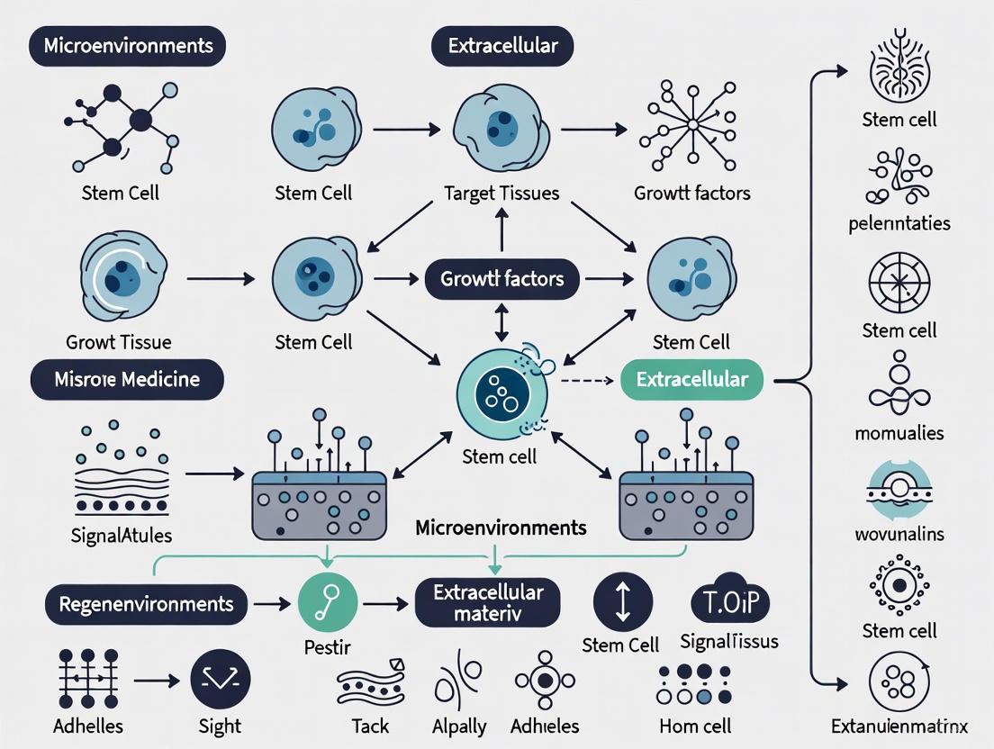
Strategic Approaches to Enhance Stem Cell Retention: From Molecular Mechanisms to Clinical Translation
This article provides a comprehensive analysis of evidence-based strategies to improve stem cell retention in target tissues, a critical challenge limiting the therapeutic efficacy of regenerative medicine.
Strategic Approaches to Enhance Stem Cell Retention: From Molecular Mechanisms to Clinical Translation
Abstract
This article provides a comprehensive analysis of evidence-based strategies to improve stem cell retention in target tissues, a critical challenge limiting the therapeutic efficacy of regenerative medicine. Targeting researchers, scientists, and drug development professionals, we synthesize foundational science on homing mechanisms with cutting-edge methodological advances in bioengineering and delivery systems. The content explores the molecular drivers of stem cell migration and adhesion, evaluates innovative retention-enhancing technologies from nanotechnology to bioreactor processes, and offers troubleshooting frameworks for overcoming physiological barriers. A critical validation segment compares administration routes, tracking methodologies, and clinical trial outcomes, providing a holistic resource for optimizing stem cell therapies from bench to bedside.
The Stem Cell Retention Imperative: Unraveling Homing Mechanisms and Pharmacokinetic Principles
Fundamental Concepts: What is Stem Cell Retention?
How is stem cell retention defined in therapeutic contexts?
Stem cell retention refers to the percentage of administered therapeutic cells that remain viable and engrafted within the target tissue over a specified period post-transplantation. It encompasses three critical phases: initial delivery and localization to the target site, short-term engraftment and survival against immediate hostile microenvironments, and long-term persistence and functional integration into host tissue.
High retention is crucial because it directly correlates with therapeutic efficacy; a higher number of functionally active cells at the injury site enhances paracrine signaling, tissue regeneration, and structural repair [1] [2]. The entire process is a race against time, where cells must survive hypoxia, inflammation, and anoikis (detachment-induced cell death) to successfully engraft [1].
What key biological barriers impede stem cell retention?
Multiple biological barriers significantly limit stem cell retention, often reducing engraftment to less than 5% in many applications [1]. The primary barriers include:
- Hostile Microenvironment: Characterized by hypoxia, nutrient deprivation, and excessive inflammatory cytokines at the target site, triggering oxidative stress and anoikis [1].
- Physical Clearance: Systemically delivered cells often become trapped in capillary beds, particularly in the lungs, liver, and spleen, preventing them from reaching the intended tissue [1].
- Immune Rejection: Both allogeneic and, in some cases, autologous cells can face immune-mediated clearance by the host's immune system [3].
- Lack of Survival Signals: The absence of appropriate ECM contact and trophic factors in the transplantation site leads to programmed cell death [4].
The following diagram illustrates the sequential biological barriers that stem cells encounter from administration to engraftment.
Troubleshooting Low Stem Cell Retention: A Practical Guide
How can I troubleshoot low cell retention in my animal models?
When facing low stem cell retention in preclinical models, systematically investigate these common failure points:
Problem: Rapid Cell Death Post-Transplantation
- Potential Causes: Hostile transplantation microenvironment (hypoxia, inflammation), inadequate preconditioning, improper cell handling during preparation.
- Solutions: Implement pharmacological preconditioning with compounds like α-ketoglutarate to enhance antioxidant defenses [1]. Use cytokine preconditioning (IL-1β, TGF-β1) to upregulate pro-survival genes [1]. Ensure cells are in optimal metabolic state pre-transplantation.
Problem: Poor Initial Engraftment
- Potential Causes: Suboptimal delivery technique, excessive injection pressure causing tissue damage and backflow, inappropriate cell carrier medium.
- Solutions: Utilize fibrin, collagen, or hyaluronic acid-based hydrogels as protective carriers to increase initial cell anchoring [1] [2]. Optimize injection parameters (volume, rate, needle gauge) for your target tissue. For systemic delivery, consider strategies to temporarily reduce pulmonary entrapment.
Problem: Inadequate Long-Term Persistence
- Potential Causes: Immune rejection, insufficient vascularization at transplant site, lack of appropriate ECM support.
- Solutions: Utilize biomaterial scaffolds that provide sustained release of angiogenic factors (VEGF, FGF-2) to promote neovascularization [2]. For allogeneic applications, consider immune modulation strategies or encapsulation approaches [3].
What quantitative methods are available for tracking stem cell retention?
Accurately quantifying stem cell retention requires multiple complementary approaches. The table below summarizes the primary methods, their applications, and limitations.
Table: Quantitative Methods for Assessing Stem Cell Retention
| Method | Application Context | Key Readouts | Technical Limitations |
|---|---|---|---|
| Bioluminescence Imaging (BLI) | Small animal tracking | Cell viability, spatial distribution over time | Semi-quantitative, limited depth penetration, requires genetic modification |
| Fluorescence Imaging | Small animal & explant analysis | Cell location, viability with vital dyes | Limited depth penetration, photo-bleaching |
| qPCR (Species-Specific) | Human cells in animal models | Absolute cell number via genomic DNA | Requires tissue sacrifice, no spatial information |
| Flow Cytometry (Cell Surface Markers) | Tissue dissociates | Cell viability, phenotype | Requires tissue processing, potential marker loss |
| Histology & Immunostaining | Tissue sections | Spatial distribution, cell-matrix interactions | Semi-quantitative, sampling bias |
For optimal results, combine one in vivo imaging method (e.g., BLI) with one endpoint quantification method (e.g., qPCR) to obtain both temporal dynamics and absolute cell numbers [1].
Engineering Solutions to Enhance Stem Cell Retention
What biomaterial strategies can improve retention?
Biomaterial-based approaches represent the most promising strategy for enhancing stem cell retention by creating protective microenvironments. The "bottom-up" design philosophy prioritizes the fundamental biological needs of stem cells when engineering these materials [4].
Table: Biomaterial Strategies for Enhancing Stem Cell Retention
| Strategy | Mechanism of Action | Target Application | Reported Efficacy |
|---|---|---|---|
| Hydrogel Encapsulation | Physical protection, ECM-mimetic signals | Local delivery to wounds, myocardial infarction | 3-5x increase in initial retention [1] |
| 3D Bioprinted Scaffolds | Spatial organization, mechanical support | Cartilage/bone repair, complex tissue engineering | Improved spatial patterning & long-term function [2] |
| Microsphere Carriers | Sustained trophic factor release | Ischemic tissue, chronic wounds | Enhanced angiogenesis & cell survival [2] |
| Nanofiber Meshes | Topographical guidance, large surface area | Nerve regeneration, skin wounds | Better cell infiltration & tissue integration [4] |
These biomaterials function by replicating critical aspects of the native stem cell niche, including mechanical cues (stiffness, topography), biochemical signals (adhesive ligands, growth factors), and spatial organization [4]. The protective effect is particularly crucial in hostile environments like chronic wounds, where biomaterials can shield MSCs from inflammatory cytokines while facilitating paracrine signaling [1] [2].
How does genetic engineering enhance cell survival and retention?
Genetic modification techniques can directly enhance stem cell resilience to hostile microenvironments. The following diagram illustrates the key genetic engineering approaches and their protective mechanisms.
These genetic approaches work synergistically with biomaterial strategies to address multiple retention barriers simultaneously. For example, MSCs engineered to overexpress VEGF not only survive better but also promote local angiogenesis, creating a more supportive niche for long-term persistence [1] [2].
Essential Reagents and Protocols for Retention Studies
What are the key reagents for stem cell retention research?
The following reagent toolkit is essential for designing and executing robust stem cell retention studies.
Table: Essential Research Reagent Solutions for Stem Cell Retention Studies
| Reagent Category | Specific Examples | Function in Retention Studies |
|---|---|---|
| Cell Tracking Dyes | CM-Dil, CFSE, PKH26, GFP/Luciferase lentivirus | Cell labeling for in vivo tracking and quantification |
| Viability Stains | Trypan Blue, Erythrosin B, Calcein-AM/EthD-1 | Assessment of cell viability pre-transplantation and in explants |
| Hydrogel Matrices | Fibrin, Collagen I, Hyaluronic acid, Matrigel | 3D cell delivery vehicles providing protective microenvironment |
| Cytokine Cocktails | IL-1β, TGF-β1, IFN-γ, TNF-α | Preconditioning to enhance stress resistance and paracrine function |
| Apoptosis Inhibitors Z-VAD-FMK, Q-VD-OPh | Testing the role of cell death pathways in poor retention | |
| Integrin-Activating Peptides | RGD, LDV, GFOGER | Enhancing cell-matrix adhesion and preventing anoikis |
For automated cell counting, which is critical for standardizing cell doses in retention studies, consider using systems like the Luna family of cell counters, which offer consistent viability assessment and reduce the subjectivity of manual counting [5].
What is a standard protocol for assessing stem cell retention?
Protocol: Quantitative Assessment of Stem Cell Retention Using Bioluminescence Imaging and qPCR
Day 1: Cell Preparation and Labeling
- Genetic Modification: Transduce stem cells with lentivirus encoding both firefly luciferase and GFP reporters 72-96 hours pre-transplantation.
- Validation: Confirm reporter expression via fluorescence microscopy and luciferase activity assay.
- Cell Preparation: Harvest cells using gentle dissociation reagent (e.g., ReLeSR) to maintain viability and minimize differentiation [6].
- Formulation: Resuspend cells in appropriate carrier (PBS for control, hydrogel for test group) at 10× the final concentration.
Day 2: Cell Transplantation
- Animal Preparation: Anesthetize and position animals for procedure.
- Cell Administration: For local delivery, inject 10-20μL containing 1×10^6 cells slowly over 60 seconds; wait 30 seconds before needle withdrawal to prevent backflow. For systemic delivery, inject 100-200μL via tail vein.
- Imaging Baseline: Acquire initial bioluminescence image 2-4 hours post-transplantation to establish 100% reference value.
Days 3-28: Longitudinal Tracking
- Image Acquisition: Anesthetize animals, administer D-luciferin substrate (150mg/kg IP), and acquire bioluminescence images at days 1, 3, 7, 14, and 28 using standardized imaging parameters.
- Quantification: Analyze images using region-of-interest (ROI) analysis to calculate total flux (photons/second) normalized to day 0 baseline.
Endpoint Analysis (Day 28)
- Tissue Collection: Harvest target tissues and immediately freeze in liquid nitrogen for DNA/RNA extraction.
- qPCR Quantification: Isolate genomic DNA and perform qPCR using human-specific Alu sequences (for human cells in rodent models) to determine absolute cell numbers.
- Histological Validation: Process adjacent tissue sections for GFP immunohistochemistry to confirm spatial distribution.
Key Calculations:
- Bioluminescence Retention (%) = (Day X flux ÷ Day 0 flux) × 100
- Genomic Retention (%) = (Human cells recovered ÷ Human cells injected) × 100
This combined approach provides both temporal dynamics (BLI) and absolute quantification (qPCR) for comprehensive retention assessment [1].
Frequently Asked Questions (FAQs)
What is the typical range of stem cell retention in current therapies?
Retention rates vary significantly by delivery method and target tissue. Local injection typically achieves 5-20% initial retention, declining to 1-5% by 1-4 weeks post-transplantation [1]. Systemic delivery results in much lower retention, often <1% in target tissues, with the majority of cells sequestered in lungs, liver, and spleen. Engineered approaches (biomaterials + preconditioning) can improve these rates by 3-5 fold, but long-term retention remains a challenge.
How does stem cell source (MSCs, iPSCs, ESCs) impact retention?
Different stem cell types face distinct retention challenges:
- MSCs: Exhibit moderate innate retention but are highly susceptible to anoikis without matrix support. Their retention benefits significantly from biomaterial encapsulation [1] [2].
- iPSCs: Face potential immune recognition despite autologous origin due to epigenetic abnormalities. Risk of teratoma formation complicates long-term retention goals [4] [7].
- ESCs: Pose significant tumorigenicity concerns, requiring complete differentiation pre-transplantation, which alters their adhesion and retention properties [3] [8].
What are the critical quality control checkpoints for maximizing retention?
Implement these checkpoints in your workflow:
- Pre-transplantation Viability: Ensure >90% viability via automated cell counting with stains like Erythrosin B [5].
- Phenotypic Characterization: Verify expression of appropriate surface markers and differentiation potential.
- Microbiological Safety: Screen for mycoplasma and endotoxins per regulatory guidelines [3].
- Functional Potency: Assess paracrine factor secretion (VEGF, IL-6) or migration capacity as retention predictors.
How do regulatory guidelines impact retention enhancement strategies?
Regulatory bodies require rigorous preclinical evaluation of safety and efficacy for substantially manipulated cells [3]. For non-homologous use (cells performing different functions than in their native tissue), comprehensive risk-benefit assessment is mandatory. Any genetic modification or combination with biomaterials typically requires additional regulatory oversight as an Advanced Therapy Medicinal Product (ATMP) [3].
What emerging technologies show promise for improving retention?
The most promising approaches include:
- Multimodal Engineering: Combining genetic modification (survival enhancement), biomaterials (physical protection), and preconditioning (metabolic priming) [1] [2].
- Dynamic Biomaterials: "Smart" scaffolds that release bioactive factors in response to microenvironmental cues [4].
- Microenvironment Reprogramming: Transient modulation of the target tissue to make it more receptive to engraftment before cell delivery.
- Advanced Imaging Technologies: Real-time tracking of cell fate and function post-transplantation to better understand retention dynamics.
The process of cellular homing is a fundamental biological mechanism that enables specific cells, such as immune cells and therapeutic stem cells, to navigate through the bloodstream and migrate into target tissues. This sophisticated journey involves a precisely coordinated sequence of molecular interactions often described as a multi-step cascade. For researchers aiming to improve stem cell retention in target tissues, a deep understanding of this cascade is paramount, as inefficiencies at any step significantly limit therapeutic efficacy.
The homing process is universally described as involving several distinct stages: (1) tethering and rolling of cells on the vascular endothelium, (2) activation by chemoattractants, (3) firm adhesion to the endothelium, (4) transmigration (or diapedesis) across the endothelial barrier, and (5) extravascular migration toward the final target within the tissue [9] [10] [11]. Each step is mediated by specific families of molecules—selectins, chemokines, and integrins—working in concert. This guide addresses the common experimental challenges researchers face when studying this process and provides targeted troubleshooting advice to enhance the reliability and translational impact of your findings.
Troubleshooting Guides
Poor Cell Rolling and Initial Tethering
Problem: Cells fail to tether and roll efficiently on the endothelial surface under flow conditions, leading to low initial capture rates.
Background: The rolling step is primarily mediated by selectins (L-, P-, and E-selectin) and their glycoprotein ligands [11] [12]. This interaction slows down circulating cells, allowing them to sense signals from the endothelium.
Solution:
- Verify Selectin Ligand Expression: Ensure your cells express functional selectin ligands (e.g., PSGL-1, CD44). For mesenchymal stem cells (MSCs), which may not express classic ligands, check for alternative ligands like galectin-1 or CD24 [9]. Use flow cytometry with specific antibodies for confirmation.
- Check Endothelial Addressins: Confirm that your activated endothelial cells or High Endothelial Venules (HEVs) express the appropriate addressins, such as Peripheral Node Addressin (PNAd) or other selectin ligands [11]. Static adhesion assays can be misleading; implement flow chamber assays to best mimic physiological shear stress.
- Optimize Shear Stress: The initial tethering is highly sensitive to shear flow. In flow chamber systems, test a range of physiological shear stresses (e.g., 0.5 - 4 dyn/cm²) to identify optimal conditions.
Inadequate Firm Adhesion Post-Activation
Problem: Cells roll but fail to arrest and form firm adhesion on the endothelium.
Background: Rolling enables cells to encounter endothelial chemokines. Chemokine binding to their G-protein coupled receptors (GPCRs) triggers intracellular "inside-out" signaling that rapidly activates integrins, shifting them to a high-affinity state for their ligands [13] [12]. Firm adhesion is primarily mediated by these activated integrins (e.g., VLA-4/α4β1, LFA-1/αLβ2) binding to immunoglobulin superfamily members (e.g., VCAM-1, ICAM-1) on the endothelium [13] [14].
Solution:
- Confirm Chemokine Receptor Expression: Validate that your cells express the relevant receptors (e.g., CXCR4 for SDF-1, CCR7 for CCL19/21) [13] [15]. Pertussis toxin, which inhibits Gi-protein coupled receptor signaling, can be used as a control to abrogate chemokine-mediated integrin activation.
- Assess Integrin Activation Status: Integrins can be present but inactive. Use conformation-specific antibodies (e.g., MEM148 for extended αLβ2) to distinguish between inactive and active states via flow cytometry [12].
- Prime Cells with Cytokines: Pre-treat (prime) MSCs with pro-inflammatory cytokines (e.g., TNF-α, IFN-γ) to upregulate the expression and affinity of adhesion molecules like VLA-4 [16].
Failure in Transmigration and Tissue Entry
Problem: Cells adhere firmly to the endothelium but do not transmigrate into the target tissue.
Background: The final step, transmigration, can occur either paracellularly (between endothelial cells) or transcellularly (through them). This process involves further integrin engagement (e.g., with JAM-A/B) and the action of matrix metalloproteinases (MMPs) to remodel the extracellular matrix and basement membrane [13] [17].
Solution:
- Check for Constitutively Active Integrins: Note that cells expressing constitutively active integrins (e.g., due to mutations in the α-subunit cytoplasmic tail) may exhibit enhanced adhesion but impaired transmigration due to failure in adhesion de-adhesion cycles [13].
- Inhibit Key Enzymes: Test the effect of broad-spectrum MMP inhibitors (e.g., GM6001) on your system. A significant reduction in transmigration indicates that MMP-mediated ECM degradation is a critical factor.
- Validate Chemokine Gradients: A stable chemokine gradient (e.g., SDF-1) is crucial for directing transmigration and parenchymal migration. Use transwell migration assays to confirm your cells' chemotactic response. Ensure that the target tissue expresses the appropriate chemokines (e.g., CCL25, CXCL12) [15] [14].
Frequently Asked Questions (FAQs)
FAQ 1: What are the key molecular pairs for tissue-specific homing? Different tissues express unique combinations of addressins and chemokines, creating a "postal code" system for homing. The table below summarizes the principal receptor-ligand pairs for major target tissues.
Table 1: Key Receptor-Ligand Pairs in Tissue-Specific Homing
| Target Tissue | Homing Receptor (Cell) | Ligand/Addressin (Endothelium) | Key Chemokine Axis |
|---|---|---|---|
| Peripheral Lymph Nodes | L-selectin (CD62L), αLβ2 (LFA-1) | PNAd, ICAM-1 | CCR7/CCL19, CCL21 [13] [18] |
| Gut & Mucosal Sites | α4β7 Integrin | MAdCAM-1 | CCR9/CCL25, CXCR3/CXCL10 [13] [18] |
| Inflamed Tissues | α4β1 (VLA-4), LFA-1 | VCAM-1, ICAM-1 | Multiple (e.g., CXCR2/CXCL1) [13] [14] |
| Bone Marrow | CD44, VLA-4, LFA-1 | Hyaluronic acid, VCAM-1, ICAM-1 | CXCR4/CXCL12 (SDF-1) [15] [17] |
FAQ 2: Why do systemically administered MSCs have low homing efficiency? Despite their therapeutic potential, a major bottleneck in MSC therapy is that after systemic infusion, only a small percentage (1-2%) of cells successfully home to the target site [10] [16]. This is due to a combination of factors: (1) physical trapping in the lung capillaries, (2) lack of expression of critical homing receptors like L-selectin or specific integrins, (3) exposure to shear forces that can cause anoikis, and (4) the absence of a strong inflammatory signal in the target tissue to upregulate the necessary adhesion molecules and chemokines [9] [10] [16].
FAQ 3: What are the main strategies to improve homing efficiency for cell therapies? Researchers are developing multiple strategies to overcome homing barriers, including:
- In vitro Priming: Pre-incubating cells with cytokines (e.g., TNF-α, IFN-γ) or growth factors to upregulate the expression of homing receptors (e.g., CXCR4, VLA-4) [9] [16].
- Genetic Modification: Engineering cells to overexpress specific homing receptors (e.g., CXCR4) or adhesion molecules (e.g., α4 integrin) to enhance their targeting capability [9] [10] [16].
- Target Tissue Modification: Using ultrasound or local irradiation to induce a localized inflammatory state that upregulates addressins and chemokines (e.g., SDF-1) in the target tissue, creating a stronger homing signal [10] [15].
- Cell Surface Engineering: Chemically modifying the cell surface to conjugate ligands or antibodies that direct them to specific vascular markers [10].
Experimental Protocols & Methodologies
In Vitro Flow Chamber Adhesion Assay
This protocol is critical for quantitatively analyzing the rolling and firm adhesion steps under physiological shear conditions.
Key Reagents and Materials:
- Ibidi or GlycoTech parallel plate flow chamber system
- Peristaltic or syringe pump
- Phase-contrast or fluorescence microscope with high-speed camera
- Recombinant adhesion proteins (e.g., ICAM-1-Fc, P-/E-selectin)
- Recombinant chemokines (e.g., CXCL1, SDF-1)
Procedure:
- Surface Coating: Coat a culture dish or ibidi slide by immobilizing a combination of selectins (e.g., 1-10 µg/mL P-selectin) and chemokines (e.g., 0.1-1 µg/mL CXCL1) on a background of ICAM-1-Fc (e.g., 10 µg/mL). Incubate overnight at 4°C [12].
- Cell Preparation: Resuspend your cells (e.g., MSCs, T cells) in a defined assay buffer (e.g., HBSS with Ca²⁺/Mg²⁺ and 0.5% HSA) at a concentration of 0.5-1 × 10⁶ cells/mL.
- Perfusion and Data Acquisition: Mount the coated slide in the flow chamber. Perfuse the cell suspension at a defined wall shear stress (e.g., 1-2 dyn/cm²). Record multiple fields of view for at least 5-10 minutes.
- Data Analysis:
- Rolling Velocity: Track the distance moved by individual cells over time.
- Firm Adhesion: Count the number of cells that remain stationary for a defined period (e.g., >10 seconds) at the end of the perfusion period.
Analyzing Integrin Activation by Flow Cytometry
Monitoring the conformational change of integrins is essential for diagnosing firm adhesion failures.
Key Reagents:
- Conformation-specific antibodies (e.g., KIM127 for β2 extension, mAb24 for high-affinity αLβ2)
- Isotype control antibodies
- Flow cytometry buffer (PBS + 1% BSA + Ca²⁺/Mg²⁺)
- Chemokine stimulant (e.g., 100 ng/mL CXCL1)
Procedure:
- Stimulation: Divide your cells into aliquots. Stimulate one aliquot with a relevant chemokine for 5-10 minutes at 37°C. Keep another aliquot unstimulated as a control.
- Staining: Immediately after stimulation, fix the cells with a low concentration of paraformaldehyde (e.g., 1-2%) to preserve integrin conformation. Avoid over-fixation.
- Antibody Incubation: Stain the fixed cells with the fluorochrome-conjugated conformation-specific antibody and corresponding isotype control on ice for 30-60 minutes.
- Analysis: Analyze by flow cytometry. A positive shift in fluorescence in the stimulated sample compared to the unstimulated control indicates successful integrin activation [12].
Signaling Pathways in the Homing Cascade
The following diagram illustrates the core signaling pathways that are activated during the chemokine-mediated activation step, leading to integrin activation and firm adhesion. This is a consolidated pathway relevant to neutrophils and lymphocytes, and aspects are applicable to MSCs.
Diagram 1: Core signaling for integrin activation during homing. Chemokine and selectin engagement trigger parallel Rap1a and PIP5Kγ90 pathways that converge to recruit and activate Talin-1. Talin binding to the integrin β-tail induces a conformational shift to a high-affinity state, enabling firm adhesion. PI3Kγ cooperates with Rap1a specifically in chemokine signaling.
The Scientist's Toolkit: Essential Research Reagents
Table 2: Key Reagents for Investigating the Homing Cascade
| Reagent / Tool | Primary Function | Example Use Case | Considerations |
|---|---|---|---|
| Recombinant Selectins & ICAM-1/VCAM-1 | Coating for static/flow adhesion assays. Mimics endothelial surface. | Testing the rolling and firm adhesion potential of cells in vitro. | Ensure proper glycosylation of ligands for functional selectin interactions. |
| Recombinant Chemokines (SDF-1, CCL19, CXCL1) | Activate chemokine receptors to trigger inside-out signaling. | Priming cells or creating gradients in migration/transwell assays. | Use a range of concentrations; check receptor expression on target cells first. |
| Conformation-Specific Anti-Integrin Antibodies | Detect active vs. total integrin levels via flow cytometry. | Diagnosing activation deficits after chemokine stimulation. | Compare to isotype control and unstimulated cells. Requires non-permeabilizing conditions. |
| Pertussis Toxin (PTx) | Inhibits Gi-protein coupled receptor signaling. | Negative control to confirm chemokine signaling is Gi-dependent. | Can have broad effects; use at validated concentrations. |
| CXCR4 Antagonist (e.g., AMD3100) | Blocks SDF-1/CXCR4 axis. | Validating the role of this key homing axis in vivo or in vitro. | Can mobilize stem cells from bone marrow, affecting circulating numbers. |
| Rap1a GTPase Inhibitors | Interfere with a central signaling node for integrin activation. | Probing the role of Rap1a in the adhesion cascade. | Specificity can be an issue; consider genetic knockdown/knockout as alternative. |
| Function-Blocking mAbs (Anti-α4, Anti-LFA-1, Anti-VCAM-1) | Block specific receptor-ligand interactions. | Identifying which molecular pair is critical for adhesion in your system. | Use isotype-matched antibody controls. Effects can be context-dependent. |
| MMP Inhibitors (e.g., GM6001) | Block matrix metalloproteinase activity. | Assessing the role of ECM degradation in transmigration. | Can be broad-spectrum; newer, more specific inhibitors are available. |
For researchers focused on improving stem cell retention in target tissues, cellular senescence presents a fundamental barrier. The gradual decline of stem cell function is a hallmark of aging, directly impacting the efficacy of regenerative therapies. Two core biological mechanisms are implicated: the erosion of telomeres, the protective caps at chromosome ends that serve as a mitotic clock, and the accumulation of senescent cells secreting a potent mix of factors known as the Senescence-Associated Secretory Phenotype (SASP) [19] [20] [21]. This technical support article provides a targeted guide for scientists to troubleshoot the specific issues these processes create in stem cell research, offering practical protocols and solutions to enhance the translational potential of their work.
Core Concepts: SASP and Telomere Dynamics
What is the Senescence-Associated Secretory Phenotype (SASP)?
Cellular senescence is a state of stable cell cycle arrest. However, senescent cells remain metabolically active and secrete a complex mixture of factors collectively known as the SASP [19] [22]. This phenotype is highly pleiotropic, meaning it can have diverse and often contradictory effects depending on the context.
- Composition: The SASP includes pro-inflammatory cytokines (e.g., IL-6, IL-1β), chemokines (e.g., IL-8), growth factors, and proteases (e.g., MMPs) [22].
- Dual Nature: The SASP can have beneficial roles, such as in wound healing and tumor suppression, but its chronic presence in aging tissues is largely deleterious [19] [22]. It can transmit senescence to neighboring healthy cells, create a chronic inflammatory microenvironment, and alter stem cell fate decisions, thereby impairing tissue regeneration and stem cell function [19] [23].
What are Telomere Dynamics?
Telomeres are nucleoprotein structures comprising repetitive TTAGGG sequences that protect the ends of chromosomes from degradation and fusion [20] [21].
- Telomere Attrition: With each somatic cell division, telomeres shorten by 50–200 base pairs due to the "end-replication problem," where DNA polymerase cannot fully replicate the lagging strand [20] [21]. Oxidative stress can also directly damage telomeric DNA, accelerating this shortening [21].
- Cellular Senescence: When telomeres reach a critically short length, they trigger a persistent DNA damage response, leading to irreversible cell cycle arrest known as replicative senescence [20] [21].
- Telomerase: The enzyme telomerase, composed of TERT and TERC, can counteract attrition by adding telomeric repeats de novo. However, its activity is low or absent in most human somatic cells, leading to gradual telomere erosion with age [21].
The Scientist's Toolkit: Key Research Reagents
The following table details essential reagents for studying senescence and telomere biology in the context of stem cell retention.
Table 1: Key Research Reagents for Senescence and Telomere Studies
| Reagent Category | Specific Examples | Primary Function in Research |
|---|---|---|
| SASP Neutralizing Antibodies | Anti-IL-6, Anti-IL-1β | Block the activity of specific SASP factors to dissect their individual contributions to stem cell dysfunction [22]. |
| Senolytics | Dasatinib, Quercetin, Fisetin | Selectively induce apoptosis in senescent cells, allowing researchers to clear them from cultures or in vivo models [22]. |
| JAK/STAT Inhibitors | Ruxolitinib (JAK1/2 inhibitor) | Suppress the expression of multiple SASP components, mitigating the inflammatory secretome's paracrine effects [22]. |
| Telomerase Activators | TERT gene therapy, small-molecule activators (e.g., TA-65) | Enhance telomerase activity to lengthen or maintain telomeres, potentially delaying replicative senescence in stem cells [21]. |
| Pharmacological Preconditioning Agents | α-ketoglutarate, Caffeic acid, Lipopolysaccharide (LPS) | Enhance MSC viability, paracrine activity, and stress resistance under hostile conditions like hypoxia [24]. |
| Cytokine Preconditioning Cocktails | IL-1β, IFN-γ, TNF-α, TGF-β1 | Modulate MSC gene expression to improve migratory capacity, immunomodulation, and post-transplantation survival [24]. |
| Biomaterial Scaffolds | Collagen-based hydrogels, 3D-bioprinted constructs | Provide structural and biochemical support for delivered stem cells, improving engraftment and retention at the target site [24] [2]. |
Troubleshooting Guides & FAQs
Troubleshooting Poor Stem Cell Retention and Engraftment
Table 2: Troubleshooting Poor Stem Cell Retention and Engraftment
| Problem | Potential Cause | Recommended Solution |
|---|---|---|
| Low cell survival post-delivery | Hostile target microenvironment (hypoxia, inflammation); Anoikis (detachment-induced death). | Preconditioning: Use hypoxic or cytokine preconditioning (e.g., with TGF-β1) to enhance stress resistance [24]. Scaffold Delivery: Utilize hydrogels or biomaterial scaffolds to provide 3D support and mimic the ECM [24] [2]. |
| Rapid loss of stemness & proliferative capacity | Replicative senescence due to telomere shortening in vitro; Exposure to SASP from resident senescent cells. | Telomere Monitoring: Regularly assess telomere length (e.g., via qPCR) in your cell stocks. Use low-passage cells. Senolytic Treatment: Pre-treat the target site or co-deliver senolytics to clear the host SASP microenvironment [22] [21]. |
| Inadequate migration to target site | Impaired homing ability due to suboptimal cell source or culture; Inflammatory barriers. | Source Selection: Use MSCs with higher inherent motility (e.g., UC-MSCs over older donor AT-MSCs) [2]. Cytokine Priming: Precondition with IL-1β to upregulate homing-related genes like MMP-3 [24]. |
| Paracrine therapeutic failure | Diminished secretome potency due to donor age or senescence. | SASP Suppression: Culture cells with a JAK inhibitor to reduce secretion of inflammatory factors that impair paracrine signaling [22]. Exosome Therapy: Shift to using MSC-derived exosomes, which retain therapeutic cargo and are resistant to senescence-related issues [25]. |
Frequently Asked Experimental Questions
Q1: How can I quantitatively assess the impact of the local microenvironment on the senescence of my delivered stem cells?
A: A robust protocol involves:
- In Vivo Senescence Detection: After delivering your stem cells, retrieve the target tissue at various time points.
- Histological Staining: Perform co-staining for senescence biomarkers on tissue sections.
- Quantification: Use image analysis software to quantify the percentage of delivered cells (e.g., via a fluorescent cell tracker) that are positive for these senescence markers. An increase over time directly links the microenvironment to induced senescence.
Q2: My MSCs show reduced proliferation and altered morphology in later passages. Is this telomere-driven senescence, and how can I confirm it?
A: This is a classic sign of replicative senescence. To confirm telomere involvement:
- Measure Telomere Length:
- Assay for Telomerase Activity:
- Method: Use the Telomeric Repeat Amplification Protocol (TRAP) assay.
- Expected Outcome: Most adult somatic MSCs have low or undetectable telomerase activity. Consistent absence confirms that shortening is due to the end-replication problem.
- Correlate with Functional Assays: Link telomere length data with functional outcomes like population doubling time and SA-β-gal activity to build a comprehensive picture.
Q3: What is the most effective strategy to protect stem cells from the paracrine effects of SASP in an aged animal model?
A: A combination strategy is often most effective, targeting both the host environment and the therapeutic cells.
- Strategy 1: Host Pre-conditioning. Administer a senolytic cocktail (e.g., Dasatinib + Quercetin) to the animal model for 1-2 weeks prior to stem cell delivery. This clears a significant portion of host senescent cells and reduces the systemic SASP load [22].
- Strategy 2: Cell Engineering. Engineer your stem cells to overexpress anti-apoptotic genes (e.g., BCL-2) or use pharmacological preconditioning (e.g., with α-ketoglutarate) to enhance their resistance to SASP-induced stress and apoptosis [24].
- Strategy 3: Localized SASP Suppression. Deliver the stem cells encapsulated in a hydrogel loaded with a JAK/STAT inhibitor (e.g., Ruxolitinib). This allows for localized, sustained release of the drug, neutralizing the SASP in the immediate vicinity of the graft without systemic effects [22].
Key Experimental Protocols & Data
Protocol: Cytokine Preconditioning to Enhance MSC Resilience
Objective: To enhance MSC survival, migratory capacity, and paracrine function prior to transplantation by mimicking inflammatory signals [24].
Materials:
- Human MSCs (e.g., BM-MSCs or UC-MSCs) at 70-80% confluence.
- Standard MSC growth medium.
- Preconditioning medium: Growth medium supplemented with a cytokine cocktail (e.g., 10 ng/mL IFN-γ + 10 ng/mL TNF-α) [24].
- Control medium: Growth medium with an equivalent volume of PBS.
Method:
- Cell Preparation: Seed MSCs at a standardized density (e.g., 5,000 cells/cm²).
- Preconditioning: After 24 hours, replace the medium with either preconditioning medium or control medium.
- Incubation: Incubate cells for 24-48 hours under standard culture conditions (37°C, 5% CO₂).
- Harvesting: After incubation, wash cells with PBS and harvest using standard trypsinization.
- Validation: Before transplantation, validate efficacy via:
Table 3: Quantitative Effects of Preconditioning on MSC Properties
| Preconditioning Agent | Effect on Migration | Effect on Anti-inflammatory Gene Expression | Key Signaling Pathways Involved |
|---|---|---|---|
| IL-1β (10 ng/mL) | ↑↑ (via MMP-3 upregulation) [24] | Moderate Increase | NF-κB |
| IFN-γ + TNF-α (10 ng/mL each) | Moderate Increase | ↑↑ (CCL2, IL-6) [24] | JAK/STAT, NF-κB |
| TGF-β1 (5-10 ng/mL) | Variable | ↑ (Tissue Repair Factors) [24] | SMAD |
| Hypoxia (1-3% O₂) | ↑ (Homing) [24] | ↑ (VEGF, HIF-1α) [24] | HIF-1α |
Visualizing the Senescence-Stem Cell Retention Axis
The following diagram illustrates the core mechanisms by which SASP and telomere attrition converge to impair stem cell retention and function.
Diagram Title: SASP and Telomere Pathways Impair Stem Cell Retention
Overcoming the barriers imposed by cellular senescence and telomere dynamics requires a multi-faceted approach. Success in improving stem cell retention hinges on integrating insights from both cell-intrinsic replicative limits and cell-extrinsic microenvironmental pressures. By employing the troubleshooting guides, utilizing preconditioning protocols, and strategically deploying senolytics and targeted inhibitors, researchers can design more resilient stem cell-based therapies. The future of this field lies in creating "senescence-resistant" therapeutic cells and "senescence-cleared" recipient environments to fully unlock the regenerative potential of stem cells for treating age-related diseases.
Understanding the journey of Mesenchymal Stem Cells (MSCs) after administration—where they go (biodistribution), how long they survive (persistence), and how they are removed (clearance)—is fundamental to developing effective regenerative treatments. These pharmacokinetic properties directly influence the safety and efficacy of MSC-based therapies. The central challenge in the field is that while MSCs are known for their immunomodulatory abilities and potential to differentiate into various tissue types, efficacy in clinical trials has been inconsistent. [26] A significant factor contributing to this inconsistency is poor cell retention and survival in the target tissues after transplantation. This technical support center provides targeted guidance to help researchers troubleshoot the critical experimental hurdles in tracking and improving the pharmacokinetic profiles of MSCs.
Troubleshooting Guides & FAQs
Tracking and Quantification
Q: What are the primary methods for tracking MSC biodistribution in vivo, and how do I choose?
The choice of tracking method depends on your research question, model system, and required sensitivity. The table below compares the most common techniques.
| Method | Principle | Key Advantage | Key Limitation | Optimal Use Case |
|---|---|---|---|---|
| Imaging Flow Cytometry [27] | Combines high-throughput flow cytometry with single-cell image acquisition. | Provides spatial information and morphology for cells in suspension. | Limited to ex vivo analysis of dissociated tissues. | Quantifying and visualizing MSCs in heterogeneous cell suspensions from blood or organs. |
| LC-MS/MS Proteomics [28] | Detects and quantifies unique human proteins from transplanted MSCs in animal tissues. | High sensitivity and specificity; avoids label-dependent signal loss. | Requires specialized instrumentation and complex sample preparation. | Highly sensitive, quantitative tracking of MSC persistence in specific organs over time. |
| Bioluminescence Imaging (BLI) | Measures light emission from cells expressing luciferase enzymes. | Enables non-invasive, whole-body longitudinal tracking. | Signal is attenuated in deep tissues and requires genetic modification of cells. | Real-time, non-invasive monitoring of overall MSC biodistribution and clearance. |
| Quantitative PCR (qPCR) | Amplifies species-specific DNA sequences. | Highly sensitive and quantitative; does not require cell viability. | Cannot distinguish between live and dead cells. | Detecting low levels of human MSCs in animal models using human-specific Alu repeats. |
Troubleshooting Common Tracking Issues:
- Problem: Rapid loss of tracking signal.
- Solution: This often indicates rapid cell death post-transplantation. Focus on improving delivery methods (see Section 2) and ensure your tracking method (e.g., LC-MS/MS for proteins [28]) is not dependent on cell viability if quantifying early clearance.
- Problem: High background noise in imaging.
- Solution: For imaging flow cytometry, ensure proper compensation for spectral cross-talk at a pixel level and titrate your antibodies or dyes carefully to maximize the signal-to-noise ratio. [27]
Q: How can I improve the sensitivity of detecting MSCs in low-abundance tissues?
- Sample Preparation is Key: When using mass spectrometry, employ sample pre-processing to deplete high-abundance host proteins and enrich for low-abundance targets. This dramatically improves the depth and reliability of analysis. [28]
- Use Targeted Assays: In the validation phase, transition from untargeted discovery to targeted MS techniques like Parallel Reaction Monitoring (PRM), which offers high precision, low limits of detection, and absolute quantification using stable isotope-labeled standards. [28]
Cell Retention and Survival
Q: My MSCs show low retention in the target tissue. What strategies can improve this?
Low retention is frequently due to cell death or washout from the injection site. Consider these material-based and engineering solutions.
| Strategy | Mechanism | Application Note |
|---|---|---|
| Cell Sheet Engineering [29] | Creates scaffold-free, contiguous layers of MSCs with preserved extracellular matrix (ECM) and cell-cell junctions. | Retained ECM enhances engraftment and function. Avoids enzymatic damage and inflammatory reactions from synthetic scaffolds. |
| Hydrogel Encapsulation | Embeds MSCs in a biocompatible, often bioactive, polymer network. | Protects cells from shear forces and immune clearance; can be tailored to provide pro-survival signals. |
| Biomaterial Scaffolds [26] | Provides a 3D structural support for cells to adhere to and organize. | Ideal for repairing tissues like bone and cartilage. Opt for degradable materials that are replaced by native tissue over time. |
Troubleshooting Low Retention:
- Problem: Cells disperse from the injection site.
- Solution: Utilize cell sheet engineering. The preserved ECM and fibronectin in cell sheets help the graft adhere firmly to the transplant site without additional sutures, mediating appropriate tissue regeneration. [29]
- Problem: Rapid cell death post-transplantation (Anoikis).
- Solution: Cell sheets prevent anoikis (detachment-induced cell death) because cells are transplanted in their natural, adherent state rather than as a single-cell suspension. This maintains intercellular signaling and improves survival. [29]
Q: How can I enhance the persistence and longevity of MSCs in vivo?
- Pre-conditioning: Expose MSCs to a hypoxic or inflammatory environment in vitro before transplantation. This can prime the cells to better withstand the harsh in vivo conditions of the target tissue.
- Genetic Modification: Engineer MSCs to overexpress pro-survival genes (e.g., Akt, Bcl-2) or chemokine receptors (e.g., CXCR4) that enhance homing to injured sites.
- Use 3D Culture Systems: Culture MSCs using 3D systems like Alvetex Advanced to generate more physiologically relevant and robust cell models that may withstand transplantation stress better. [30]
Experimental Models and Translation
Q: What are the critical considerations for choosing an animal model for MSC pharmacokinetic studies?
The choice of animal model is crucial for preclinical research. Immunocompetent models are necessary to study the impact of the host immune system on MSC clearance, while immunodeficiency models are used to isolate the pharmacokinetics of the cells themselves. [29]
Troubleshooting Model Selection:
- Problem: Translational gap between small and large animal data.
- Solution: While mice and rats are common and cost-effective, the anatomy and physiology of pigs are more similar to humans, making them an ideal model for later-stage preclinical studies, especially for cardiovascular diseases. [29]
- Problem: Rejection of human MSCs in immunocompetent models.
- Solution: Use immunodeficiency animal models such as SCID mice or nude rats to study the pharmacokinetics of human MSCs without the confounding factor of immune rejection. [29] For larger mammals, CRISPR/Cas9 technology is enabling the development of immunodeficient pig models, which are emerging as valuable candidates. [29]
Experimental Protocols & Data
This protocol is ideal for quantifying and visualizing MSCs from blood or homogenized tissues.
1. Sample Preparation:
- Prepare a single-cell suspension from the tissue or blood sample.
- Stain with optimized concentrations of surface marker antibodies (e.g., CD45, CD90, CD105) to identify MSCs.
- After washing, fix cells with 2–5% formaldehyde and permeabilize with a detergent (e.g., TritonX-100).
- Stain for intracellular markers if needed. A nuclear dye (e.g., DAPI) can be added last.
- Critical Step: Concentrate the sample to ~20-30 million cells per mL in a maximum volume of 50 µL to ensure efficient data acquisition.
2. Data Acquisition on ImageStream System:
- Use INSPIRE software for acquisition.
- Set up lasers and channels based on your fluorochrome panel.
- Use plots showing "raw maximum pixel" intensity to ensure signals are not saturated.
- Acquire data at a rate suitable for your cell concentration, typically up to 5,000 objects per second.
3. Data Analysis:
- Compensation: Perform compensation using single-stained controls to correct for spectral cross-talk at the pixel level.
- Gating & Phenotyping: Use image-based features to identify your cell population.
- Create a gate for focused cells using the Gradient RMS feature.
- Gate on singlets using Aspect Ratio vs. Area.
- Identify MSCs based on marker expression (e.g., brightfield area and positivity for MSC markers).
- Advanced Analysis: Use features like Similarity to quantify the co-localization of a marker (e.g., a labeled MSC) with a reference channel (e.g., a nuclear stain).
Quantitative Data on MSC Pharmacokinetics
The table below summarizes key quantitative findings from the literature to provide reference points for your experiments. Always interpret data in the context of the specific administration route and model used.
| Parameter | Typical Range / Value | Key Influencing Factors | Relevant Context |
|---|---|---|---|
| Initial Tissue Retention | Often <10% of injected dose within 24 hours | Delivery method, cell preparation (suspension vs. sheet), target tissue vascularity. | Cell sheet engineering dramatically improves initial retention compared to suspension injection. [29] |
| Major Clearance Organs | Lungs, Liver, Spleen | Cell size, surface markers, injection route (intravenous leads to lung entrapment). | Imaging flow cytometry can quantify MSC accumulation in these organs. [27] |
| In Vivo Persistence | Days to several weeks | Immune compatibility, pro-survival interventions (e.g., genetic modification, hydrogel encapsulation). | LC-MS/MS can track specific human proteins in animal models for weeks. [28] |
| Key Efficacy Metric | Inconsistent in clinical trials [26] | Cell dose, timing, route of delivery, patient selection. | Optimization of the transplant regimen is a major research focus. |
The Scientist's Toolkit: Research Reagent Solutions
| Item | Function | Application Note |
|---|---|---|
| Temperature-Responsive Culture Dishes | Allows for enzyme-free harvest of contiguous cell sheets by reducing temperature. [29] | Core tool for Cell Sheet Engineering; preserves ECM and cell junctions. |
| Liquid Chromatography-Tandem Mass Spectrometry (LC-MS/MS) [28] | Untargeted discovery and targeted quantification of MSC-specific proteins in complex tissue samples. | Used for highly sensitive, label-free tracking of MSC persistence and clearance. |
| Alvetex Advanced 3D Cell Culture System [30] | Provides a scaffold for more physiologically relevant 3D cell culture. | Can be used to pre-condition MSCs, potentially enhancing their robustness post-transplantation. |
| Stable Isotope-Labeled Internal Standards [28] | Enables absolute quantification of proteins/peptides in targeted MS assays. | Essential for validating biomarker panels and generating reproducible, clinical-grade pharmacokinetic data. |
| Hypoimmune hiPSC Lines [30] | Gene-edited stem cell lines with reduced immunogenicity. | A potential source for universally compatible MSCs, which could evade immune clearance and exhibit prolonged persistence. |
Workflow Visualization
Diagram: MSC Pharmacokinetic Study Workflow
This diagram outlines the core experimental workflow for a typical MSC pharmacokinetic study, integrating the tools and methods discussed.
Diagram: Strategies to Enhance MSC Retention
This diagram visualizes the logical relationship between the challenge of poor retention and the various strategies to address it.
Frequently Asked Questions (FAQs)
Capillary Entrapment
Q1: What is capillary entrapment, and why does it reduce stem cell retention in target tissues?
Capillary entrapment occurs when intravenously administered stem cells, particularly Mesenchymal Stem Cells (MSCs), become physically lodged in the narrow capillary beds of the first major organ they encounter, typically the lungs [24]. This prevents a significant proportion of cells from reaching the intended target tissue, drastically reducing therapeutic efficacy. Systemic delivery often results in their accumulation in the lungs, whereas localized delivery improves targeting but is hindered by the adverse wound microenvironment [24].
Q2: What engineering strategies can mitigate capillary entrapment and improve delivery to target sites?
Several engineering strategies have been developed to enhance cell survival and navigation. The table below summarizes key approaches:
Table: Engineering Strategies to Overcome Capillary Entrapment
| Strategy | Mechanism of Action | Key Findings/Examples |
|---|---|---|
| Biomaterial Scaffolds & Hydrogels [24] | Provides a protective, supportive 3D structure for cells, improving retention at the target site. | Creates a hydrated, supportive microenvironment; improves cell proliferation, differentiation, and secretion of therapeutic factors [24]. |
| Cell Preconditioning [24] | Enhances cell resilience to stress factors encountered during and after transplantation. | Hypoxic preconditioning maintains self-renewal and migratory capacity; pharmacological preconditioning (e.g., with α-ketoglutarate) improves survival and angiogenic factor expression [24]. |
| Genetic Modification [24] [2] | Modifies cells to overexpress proteins that enhance homing, survival, or integration. | Genetic modifications can enhance the therapeutic potential of MSCs by improving their survival, proliferation, and migration [24]. |
Host Immune Responses
Q3: How does the host immune system recognize and reject allogeneic stem cells?
The host immune system primarily recognizes allogeneic (non-self) cells via Major Histocompatibility Complex (MHC) molecules, known in humans as Human Leukocyte Antigens (HLA). A mismatch in HLA proteins can trigger a robust immune response, leading to the clearance of transplanted cells [31]. This is a central challenge highlighted in recent clinical trials [31].
Q4: What are the primary mechanisms of immune rejection, and how can they be overcome?
The two primary mechanisms are cell-mediated rejection by host T cells and antibody-mediated rejection. The following table outlines the challenges and potential solutions.
Table: Strategies to Modulate Host Immune Responses
| Immune Challenge | Troubleshooting Strategy | Experimental Evidence |
|---|---|---|
| HLA Mismatch & T-cell Activation [31] | Use of low immunogenicity cell sources; Immunosuppressive drugs. | Umbilical cord-derived MSCs (UC-MSCs) are known for their lower immunogenicity [32]. Immunological analyses from trials suggest strategies to mitigate these risks [31]. |
| Inflammatory Microenvironment [24] [33] | Leverage inherent immunomodulatory properties of MSCs; Preconditioning. | MSCs secrete factors (PGE2, TGF-β, HLA-G5, IL-10) that suppress immune cells [33]. Cytokine preconditioning (e.g., with IFN-γ and TNF-α) can enhance this effect [24]. |
Experimental Protocols for Key Investigations
Protocol 1: Assessing Cell Retention and Homing In Vivo
Aim: To quantify the retention and distribution of engineered versus non-engineered stem cells in a target tissue.
- Cell Labeling: Label your test stem cells (e.g., engineered MSCs) and control cells with a fluorescent dye (e.g., DiR or GFP) or a luciferase reporter gene for bioluminescence imaging.
- Animal Model: Induce the target disease or injury in an immunocompetent rodent model.
- Cell Administration: Administer a standardized number of labeled cells via the intended route (e.g., intravenous, local injection).
- In Vivo Imaging: At predetermined time points (e.g., 1, 24, 72 hours post-injection), image the animals using an in vivo imaging system to track the location and intensity of the signal.
- Ex Vivo Analysis: At the endpoint, harvest major organs (lungs, liver, spleen, and target tissue). Quantify the fluorescent or bioluminescent signal from each organ to determine the percentage of cells retained.
- Histology: Process tissues for histology. Use fluorescence microscopy or immunohistochemistry to visualize the precise location of the cells within the tissue architecture.
Protocol 2: Evaluating Immunomodulatory Function In Vitro
Aim: To test the efficacy of preconditioning or genetic modification on the immunomodulatory capacity of stem cells.
- Stem Cell Preparation: Culture your test MSCs (e.g., preconditioned with IFN-γ/TNF-α or genetically modified) and control MSCs.
- Immune Cell Isolation: Isolate peripheral blood mononuclear cells from human blood or mouse spleen.
- Co-culture Setup: Establish a co-culture system where MSCs are cultured with immune cells (e.g., T-cells) in a transwell system or direct contact, stimulated with a mitogen like anti-CD3/CD28.
- Proliferation Assay: After 72-96 hours, measure T-cell proliferation using a flow cytometry-based assay.
- Cytokine Profiling: Collect the co-culture supernatant and analyze the levels of key cytokines using an ELISA or multiplex assay.
Key Signaling Pathways in Stem Cell Retention
The following diagrams illustrate critical pathways involved in stem cell homing and the immune response, which are prime targets for engineering strategies.
Stem Cell Homing and Entrapment
Host Immune Recognition of Stem Cells
The Scientist's Toolkit: Research Reagent Solutions
Table: Essential Reagents for Investigating Stem Cell Retention
| Research Reagent / Material | Function / Application | Key Details |
|---|---|---|
| Transwell Migration Assay | To study stem cell homing and migration capacity in vitro. | Measures cell movement toward a chemoattractant gradient (e.g., SDF-1). Used to test the effect of genetic modifications or preconditioning on migratory ability [34]. |
| In Vivo Imaging System | To non-invasively track the location, distribution, and persistence of administered cells in live animal models. | Requires cells to be pre-labeled with fluorescent or bioluminescent reporters. Critical for quantifying cell retention over time [2]. |
| Flow Cytometry Antibody Panel | To characterize stem cell surface markers and analyze co-cultured immune cell populations. | Essential markers for MSCs: CD73, CD90, CD105 (positive); CD34, CD45, HLA-DR (negative). For immune cells: CD3 (T-cells), CD19 (B-cells), CD14 (Monocytes) [32]. |
| Recombinant Human Cytokines | For preconditioning stem cells to enhance their survival and immunomodulatory function. | Common preconditioning cytokines include Interferon-gamma and Tumor Necrosis Factor-alpha [24]. |
| ELISA Kits | To quantify the secretion of specific proteins and cytokines in cell culture supernatants. | Used to measure immunomodulatory factors (e.g., PGE2, IL-10) or angiogenic factors (e.g., VEGF) produced by engineered stem cells [24] [33]. |
Engineering Retention: Bioengineering, Nanotechnology, and Advanced Delivery Systems
A major bottleneck in stem cell-based regenerative medicine is the low survival and inefficient homing of transplanted cells to target tissues. Preconditioning is a strategy designed to address this by exposing stem cells to sub-lethal insults in vitro to enhance their resilience and function in vivo [35]. This process activates endogenous defense mechanisms, preparing the cells to survive the harsh inflammatory and ischemic microenvironment of the transplantation site [36]. The ultimate goal is to improve cell retention, engraftment, and the subsequent therapeutic efficacy of the cell product [37].
The homing process of Mesenchymal Stromal Cells (MSCs) is a multi-step cascade analogous to that of leukocytes. It involves initial tethering and rolling along the endothelium, activation by chemokines, firm arrest on the endothelial wall, transmigration across the endothelium (diapedesis), and final migration through the extracellular matrix toward the injury signal [37]. Preconditioning strategies aim to enhance the cells' capacity to complete each of these critical steps successfully.
Foundational Preconditioning Strategies & Protocols
Several core preconditioning triggers have been established to enhance the homing potency and therapeutic profile of stem cells. The following table summarizes the key strategies, their effects, and applicable cell types.
Table 1: Core Preconditioning Strategies for Stem Cells
| Preconditioning Trigger | Key Effects on Stem Cells | Applicable Cell Types | Reported Outcomes in Vivo |
|---|---|---|---|
| Hypoxia (e.g., 2% O₂) [35] [36] | Improves survival; Increases secretion of trophic factors (VEGF, EPO); Enhances homing molecule expression; Improves immunosuppressive properties. | MSCs, Neural Stem Cells, Endothelial Progenitor Cells [35] | Reduced infarct size; Enhanced angiogenesis/neurogenesis; Improved functional recovery [35] [36] |
| Inflammatory Cytokines (e.g., IFN-γ, TNF-α, IL-1β) [36] | Upregulates immunomodulatory factors (IDO, PGE2, TSG-6); Enhances immunosuppressive capacity; Promotes anti-apoptotic signaling. | MSCs (Umbilical Cord, Bone Marrow, Adipose) [36] | Inhibition of T-cell and NK cell proliferation; Improved immune disease pathology [36] |
| Pharmacological Agents (e.g., Diazoxide, LPS, CoPP) [35] | Activates survival pathways (Akt, ERK); Increases paracrine factor release; Improves cell migration. | MSCs, Cardiac Progenitor Cells, Skeletal Myoblasts [35] | Improved survival; Reduced infarct size; Recovery of cardiac function [35] |
| Physical Stimulation (e.g., Low-Intensity Ultrasound) [38] | Promotes proliferation; Reinforces anti-apoptotic attributes; Improves homing ability. | Bone Marrow MSCs (BMSCs) [38] | Accelerated wound healing; Reduced scar formation [38] |
Detailed Experimental Protocols
Protocol 1: Combined Hypoxic and Inflammatory Preconditioning of UC-MSCs This protocol simulates the injury site environment to enhance the immunomodulatory capacity and survival of MSCs [36].
- Cell Culture: Culture human Umbilical Cord MSCs (UC-MSCs) in standard normoxic conditions (5% CO₂, 95% air, 37°C) until 70-80% confluent.
- Preconditioning Medium Preparation: Supplement the standard growth medium with a cocktail of inflammatory cytokines:
- Interferon-γ (IFN-γ)
- Tumor Necrosis Factor-α (TNF-α)
- Interleukin-1β (IL-1β)
- Application of Triggers: Replace the culture medium with the preconditioning medium and immediately transfer the cells to a triple-gas incubator set to hypoxic conditions (2% O₂, 5% CO₂, 93% N₂ at 37°C).
- Incubation Duration: Maintain the cells under these combined stress conditions for 24 hours.
- Harvesting: After 24 hours, the cells, now termed "primed UC-MSCs (PUC-MSCs)," are ready for harvest and subsequent transplantation or analysis [36].
Protocol 2: Low-Intensity Ultrasound (LIUS) Preconditioning of BMSCs This physical priming method enhances the transplantation efficacy of BMSCs in a skin trauma model [38].
- Cell Preparation: Culture Bone Marrow MSCs (BMSCs) using standard methods.
- Ultrasound Application: Expose the BMSCs to Low-Intensity Ultrasound (LIUS). The specific parameters (frequency, intensity, duration) must be optimized for the specific experimental setup.
- Transplantation: Following LIUS pretreatment, the cells are transplanted into the animal model.
- Outcome Assessment: The therapeutic effect is evaluated by measuring wound healing rates, scar formation, and homing ability through various histological and molecular assays [38].
The Scientist's Toolkit: Essential Research Reagents
Successful implementation of preconditioning strategies requires specific reagents and equipment. The table below lists key materials and their functions.
Table 2: Essential Reagents and Equipment for Preconditioning Experiments
| Item | Function / Application | Example / Note |
|---|---|---|
| Triple-Gas Incubator | Provides precise control over O₂, CO₂, and N₂ levels for hypoxic preconditioning. | Essential for creating 2% O₂ environments [36]. |
| Inflammatory Cytokines | Used to prime MSCs to enhance their immunomodulatory capacity. | IFN-γ, TNF-α, IL-1β [36]. |
| Pharmacological Preconditioning Agents | Chemical triggers to activate specific survival and paracrine signaling pathways. | Diazoxide, Lipopolysaccharide (LPS), Cobalt Protoporphyrin (CoPP) [35]. |
| Flow Cytometry Antibodies | To verify phenotype and analyze changes in homing/immunomodulatory marker expression. | CD105, CD90, CD73 for phenotype; CD142 (TF) for safety; CXCR4 for homing [37] [36]. |
| ELISA Kits | To quantify the secretion of paracrine factors in conditioned medium. | For VEGF, PGE2, TSG-6, IDO activity, etc. [36]. |
| Low-Intensity Ultrasound Setup | Instrument for applying physical preconditioning stimuli. | Parameters require optimization for different cell types [38]. |
Signaling Pathways in Preconditioning
Preconditioning stimuli activate a network of interconnected signaling pathways that collectively enhance cell survival, paracrine function, and homing. The core pathway involves the stabilization of Hypoxia-Inducible Factor-1α (HIF-1α) under low oxygen conditions. HIF-1α then translocates to the nucleus, dimerizes with HIF-1β, and activates the transcription of a vast array of target genes. These include:
- Survival and Trophic Factors: VEGF (angiogenesis), EPO (cell survival).
- Metabolic Regulators: PDK-1, LDHA (glycolytic shift).
- Homing and Migration Receptors: CXCR4, SDF-1 [35].
Inflammatory priming, particularly with IFN-γ, potently upregulates the enzyme Indoleamine 2,3-Dioxygenase (IDO), which is a critical mediator of immunomodulation through tryptophan catabolism [36]. Pharmacological agents like diazoxide can activate the Akt and ERK signaling pathways, which are central regulators of cell growth and survival [35].
Diagram 1: Preconditioning activates multiple protective pathways.
Troubleshooting Guide: Common Experimental Challenges
Problem 1: Preconditioned cells show excessive apoptosis or senescence after priming.
- Potential Cause: The preconditioning stimulus is too severe (e.g., oxygen concentration too low, cytokine concentration too high, or exposure duration too long).
- Solution: Perform a dose-response and time-course experiment to find the sub-lethal "sweet spot" for your specific cell type and source. For example, while 24 hours at 2% O₂ with cytokines was effective for UC-MSCs [36], other MSC sources may require milder conditions. Monitoring apoptosis and senescence markers post-priming is crucial for protocol optimization [36].
Problem 2: Preconditioned MSCs do not show improved homing in my animal model.
- Potential Cause 1: The homing molecules are not adequately upregulated. The expression of key homing receptors like CXCR4 is highly variable and not guaranteed by all preconditioning regimens [37].
- Solution: Use flow cytometry to validate the surface expression of homing receptors (e.g., CXCR4, CXCR7) on your preconditioned cells in vitro before transplantation. If expression is low, consider alternative priming methods or genetic modification to overexpress the required receptor [37].
- Potential Cause 2: The animal model does not create a sufficient chemotactic gradient.
- Solution: Ensure the disease/injury model generates a strong signal for homing. Confirm local upregulation of ligands like SDF-1 at the target site. The homing process relies on a chemokine gradient for directed migration [37].
Problem 3: Preconditioning alters MSC surface marker phenotype, risking loss of identity.
- Potential Cause: Certain harsh preconditioning triggers can negatively affect the expression of standard MSC surface markers (CD105, CD90, CD73) or induce unwanted markers like tissue factor (TF/CD142), which is associated with pro-coagulant activity [36].
- Solution: Always perform a full phenotypic characterization by flow cytometry post-priming. Ensure the cells still meet ISCT criteria. Specifically, check for CD142 expression to assess thrombosis risk before in vivo administration. The ideal preconditioning strategy should not increase CD142 [36].
Problem 4: Inconsistent results between different batches of primed cells.
- Potential Cause: Uncontrolled variability in cell confluency, passage number, or serum lot during the preconditioning process.
- Solution: Standardize the protocol rigorously. Use cells at a consistent confluence (e.g., 70-80%) and low passage number. Use the same lot of serum or, better yet, transition to a defined, serum-free medium if possible. Record all parameters meticulously for each batch.
Frequently Asked Questions (FAQs)
Q1: What is the main advantage of combined preconditioning (e.g., hypoxia + cytokines) over a single trigger? Combined preconditioning more effectively mimics the complex, multi-faceted environment of an injury site (e.g., ischemia and inflammation). This synergistic approach can activate a broader set of protective and functional pathways, potentially leading to greater resilience and therapeutic efficacy than a single stimulus alone [36]. For instance, hypoxia primarily targets HIF-1α-mediated survival and angiogenic pathways, while inflammatory cytokines directly boost immunomodulatory machinery like IDO.
Q2: Does pharmacological preconditioning with LPS pose a risk for clinical translation? Yes, while LPS is a potent research tool for activating survival pathways like Akt, its use as a priming agent in clinical therapies is problematic due to its inherent toxicity and pyrogenic nature. The focus of translational research is shifting toward identifying safer pharmacological mimetics that can activate the desired protective pathways without the associated risks.
Q3: How does preconditioning specifically enhance the "homing" of stem cells? Preconditioning enhances homing by making stem cells more adept at the multi-step homing process. It does this by:
- Upregulating Homing Receptors: Increasing the expression of chemokine receptors like CXCR4, allowing cells to better sense gradients from the injury site (e.g., SDF-1) [37].
- Enhancing Adhesion: Modulating the expression and affinity of integrins (e.g., VLA-4) for adhesion molecules (e.g., VCAM-1) on the activated endothelium [37].
- Increasing Matrix Remodeling Capacity: Upregulating enzymes like matrix metalloproteinases (MMPs) to facilitate transmigration through physical barriers [35].
- Promoting Survival: Ensuring more transplanted cells survive the shear stress of circulation and the inflammatory environment to reach the target [35] [36].
Q4: Can preconditioning be applied to stem cell-derived extracellular vesicles (EVs)? Absolutely. Preconditioning the parent stem cells (e.g., with hypoxia or cytokines) can alter the cargo (proteins, miRNAs) and yield of the EVs they secrete. These modified EVs can then inherit enhanced immunomodulatory and regenerative properties, offering a cell-free therapeutic alternative. For example, EVs from hypoxically-primed MSCs have shown improved therapeutic effects in inflammatory arthritis models [39].
For researchers developing stem cell-based therapies, a central and often frustrating challenge is the poor retention of administered cells within the target tissue. A significant proportion of transplanted cells are lost due to washout, diffusion, or anoikis, drastically reducing therapeutic efficacy. This technical support center is designed within the context of a broader thesis on strategies to enhance stem cell retention. It focuses on the genetic engineering of homing receptors and adhesion molecules, providing targeted troubleshooting guides and FAQs to address the specific experimental hurdles you may encounter.
FAQs: Core Concepts
Q1: Why is genetic modification of adhesion molecules a promising strategy for improving stem cell retention?
Genetic engineering directly addresses the fundamental mechanism of cell-anchoring. By enhancing the expression of specific adhesion molecules on the stem cell surface, you can strengthen the cell's interaction with the extracellular matrix (ECM) or other cells in the target tissue niche. This is crucial because the stem cell niche relies on adhesion molecules like cadherins for cell-cell contact and integrins for cell-ECM attachment to retain stem cells and regulate their division and fate [40]. Modifying these molecules helps transplanted stem cells "engage" with their new environment, mimicking natural retention signals.
Q2: What are the key functional differences between targeting integrins versus cadherins?
The choice between targeting integrins or cadherins depends on your target tissue's structure.
- Integrins (e.g., α6, β1, ITGAV): Primarily mediate cell-to-ECM adhesion. They are ideal for targeting stem cells to the basal lamina or specific ECM components in the tissue niche. Research shows high expression of α6 and β1 integrins is a feature of stem cells in various tissues, including the skin and brain [40] [41].
- Cadherins (e.g., N-cadherin, E-cadherin): Mediate cell-to-cell adhesion. They are critical for integrating into stromal niches where stem cells interact with support cells like osteoblasts or hub cells. Studies in Drosophila gonads demonstrate that E-cadherin is essential for anchoring germline stem cells to their niche support cells [40].
Q3: My ICAM-1 knockout successfully diminished immune cell binding, but the graft survival in vivo is still suboptimal. What could be missing?
While ICAM-1 knockout is a powerful strategy to evade immune rejection—as it diminishes binding of immune cells like T cells and monocytes to grafts—it primarily addresses the innate and adaptive immune barriers [42]. Suboptimal retention can also be due to:
- Poor initial homing: The cells may not be reaching the site effectively.
- Lack of pro-adhesive signals: Knocking out a negative signal (immune adhesion) is helpful, but you may also need to introduce a positive adhesive signal (e.g., overexpression of a beneficial integrin) to actively anchor the cell.
- MHC-mediated rejection: In allogeneic settings, the first-generation hypoimmune approach often includes knocking out Major Histocompatibility Complex (MHC) class I and II to prevent T cell recognition. Combining ICAM-1 knockout with MHC modification may be necessary for full immune evasion [42].
Troubleshooting Guides
Problem 1: Low Editing Efficiency in Stem Cells
Potential Cause: The designed gRNA has low on-target activity. Solution:
- Follow validated gRNA design rules: Use modern design tools that implement machine learning-based scoring algorithms (e.g., like those from IDT or Synthego) to predict gRNAs with high on-target activity [43] [44].
- Prioritize location and sequence: For knockout experiments, target exons critical for protein function and avoid regions too close to the N- or C-terminus. The gRNA sequence should have high sequence complementarity to its genomic target [44].
- Validate your design: Use a "checker" tool to analyze the on-target and off-target potential of your chosen gRNA sequence before synthesizing it [43].
Potential Cause: The CRISPR delivery system is inefficient or toxic to your stem cell type. Solution:
- Optimize delivery method: Test different methods (e.g., electroporation, lipofection, viral delivery) to find the one with the best efficiency and viability for your specific MSC line.
- Consider nuclease variant: Use high-fidelity Cas9 variants (e.g., Alt-R S.p. HiFi Cas9 Nuclease) to reduce off-target effects, which can be particularly detrimental to sensitive stem cells [43].
Problem 2: Successful Knockout, but No Functional Improvement in Adhesion
Potential Cause: Functional redundancy from other adhesion molecules. Solution:
- Perform a literature and expression analysis: Investigate which other adhesion molecules (e.g., VCAM-1, other integrins) are expressed in your cell type and might be compensating for the lost molecule [32].
- Consider a multiplexed approach: Design gRNAs to target multiple redundant adhesion pathways simultaneously. Using multiple gRNAs targeting the same gene can also improve knockout efficiency [44].
Potential Cause: The knockout adversely affects cell viability or differentiation capacity. Solution:
- Thoroughly characterize the edited clone: After editing, you must confirm that the knockout does not impair fundamental stem cell properties. Perform assays for:
- Pluripotency: Check standard surface markers (e.g., CD73, CD90, CD105) and conduct teratoma assays [42] [32].
- Differentiation Potential: Ensure the cells can still differentiate into the desired lineages (e.g., osteogenic, chondrogenic) [42].
- Karyotype Stability: Verify genetic stability post-editing via g-banding [42].
Problem 3: High Off-Target Editing Effects
Potential Cause: The gRNA has high similarity to other genomic sequences. Solution:
- Use a rigorous off-target scoring tool: Select gRNAs with high "off-target scores," which indicate a lower likelihood of binding to unintended sites in the genome [43] [44].
- Perform off-target analysis: After editing, use methods like Sanger sequencing of the top predicted off-target sites (as predicted by tools like CRISPOR) to validate the specificity of your editing [42].
Data Presentation: Key Adhesion Targets
Table 1: Selected Adhesion Molecules for Genetic Engineering to Improve Retention
| Target Molecule | Primary Function | Experimental Outcome of Modulation | Key Considerations |
|---|---|---|---|
| ICAM-1 (CD54) | Binds to LFA-1/MAC-1 on immune cells; mediates immune synapse stabilization [42]. | Knockout in hPSCs significantly diminished T cell and monocyte binding, prolonged in vivo graft retention in humanized mice [42]. | A pleiotropic target affecting both innate and adaptive immune responses. Combining with MHC KO may be required for full immune evasion. |
| Integrin α6 (CD49f) | Heterodimerizes with β1; receptor for laminins in the ECM [41] [40]. | High expression enriches for cancer stem cells; critical for maintaining stem cells in their niche [41] [40]. | Essential for interaction with the basal lamina. Overexpression may enhance niche retention but requires controlled signaling. |
| Integrin β1 (CD29) | Common subunit pairing with multiple α-integrins (e.g., α6, α5); key for ECM adhesion and signaling [41] [40]. | Blocking attenuates sphere formation; essential for stem cell maintenance in various niches [41] [40]. | A central player in adhesion; potential for functional redundancy with other β subunits. |
| E-cadherin | Mediates homophilic cell-cell adhesion; key for epithelial niches and stromal support cell interaction [40]. | In Drosophila ovaries, loss leads to stem cell displacement from the niche; higher expression confers competitive advantage for niche occupancy [40]. | Ideal for therapies where integration into a specific cellular niche (e.g., via support cells) is the goal. |
Table 2: Quantitative Data from Key In-Vitro Functional Assays
| Study Focus | Experimental Group | Result & Metric | Significance |
|---|---|---|---|
| ICAM-1 Blocking in hPSC-ECs [42] | Anti-ICAM-1 Antibody | Significantly diminished binding of U937 monocytic cells. | Confirms ICAM-1 as a critical mediator of immune cell adhesion to therapeutic cells. |
| ICAM-1 Genetic KO [42] | ICAM-1 KO hPSCs | No detectable surface ICAM-1 by flow cytometry post-inflammatory stimulation. | Validates complete functional knockout and confirms ablation of inducible expression. |
| Fibrin Scaffold Effect [45] | USSCs on 3D Fibrin | Significantly increased ITGAV expression; Downregulated ICAM-1. | Shows the substrate (e.g., fibrin) can directly modulate adhesion molecule expression to favor pro-hematopoietic interactions. |
Experimental Protocols
Protocol 1: CRISPR/Cas9-Mediated Knockout of ICAM-1 in hPSCs
This protocol is adapted from a recent Nature Communications study demonstrating the generation of hypoimmune ICAM-1 KO hPSCs [42].
1. gRNA Design and Synthesis:
- Design: Design a gRNA sequence targeting an early exon of the ICAM1 gene to introduce a frameshift mutation and premature stop codon. Use a design tool (e.g., IDT Custom Alt-R CRISPR-Cas9 guide RNA design tool) and select a gRNA with high on-target and high off-target scores [43].
- Synthesis: Order the crRNA and tracrRNA as synthetic, chemically modified RNAs (e.g., Alt-R CRISPR-Cas9 system) for enhanced stability and reduced immune response in cells.
2. Delivery of RNP Complex into hPSCs:
- Complex Formation: Form the ribonucleoprotein (RNP) complex by mixing the following and incubating at room temperature for 10-20 minutes:
- 10 µL of 40 µM Alt-R Cas9 nuclease
- 10 µL of 40 µM crRNA:tracrRNA duplex (from step 1)
- 30 µL of Opti-MEM medium
- Cell Preparation: Culture your hPSCs to ~70-80% confluence. Dissociate them into a single-cell suspension using a gentle cell dissociation reagent. Wash and resuspend the cells in an appropriate electroporation buffer.
- Electroporation: Mix the RNP complex with the cell suspension and electroporate using a specialized stem cell nucleofector system (e.g., Neon, Amaxa). Use manufacturer-recommended settings for your specific hPSC line.
3. Clonal Selection and Expansion:
- After electroporation, plate the cells at a low density in a culture vessel with RevitaCell supplement to enhance single-cell survival.
- After 7-14 days, manually pick individual, healthy colonies and expand them in 96-well plates.
4. Validation of Knockout:
- Genotyping: Perform genomic PCR on the target region followed by Sanger sequencing to confirm the presence of indels.
- Flow Cytometry: Differentiate a portion of the candidate clones into the relevant cell type (e.g., endothelial cells). Stimulate with TNFα (10 ng/ml) and IFNγ (50 ng/ml) for 48 hours to induce ICAM-1 expression. Stain cells with an anti-ICAM-1 antibody and analyze by flow cytometry. Successful KO clones will show no detectable surface ICAM-1 compared to wild-type controls [42].
- Functional Assay: Perform a immune cell binding assay (see Protocol 2) to confirm diminished adhesion.
Protocol 2: In-Vitro Immune Cell Binding Assay
This protocol is used to functionally validate that ICAM-1 knockout diminishes immune cell adhesion [42].
1. Co-culture Setup:
- Differentiate and Plate Target Cells: Differentiate your engineered hPSCs (e.g., WT and ICAM-1 KO) into the desired therapeutic cell type (e.g., endothelial cells, cardiomyocytes). Plate them in a multi-well plate and stimulate with TNFα (10 ng/ml) and IFNγ (50 ng/ml) for 48 hours to create an inflammatory microenvironment.
- Prepare Immune Cells: Culture a monocytic cell line like U937. Label the cells with a fluorescent cell tracker dye (e.g., CFSE, Calcein-AM) according to the manufacturer's protocol.
2. Binding Assay Execution:
- Blocking (Optional Control): For a positive control, pre-treat some wells of WT cells with an ICAM-1 blocking antibody (0.5 mg/µl) for 1 hour at 37°C [42].
- Co-culture: Add the labeled U937 cells to the plated and stimulated target cells at a predetermined ratio (e.g., 5:1). Centrifuge the plate briefly (200-300 x g for 1 min) to initiate contact.
- Incubate: Incubate the co-culture for 30-60 minutes at 37°C.
3. Washing and Quantification:
- Gently Wash: Carefully aspirate the medium and wash the wells 2-3 times with warm PBS to remove non-adherent U937 cells.
- Quantify Binding:
- Method A (Fluorescence): Lys the cells and measure the fluorescence on a plate reader. The signal is proportional to the number of adhered immune cells.
- Method B (Imaging): Fix the cells and image them under a fluorescence microscope. Count the number of adherent fluorescent immune cells per field of view.
4. Analysis:
- Compare the number of bound immune cells between WT, ICAM-1 KO, and antibody-blocked groups. A successful ICAM-1 KO should show a significant reduction in immune cell binding, comparable to the antibody-blocked control [42].
The Scientist's Toolkit
Table 3: Research Reagent Solutions for Adhesion Molecule Engineering
| Reagent / Tool | Function | Example & Notes |
|---|---|---|
| CRISPR gRNA Design Tool | Predicts gRNA sequences with high on-target activity and low off-target effects. | IDT Custom Alt-R Design Tool [43], Synthego CRISPR Design Tool [44]. Use for any species; includes pre-designed gRNAs for common models. |
| High-Fidelity Cas9 Nuclease | Improves editing specificity by reducing off-target cleavage. | Alt-R S.p. HiFi Cas9 Nuclease [43]. Recommended for sensitive stem cell lines where off-target effects are a major concern. |
| Flow Cytometry Antibodies | Validates surface expression of target adhesion molecules pre- and post-editing. | Anti-ICAM-1, Anti-Integrin α6 (CD49f), Anti-Integrin β1 (CD29). Confirm knockout efficiency or successful overexpression. Use after inflammatory stimulation (e.g., TNFα/IFNγ) [42]. |
| Pro-inflammatory Cytokines | Mimic the inflammatory transplant microenvironment and upregulate adhesion molecule expression. | Recombinant Human TNFα and IFNγ. Use at 10 ng/ml and 50 ng/ml, respectively, for 48 hours to induce expression for validation assays [42]. |
| 3D Scaffolds | Provides a physiologically relevant substrate to test adhesion and retention. | Fibrin Scaffolds. Can directly influence adhesion molecule expression (e.g., increases ITGAV) [45]. |
Signaling Pathway & Experimental Workflow
Diagram 1: Genetic engineering and validation workflow.
Diagram 2: ICAM-1 knockout mechanism for immune evasion.
Troubleshooting Guide: Common Challenges in Scaffold-Based Stem Cell Delivery
The following table outlines frequent issues encountered when using biomaterial scaffolds to improve stem cell retention and survival, along with their potential causes and solutions.
| Challenge | Potential Causes | Troubleshooting Solutions |
|---|---|---|
| Poor Cell Retention Post-Implantation | • Rapid scaffold degradation• Low cell adhesion to material• Inadequate pore size/interconnectivity• Mismatch between material mechanics and target tissue | • Functionalize scaffold with RGD peptides to enhance integrin-mediated cell adhesion [46]• Optimize scaffold porosity (typically >80%) and pore interconnectivity for cell colonization [46]• Tune degradation rate to match new tissue formation [46] |
| Low Cell Survival/Viability | • Hostile implantation microenvironment (e.g., hypoxia, inflammation)• Lack of bioactive signals in scaffold• Poor nutrient/waste diffusion | • Use hydrogels (e.g., chitosan, HA) to create a hydrated, protective microenvironment [47]• Incorporate sustained-release systems for cytoprotective factors (e.g., SDF-1α, BDNF) [46] [47]• Design highly porous, interconnected architectures to facilitate vascular ingrowth and diffusion [46] |
| Insufficient Integration with Host Tissue | • Fibrous capsule formation• Lack of host cell infiltration• Mismatched mechanical properties | • Use biocompatible, immunomodulatory materials (e.g., MSC secretome-loaded hydrogels) to minimize foreign body reaction [47] [4]• Incorporate chemotactic signals (e.g., SDF-1α, HGF) to recruit host stem/progenitor cells [46] |
| Inconsistent or Uncontrolled Differentiation | • Uncontrolled release of differentiation factors• Inability of scaffold to maintain stem cell "stemness" | • Employ "bottom-up" design to replicate lineage-specific mechanical, chemical, and spatial cues [4]• Use native ECM-embedded scaffolds that provide a cell-instructive framework for controlled differentiation [48] |
Frequently Asked Questions (FAQs)
Q1: What are the most critical architectural properties of a biomaterial scaffold for ensuring stem cell retention?
The most critical properties are porosity, pore interconnectivity, and pore size.
- High Porosity and Interconnectivity: Scaffolds require high porosity (often >80%) with fully interconnected pores. This architecture allows for adequate cell penetration, uniform nutrient supply throughout the entire structure, and efficient removal of metabolic waste. Without this, the center of the scaffold can suffer from core degradation, leading to necrotic cell death and failure of the construct [46].
- Optimal Pore Size: The ideal pore size is cell- and tissue-specific. It must be large enough to allow for cell migration and infiltration, yet small enough to provide a high surface area for cell attachment and sufficient cell-to-cell communication pathways. A poorly chosen pore size can physically block cells from entering the scaffold or fail to support the formation of a functional tissue network [46].
Q2: How can I design a scaffold to protect stem cells from a harsh inflammatory microenvironment in vivo?
A multi-pronged strategy is required to shield cells from inflammatory and hypoxic conditions.
- Use Protective Biomaterials: Natural hydrogels like chitosan or hyaluronic acid can create a physical barrier. Their high water content establishes a hydrated biological microenvironment that shields cells from the initial inflammatory storm [47].
- Incorporate Bioactive Cues: The scaffold can be engineered to actively modulate the microenvironment. This includes:
- Sustained Drug Delivery: Releasing anti-inflammatory drugs (e.g., dexamethasone) directly at the site to calm the immune response [47].
- Trophic Factor Support: Delivering cytoprotective growth factors like Brain-Derived Neurotrophic Factor (BDNF) or Stromal Cell-Derived Factor-1α (SDF-1α) to promote stem cell survival and angiogenesis [46] [47].
- Genetic Engineering: Using stem cells genetically modified to overexpress protective factors like BDNF, creating a self-protecting niche [47].
Q3: Beyond structural support, how can a scaffold actively instruct stem cell behavior?
Modern scaffolds are designed as "cell-instructive" platforms, not just passive 3D structures. This "bottom-up" approach involves designing materials from the molecular level to provide specific cues [4].
- Biochemical Instruction: Immobilizing specific signaling molecules (e.g., RGD peptides for adhesion) or incorporating controlled-release systems for growth factors to direct differentiation and proliferation [46] [48].
- Mechanical Instruction: Tuning the scaffold's stiffness (elastic modulus) to mimic the target tissue, as mechanical properties are a powerful cue that can direct stem cell lineage (e.g., soft for neural tissue, stiffer for bone) [4].
- Topographical Instruction: Patterning the scaffold surface with nano- or micro-scale features to guide cell alignment, migration, and organization [49].
Q4: What is a "biomimetic ECM" scaffold and what are its advantages?
A biomimetic extracellular matrix (ECM) scaffold is engineered to closely replicate the composition and biofunctionality of the native cellular environment in a specific tissue [48].
- Advantages:
- Directs Cell Fate: These scaffolds can directly control cell behavior and support specific differentiation pathways, such as osteogenic differentiation in bone repair, by presenting the correct combination of native ECM proteins [48].
- Overcomes Limitations of Simple Scaffolds: They avoid the inconsistent release kinetics and loss of bioactivity associated with simply adding soluble factors to a generic scaffold. The native ECM is a complex entity comprising structural proteins (e.g., collagen), proteoglycans, and multi-adhesive proteins that work together to transmit biochemical signals in a spatially defined manner [48].
Key Experimental Protocols
Protocol: Fabrication of a Native ECM-Embedded Biomimetic Scaffold
This protocol details the creation of a cell-instructive scaffold that incorporates the native extracellular matrix secreted by differentiating cells, based on the methodology of Ganesh et al. (2011) [48].
Objective: To fabricate a three-dimensional collagen/chitosan scaffold embedded with a native osteogenic ECM for directing mesenchymal stem cell differentiation.
Materials:
- Human Marrow Stromal Cells (HMSCs) [48]
- Type I Collagen (1 mg/mL stock solution) [48]
- Chitosan (1 mg/mL stock solution) [48]
- Osteogenic Differentiation Media: Base medium supplemented with 100 μg/mL ascorbic acid, 10 mM β-glycerophosphate, and 10 mM dexamethasone [48]
- Decellularization Buffers:
- DNAse I solution [48]
Methodology:
- Scaffold Fabrication & Cell Seeding: Mix Type I collagen and chitosan solutions in a 1:1 ratio to form a copolymer matrix. Embed HMSCs at a density of ( 2 \times 10^6 ) cells/mL within this matrix and culture in growth media for 48 hours [48].
- ECM Deposition: Switch the culture to osteogenic differentiation media. Maintain the cell-seeded constructs in this medium for 2 weeks to allow the cells to synthesize and deposit a native, osteogenic ECM throughout the scaffold [48].
- Decellularization: To create an acellular, ECM-embedded scaffold for later use:
- Treat constructs with Buffer 1 for 30 minutes at 37°C.
- Replace with Buffer 2 and incubate for 20 minutes at 37°C.
- Wash thoroughly with HBSS (without calcium or magnesium).
- Scaffold Preservation: Subject the decellularized scaffolds to three freeze-thaw cycles (alternating between liquid nitrogen and a 37°C incubator). Treat with DNAse I for 30 minutes at 37°C to remove residual DNA. Perform final washes and store at 4°C in HBSS with antibiotics [48].
Protocol: Functionalization of Scaffolds with Chemotactic Cues for Endogenous Cell Recruitment
Objective: To functionalize a biodegradable polymeric scaffold for the sustained release of the chemokine SDF-1α to recruit host stem cells to a target tissue defect [46].
Materials:
- Biodegradable Polymer Scaffold: e.g., PLA, PGA, or PEG-based hydrogel [46].
- Recombinant SDF-1α (CXCL12) chemokine [46].
- Encapsulation System: e.g., a microparticle or nanoparticle system made from a biodegradable polymer like PLGA, or the scaffold material itself for bulk incorporation [46].
Methodology:
- Factor Incorporation: Incorporate SDF-1α into the scaffold using a method that ensures sustained release. This can be achieved by:
- Bulk Incorporation: Mixing SDF-1α directly into the polymer solution or hydrogel precursor before cross-linking or solidification.
- Microparticle Loading: First encapsulating SDF-1α within PLGA microparticles, then embedding these loaded microparticles within the main scaffold structure.
- Release Kinetics Profiling: Characterize the release profile of SDF-1α from the functionalized scaffold in vitro by incubating it in phosphate-buffered saline (PBS) at 37°C. Collect release medium at predetermined time points and use an ELISA to quantify the amount of SDF-1α released, ensuring sustained delivery over days to weeks.
- Functional Validation: Evaluate the bioactivity and efficacy of the released SDF-1α using a cell migration (chemotaxis) assay, such as a transwell system, to confirm its ability to recruit target stem cells (e.g., MSCs).
The Scientist's Toolkit: Research Reagent Solutions
The following table details essential materials and their functions for developing advanced biomaterial scaffolds.
| Research Reagent | Function / Rationale |
|---|---|
| RGD Peptide Sequence | A key integrin-binding motif (Arginine-Glycine-Aspartic acid) used to functionalize scaffold surfaces. Enhances cell adhesion, migration, and survival by mediating interactions with cell surface receptors [46]. |
| SDF-1α (CXCL12) | A potent chemokine for recruiting endogenous stem cells, including hematopoietic and mesenchymal stem cells, to the scaffold site. Crucial for in situ tissue regeneration strategies [46]. |
| Chitosan | A natural, cationic polysaccharide. Forms biocompatible and biodegradable hydrogels that provide a protective, hydrated microenvironment for cells, helping to shield them from inflammatory conditions [47]. |
| Type I Collagen | The most abundant protein in the mammalian ECM. Serves as a fundamental structural and biological component of biomimetic scaffolds, promoting excellent cell attachment and tissue integration [48]. |
| PLGA ([poly(lactic-co-glycolic acid)]) | A synthetic, biodegradable copolymer widely used to create microparticles or nanoparticles for the sustained release of bioactive molecules (e.g., growth factors, drugs) from within a scaffold [46]. |
| Mesenchymal Stem Cell (MSC) Secretome | The collective set of factors (cytokines, growth factors, extracellular vesicles) secreted by MSCs. When incorporated into scaffolds, it can modulate immune responses, promote vascularization, and enhance tissue repair [4]. |
Signaling Pathways in the Stem Cell Niche
The bone marrow niche provides a blueprint of key signaling pathways that can be replicated in scaffold design to control stem cell fate. The following diagram summarizes the core biochemical signaling within hematopoietic stem cell (HSC) niches [49].
This technical support center is established within the broader research context of developing strategies to improve stem cell retention and efficacy in target tissues. A promising approach involves leveraging exosomes—natural, nano-sized extracellular vesicles (30-150 nm)—as delivery vehicles for stem cell-derived therapeutic cargo [50] [51]. Unlike synthetic nanocarriers, exosomes offer inherent advantages for this role, including low immunogenicity, high biocompatibility, and a natural ability to cross biological barriers like the blood-brain barrier (BBB) [52] [53]. Their surface can be engineered to enhance targeting specificity to diseased tissues, thereby increasing the localized retention and action of therapeutic molecules, such as nucleic acids, proteins, and drugs, released from stem cells [54] [55].
The following guide addresses frequent experimental challenges and provides standardized protocols to facilitate robust and reproducible research in this rapidly advancing field.
Frequently Asked Questions (FAQs)
Q1: Why are exosomes considered superior to synthetic liposomes for targeted drug delivery?
Exosomes, as natural nanocarriers, possess several intrinsic properties that can outperform conventional synthetic systems like liposomes.
- Enhanced Bio-Distribution and Targeting: They can escape metabolic destruction in the liver, have a longer systemic half-life due to a slightly negative zeta potential, and possess natural tropism (homing ability) to specific tissues, which can be further enhanced through surface engineering [50].
- Superior Delivery Efficiency: Studies have shown that exosomes loaded with doxorubicin (Exo-Dox) induced rapid apoptotic cell death at concentrations 20- to 80-fold lower than those required for free doxorubicin or liposomal doxorubicin (e.g., Myocet, Doxil). Exosomal cargo is also more rapidly taken up and released inside recipient cells [50].
- Blood-Brain Barrier Penetration: A critical advantage for neurological applications is the demonstrated ability of exosomes to cross the blood-brain barrier, delivering cargo like nerve growth factor to the ischemic cortex, which is unfeasible for most synthetic delivery systems [50] [52].
Q2: What are the primary strategies for loading therapeutic cargo into exosomes?
There are two main engineering strategies for developing therapeutic exosomes, each with a defined goal [54].
- Cargo Loading: This involves incorporating therapeutic molecules (e.g., siRNA, miRNA, mRNA, chemotherapeutic drugs) into the exosome's lumen or membrane. The goal is to improve the bioavailability and stability of these agents, shielding them from enzymatic degradation and ensuring efficient delivery to intracellular targets.
- Surface Modification: This strategy involves adding targeting ligands (e.g., peptides, antibodies) to the exosome membrane. The purpose is to enhance tissue-specific targeting, which increases accumulation at the disease site and reduces off-target effects and systemic toxicity.
Q3: Our team is encountering low yields of exosomes from stem cell cultures. What are the key factors to optimize?
Low exosome yield is a common hurdle in scaling up research. Key factors to optimize include:
- Cell Source and Health: Use early-passage, healthy stem cells (e.g., Mesenchymal Stem Cells - MSCs) as the source. The properties and yield of exosomes can vary with the specific MSC tissue origin (e.g., bone marrow, adipose) [51].
- Culture Conditions: Implement a high-cell density and use serum-free media or media supplemented with exosome-depleted fetal bovine serum (FBS) to avoid contaminating bovine exosomes in your harvest [53].
- Induction Strategies: Certain stressors or inducing agents can boost exosome release. The activation of specific pathways, such as the Rab27 GTPase pathway, is known to regulate exosome secretion [51].
Troubleshooting Guides
Low Cargo Loading Efficiency
| Symptom | Possible Cause | Solution |
|---|---|---|
| Low encapsulation of nucleic acids (siRNA/miRNA) or small molecule drugs. | Passive diffusion is inefficient for large or charged molecules. | Use active loading techniques such as electroporation for nucleic acids or saponin-assisted incubation for small molecules. Electroporation creates temporary pores in the exosome membrane [53]. |
| Cargo degradation before cellular delivery. | The loading process may be too harsh, damaging the exosome membrane. | Optimize electroporation parameters (voltage, pulse length). As a gentler alternative, use co-incubation with transfection reagents or permeabilizing agents like saponin [54]. |
| Inconsistent results between batches. | Lack of standardized and purified starting materials. | Isolate exosomes using a consistent, high-purity method (e.g., size-exclusion chromatography) and use a highly concentrated exosome source for loading [51]. |
Poor Target Cell Specificity
| Symptom | Possible Cause | Solution |
|---|---|---|
| Exosomes accumulate in non-target organs (e.g., liver, spleen). | Lack of specific targeting ligands on the exosome surface. | Engineer the exosome surface by incorporating targeting peptides or antibody fragments. For example, use genetic engineering to express a targeting ligand (e.g., RVG peptide for brain targeting) fused to an exosomal surface protein like Lamp2b [52] [54]. |
| Engineered exosomes still show off-target binding. | The surface modification may interfere with exosome stability or natural tropism. | Characterize the surface charge (zeta potential) and size post-modification. Test different conjugation chemistries (e.g., click chemistry) and ensure the targeting moiety does not trigger immune recognition [54] [55]. |
Challenges in Isolation and Characterization
| Symptom | Possible Cause | Solution |
|---|---|---|
| Low purity; co-isolation of protein aggregates or other contaminants. | The isolation technique is not specific enough for the desired application. | Combine isolation techniques. For example, follow ultracentrifugation with a purity-enhancing step like size-exclusion chromatography (SEC). SEC effectively separates exosomes from soluble proteins [51]. |
| Inconsistent particle size and concentration between preparations. | High variability in cell culture conditions or isolation protocols. | Standardize the entire workflow from cell culture to isolation. Use nanoparticle tracking analysis (NTA) to consistently measure particle size and concentration. Transmission electron microscopy (TEM) can be used for morphological validation [54] [53]. |
Standardized Experimental Protocols
Protocol: Loading siRNA into MSC-Derived Exosomes via Electroporation
This protocol is critical for applying RNA-interference technology to enhance stem cell-mediated therapeutic outcomes.
- Objective: To efficiently load small interfering RNA (siRNA) into exosomes isolated from mesenchymal stem cell (MSC) culture supernatants for targeted gene silencing.
- Materials:
- Purified MSC-derived exosomes (via SEC or UC)
- siRNA (e.g., targeting a pro-apoptotic gene to improve cell survival)
- Electroporation buffer (e.g., sucrose-based isotonic buffer)
- Electroporator and corresponding cuvettes
- Step-by-Step Method:
- Isolate Exosomes: Harvest conditioned media from MSCs cultured in exosome-depleted FBS. Isolate exosomes using sequential ultracentrifugation (e.g., 10,000 × g for 30 min to remove debris, followed by 100,000 × g for 70 min to pellet exosomes). Resuspend the pellet in sterile PBS. For higher purity, purify further using size-exclusion chromatography [53] [51].
- Prepare Mixture: Mix 100 µg of exosomes with 10 µL of siRNA (10 µM) in 200 µL of ice-cold electroporation buffer.
- Electroporation: Transfer the mixture to a pre-chilled electroporation cuvette. Apply an optimized electroporation pulse (e.g., 500 V, 125 µF, ∞ Ω for a 0.4 cm cuvette). Immediately place the cuvette on ice for 30 minutes to allow the exosome membrane to reseal [53].
- Purification: To remove unencapsulated siRNA, subject the electroporated sample to another round of size-exclusion chromatography or dialysis.
- Validation: Confirm loading efficiency by quantifying siRNA concentration using a fluorescence-based assay (if using labeled siRNA) and assess exosome integrity post-electroporation via NTA.
Protocol: Surface Functionalization of Exosomes for Targeted Delivery
This protocol enables the directed homing of exosomes to specific tissues, a cornerstone for improving stem cell retention.
- Objective: To conjugate a targeting peptide (e.g., the iRGD peptide for tumor targeting) to the surface of exosomes to enhance their accumulation in specific tissues.
- Materials:
- Purified exosomes
- iRGD peptide conjugated to DBCO (dibenzocyclooctyne)
- Azide-modified phospholipid (e.g., DOPE-N3)
- Phosphate-Buffered Saline (PBS)
- Step-by-Step Method:
- Membrane Incorporation: Incubate the azide-modified phospholipid with the exosomes (e.g., 50 µM lipid with 10^10 exosomes) in PBS for 1-2 hours at 37°C. The lipid will spontaneously insert into the exosomal lipid bilayer, presenting the azide group on the surface [54] [55].
- Click Chemistry Conjugation: Add the DBCO-conjugated iRGD peptide to the azide-presenting exosomes. Incubate the mixture for 2-4 hours at room temperature. The DBCO and azide groups will undergo a copper-free "click" reaction, covalently attaching the peptide to the exosome surface.
- Purification: Remove excess, unreacted peptide by ultracentrifugation at 100,000 × g for 70 minutes or by using a size-exclusion column equilibrated with PBS.
- Validation: Confirm successful conjugation using techniques like flow cytometry (if the peptide is fluorescently labeled) or western blot for a surface tag.
The following diagram illustrates this surface functionalization workflow.
Table 1: Comparison of Nanocarrier Systems for Drug Delivery
| Parameter | Synthetic Liposomes (e.g., Doxil) | Polymeric Nanoparticles | Exosomes (Natural) | Engineered Exosomes |
|---|---|---|---|---|
| Size Range | ~80-100 nm | Varies widely | ~30-150 nm [50] | ~30-150 nm |
| Biocompatibility | Moderate (PEGylation can reduce toxicity) | Variable, toxicity concerns possible | High (Native to body) [50] [53] | High |
| Targeting Ability | Passive (EPR effect) | Can be functionalized for active targeting | Inherent tropism [50] | High (Engineered active targeting) [54] |
| BBB Penetration | Limited | Limited | Yes (Demonstrated) [50] [52] | Enhanced |
| Immunogenicity | Low to Moderate | Can be significant | Low (Especially autologous) [50] | Can be designed to be low |
| Drug Delivery Potency | Reference (1x) | Varies | 20-80x more potent than free/liposomal Dox [50] | Potency combined with specificity |
Table 2: Common Exosome Isolation Techniques
| Method | Principle | Average Yield | Purity | Throughput | Key Advantage |
|---|---|---|---|---|---|
| Ultracentrifugation (UC) | Density and size | High | Low-Medium | Low | Gold standard; handles large volumes [53] |
| Size-Exclusion Chromatography (SEC) | Size | Medium | High | Medium | Superior purity, preserves vesicle integrity [51] |
| Precipitation | Solubility | Very High | Low | High | Simple and fast; can co-precipitate contaminants |
| Immunoaffinity Capture | Surface markers | Low | Very High | Low | Highly specific for subpopulations |
Signaling Pathways & Experimental Workflows
Exosome Biogenesis and Secretion Pathway
Understanding this intrinsic cellular pathway is fundamental to manipulating exosome production from stem cells.
Experimental Workflow for Developing Therapeutic Exosomes
This comprehensive workflow outlines the key stages from conception to validation in creating a therapeutic exosome platform.
The Scientist's Toolkit: Research Reagent Solutions
| Reagent / Material | Function / Application in Exosome Research |
|---|---|
| Mesenchymal Stem Cells (MSCs) | A primary cellular source for producing therapeutic exosomes with inherent regenerative properties [50] [51]. |
| Exosome-Depleted FBS | Essential for cell culture to provide nutrients without contaminating the harvested exosomes with bovine vesicles. |
| Size-Exclusion Chromatography (SEC) Columns (e.g., qEV columns) | For high-purity isolation of exosomes from conditioned media or biofluids, separating them from proteins and other contaminants [51]. |
| Azide-Modified Lipids (e.g., DOPE-N3) | Used in bioorthogonal "click chemistry" for stable and efficient conjugation of targeting ligands (e.g., DBCO-peptides) to the exosome surface [54]. |
| Sucrose-based Electroporation Buffer | An isotonic buffer used during electroporation to maintain exosome integrity while allowing for efficient nucleic acid loading [53]. |
| Antibodies for Characterization (e.g., anti-CD63, anti-CD81, anti-TSG101) | Critical markers for identifying and validating exosome identity via Western Blot or flow cytometry [51]. |
FAQs and Troubleshooting Guides
Frequently Asked Questions
What is process intensification, and why is it important for stem cell production? Process intensification refers to strategies that make biomanufacturing processes more efficient, typically by achieving higher productivity in a smaller footprint and shorter time [56]. For stem cell therapies, where clinical doses can require billions of cells per patient, it addresses the critical challenge of producing clinically relevant quantities of cells that are both viable and functional [57]. Technologies like perfusion systems are central to this, enabling higher cell densities and automation while reducing manual handling and contamination risk [58] [57].
How does an Alternating Tangential Flow (ATF) system work? An ATF system uses a hollow fiber filter connected to a diaphragm pump [56] [59]. The pump moves fluid between the bioreactor and the filter in a bi-directional flow. This creates a back-flush action with each cycle, which continuously cleans the filter, prevents clogging, and allows for reliable long-term operation [56]. The filter retains cells inside the bioreactor while allowing spent media and waste products to be removed, facilitating continuous medium exchange [59].
What are the main applications of ATF systems in cell therapy? The two primary applications are N-1 perfusion and long-term perfusion [56]. N-1 perfusion is a short process (around 5 days) used to intensify the seed train, resulting in a much higher cell density for seeding the final production bioreactor. Long-term perfusion (or steady-state perfusion) maintains cells at a high density for extended periods (up to 60 days or more) for continuous cell expansion or production [56] [59]. ATF is also highly useful for medium exchange and washing steps prior to cell harvest in microcarrier-based processes [58] [57].
What is the difference between ATF and Tangential Flow Depth Filtration (TFDF)? While both are cell retention technologies used in perfusion, they can have different impacts on cell cultures. A proof-of-concept study for hMSC expansion on microcarriers found that an ATF system successfully constrained microcarrier aggregate size to a median diameter of 250 µm [58] [57]. In contrast, the shear forces in the TFDF recirculation loop were sufficient to strip most hMSCs from the microcarriers, leading to the formation of spheroids [58] [57]. This suggests that the choice of technology can significantly influence cell morphology and growth behavior.
Troubleshooting Common Issues in Perfusion Processes
Problem: Filter Fouling or Rapid Pressure Increase
- Potential Causes: Cell aggregation or high cell density leading to filter blockage [57].
- Solutions:
- Ensure the bi-directional flow of the ATF system is functioning correctly, as this provides the essential back-flush that cleans the filter [56].
- For TFDF systems, which can be more susceptible to fouling, optimizing the shear rate in the recirculation loop may be necessary [58].
- Monitor cell density and morphology closely, as spontaneous cell differentiation or aggregation can increase culture viscosity and fouling risk [60].
Problem: Low Cell Viability or Growth Rate
- Potential Causes: Excessive shear stress from the cell retention device or inadequate nutrient supply.
- Solutions:
- Validate that the perfusion rate is sufficient to meet nutrient demands and remove inhibitory metabolites [57].
- For sensitive cells like hMSCs, compare the impact of different retention devices. If using TFDF leads to poor growth due to shear, switching to ATF may be beneficial [58].
- Confirm that the growth surface is not limiting, as perfusion can only boost productivity if there is sufficient area for cells to expand [57].
Problem: Inconsistent Cell Quality or Spontaneous Differentiation
- Potential Causes: Inadequate control of the microenvironment or formation of large cell clusters.
- Solutions:
- Implement rigorous, automated process control to maintain stable pH, temperature, and dissolved oxygen levels [61].
- Use a bioreactor system and cell retention device that minimizes uncontrolled cluster formation. Some modern bioreactors are designed to maintain cells in a monolayer to prevent spontaneous differentiation [60].
- Perform regular sampling and offline assays to monitor cell potency and differentiation markers [62].
Experimental Protocols and Data
Key Experimental Setup for hMSC Expansion with ATF Perfusion
The following table summarizes the methodology from a proof-of-concept study investigating ATF for hMSC expansion [57].
| Parameter | Specification |
|---|---|
| Cell Line | ASC52telo hMSCs (adipose tissue-derived, immortalized) [57] |
| Bioreactor System | Stirred Tank Reactor (STR), single-use [58] [57] |
| Culture Scale | 1.8 L working volume [58] [57] |
| Growth Substrate | Microcarriers (MCs) [58] [57] |
| Culture Mode | Perfusion (with ATF) vs. Repeated-Batch (control) [58] [57] |
| Medium | Xeno-free Stemline XF MSC medium, supplemented with L-alanyl-L-glutamine [57] |
| Cell Retention Device | Repligen's ATF system [57] |
| Key Performance Outcomes | Viable cell concentration: ≈2.9 × 10⁶ cells mL⁻¹Expansion Factor: 41-57Cultivation period: 5-7 days [58] [57] |
The table below compares key quantitative findings from the referenced research, highlighting the performance of different process modes.
| Process Parameter | Repeated-Batch Control | ATF Perfusion | TFDF Perfusion |
|---|---|---|---|
| Viable Cell Concentration | ≈2.9 × 10⁶ cells mL⁻¹ [58] [57] | ≈2.9 × 10⁶ cells mL⁻¹ [58] [57] | Information missing |
| Expansion Factor | 41-57 [58] [57] | 41-57 [58] [57] | Information missing |
| Microcarrier Aggregate Size | Median diameter of 470 µm [58] [57] | Median diameter of 250 µm [58] [57] | Cells stripped from MCs, forming spheroids [58] [57] |
| Handling & Contamination Risk | Higher (daily manual medium exchanges) [58] [57] | Lower (automated perfusion) [58] [57] | Lower (automated perfusion) [58] [57] |
The Scientist's Toolkit: Essential Research Reagents and Materials
The following table details key materials used in advanced stem cell bioprocessing as cited in the research.
| Item | Function / Application |
|---|---|
| Stirred Tank Bioreactor (STR) | A scalable vessel providing controlled conditions (pH, O₂, temperature) for cell growth, typically used with microcarriers for adherent cells [58] [57] [62]. |
| Microcarriers (MCs) | Small beads (100-250 µm) that provide a high surface-to-volume ratio for the adherent growth of cells like MSCs in suspension culture within STRs [62]. |
| Xeno-Free Medium | A culture medium free of components from other species (e.g., fetal bovine serum), which is critical for clinical applications to avoid immune reactions and introduce undefined variables [57]. |
| ATF/TFDF Cell Retention Device | A system for continuously retaining cells in a perfusion bioreactor while removing spent medium. Essential for process intensification and automating harvest steps [58] [57] [56]. |
| Synthemax II-SC Substrate | A synthetic peptide acrylate surface coating used to promote cell attachment in culture vessels prior to seeding [57]. |
Process Workflows and Logical Diagrams
Workflow for Microcarrier-Based hMSC Expansion with Perfusion
Strategic Framework for Scalable Production of Potent Cells
Overcoming Retention Barriers: Strategic Solutions for Common Translational Challenges
Frequently Asked Questions (FAQs)
Q1: What is the fundamental difference between "lung entrapment" and "trapped lung"? The terms describe distinct forms of a "non-expandable lung," where the lung cannot fully expand into the pleural space [63] [64].
- Lung Entrapment: This is an active inflammatory process. It is often caused by conditions like malignant pleural disease, complicated parapneumonic effusions, or empyema. The pleural fluid analysis typically shows an exudate, and patients often present with symptoms like chest pain and dyspnea [63] [64].
- Trapped Lung: This is a chronic, remote sequelae of past inflammation. It occurs when a mature, fibrous peel forms on the visceral pleura, mechanically restricting lung expansion. The pleural fluid is typically a transudate (or a protein-discordant exudate), and patients are often asymptomatic, with the condition discovered incidentally [63] [64].
Q2: What are the primary physiological barriers that limit systemic delivery efficiency in the context of a non-expandable lung? A non-expandable lung creates a hostile environment for drug delivery by compromising two key prerequisites for efficient systemic absorption [63] [65]:
- Loss of Pleural Apposition: The restricted lung cannot expand to create normal contact between the visceral and parietal pleurae. This disrupts the large surface area essential for drug exchange [63].
- Altered Pleural Pressure Dynamics: In trapped lung syndrome, pleural pressure is paradoxically low and drops significantly with fluid removal (high pleural elastance). This negative pressure drives rapid re-accumulation of pleural fluid ("pleural effusion ex-vacuo"), which can dilute therapeutics and hinder their contact with the absorption surface [63].
Q3: How can biomaterial scaffolds be engineered to improve stem cell retention in target tissues? A leading strategy is to create biomimetic scaffolds that recruit the body's own (endogenous) stem cells, thereby bypassing the challenges of external cell transplantation [66]. Key approaches include:
- Incorporating Cell-Adhesive Signals: Modifying scaffolds with peptides (e.g., RGD) that bind to integrin receptors on stem cells to promote adhesion and recruitment [66].
- Sustained Release of Chemoattractants: Designing scaffolds to provide controlled, localized delivery of growth factors (e.g., SDF-1) to establish chemical gradients that guide stem cells to the injury site [66].
- Using Decellularized Extracellular Matrix (ECM): Naturally derived ECM is rich in inherent chemokines and bioactive molecules that can actively instruct stem cell migration and tissue repair [66].
Q4: What advanced preconditioning methods can enhance stem cell homing and retention after transplantation? Preconditioning stem cells before transplantation can significantly improve their therapeutic efficacy. A promising method is Low-Intensity Ultrasound (LIUS) Preconditioning [67].
- Mechanism: Pre-treating Bone Marrow Mesenchymal Stem Cells (BMSCs) with LIUS has been shown to enhance their proliferation capacity, reinforce anti-apoptotic attributes, and improve homing ability.
- Outcome: In a skin trauma model, LIUS-preconditioned BMSCs accelerated wound healing and reduced scar formation post-transplantation, demonstrating a significantly enhanced transplantation effect [67].
Troubleshooting Common Experimental Challenges
Table 1: Troubleshooting Systemic Delivery and Stem Cell Retention
| Problem Symptom | Potential Cause | Diagnostic Steps | Solution Strategies |
|---|---|---|---|
| Rapid re-accumulation of pleural fluid after intrapleural delivery. | High pleural elastance from a trapped lung or active lung entrapment [63]. | Perform pleural manometry during thoracentesis to measure pleural elastance; use ultrasound to check for an "absent sinusoid sign" [63]. | Consider a chronic indwelling pleural catheter for symptom management instead of repeated thoracentesis [63]. |
| Low stem cell retention and viability at the target site post-transplantation. | Hostile microenvironment, poor cell adhesion, and anoikis (detachment-induced cell death) [66] [67]. | Use in vivo imaging to track cell survival; analyze tissue sections for apoptotic markers. | Precondition cells with Low-Intensity Ultrasound (LIUS) to enhance anti-apoptotic properties and homing [67]. Engineer scaffolds with RGD peptides to improve integrin-mediated adhesion [66]. |
| Inefficient recruitment of endogenous stem cells to a biomaterial scaffold. | Lack of a strong or sustained chemoattractant signal; suboptimal scaffold surface properties [66]. | Analyze the scaffold for controlled release of homing factors (e.g., SDF-1); test stem cell adhesion in vitro. | Functionalize the scaffold with selective chemoattractant gradients of growth factors [66]. Use decellularized ECM-based scaffolds that naturally contain homing signals [66]. |
| Variable and unpredictable pulmonary deposition of inhaled therapeutics. | Incompatibility between the aerosol particle size and the patient's breathing pattern; instability of the formulation [65]. | Use cascade impaction to characterize the aerosol's aerodynamic particle size distribution. | Utilize advanced inhalers like soft mist inhalers (SMIs) or electronically-controlled DPIs for more consistent delivery [65]. Reformulate into stable, carrier-based systems like liposomes [65]. |
Key Experimental Protocols
Protocol 1: Evaluating Stem Cell Homing to a Bioengineered Scaffold In Vivo
This protocol outlines the steps to assess the effectiveness of a functionalized scaffold in recruiting endogenous or transplanted stem cells in an animal injury model.
1. Scaffold Preparation and Functionalization:
- Base Scaffold: Fabricate a porous, biocompatible scaffold from a synthetic (e.g., PLGA) or natural (e.g., collagen, decellularized ECM) polymer [66].
- Functionalization: Conjugate cell-adhesive peptides (e.g., cyclic RGD) to the scaffold surface via standard carbodiimide chemistry. Alternatively, incorporate growth factors (e.g., SDF-1α) into the scaffold using heparin-based binding or encapsulation within microspheres for sustained release [66].
2. Animal Model and Implantation:
- Establish a relevant disease model (e.g., myocardial infarction, bone defect).
- Randomize animals into groups: (1) Non-functionalized scaffold (control), (2) RGD-functionalized scaffold, (3) Growth factor-loaded scaffold.
- Surgically implant the scaffolds at the site of injury.
3. Cell Tracking and Analysis:
- For endogenous stem cells: Sacrifice animals at predetermined time points. Excise the scaffold and surrounding tissue, and process for immunohistochemistry. Stain for specific stem cell markers (e.g., c-kit for cardiac progenitors, CD34 for hematopoietic stem cells) and quantify the number of marker-positive cells per area within the scaffold [66].
- For transplanted cells: Pre-label stem cells with a fluorescent dye (e.g., DiI) or a luciferase reporter for bioluminescence imaging before transplantation. Use in vivo imaging systems (IVIS) to track the spatial and temporal distribution of cells at the implant site over time [67].
Protocol 2: Assessing Pleural Mechanics and Lung Expandability
This protocol describes the use of pleural manometry to diagnose a non-expandable lung, a key factor in lung entrapment.
1. Patient Preparation and Equipment Setup:
- Obtain informed consent. Perform the procedure under sterile conditions.
- Use a standard thoracentesis kit. Integrate a pressure transducer into the closed system, connected to a manometer or a bedside monitor capable of recording pressure.
2. Pressure Measurement and Data Collection:
- Record the initial pleural pressure after needle insertion but before fluid removal.
- Remove a known volume of pleural fluid (e.g., 200-400 mL) and record the pressure after each withdrawal.
- Continue this process until the procedure endpoint (e.g., symptom development, no more fluid, or a sharp pressure drop).
3. Data Analysis and Interpretation:
- Calculate pleural elastance as the change in pressure (ΔP) divided by the change in volume (ΔV) withdrawn. Elastance = ΔP / ΔV (cm H₂O/L).
- Interpretation: High pleural elastance (>14.5 cm H₂O/L) is highly suggestive of a non-expandable lung (trapped lung or lung entrapment). A progressively negative pressure that fails to normalize indicates visceral pleural restriction [63].
Visualization of Key Concepts
Diagram 1: Stem Cell Recruitment to Engineered Scaffold
Diagram 2: Lung Entrapment vs. Trapped Lung Pathophysiology
The Scientist's Toolkit: Research Reagent Solutions
Table 2: Essential Reagents for Stem Cell Retention and Pulmonary Delivery Research
| Item | Function / Application |
|---|---|
| RGD Peptide (cyclic) | A synthetic peptide that mimics the cell-binding domain of fibronectin. Used to functionalize biomaterial surfaces to promote integrin-mediated stem cell adhesion and survival [66]. |
| Stromal Cell-Derived Factor-1 (SDF-1/CXCL12) | A potent chemoattractant for CXCR4-positive stem cells. Can be encapsulated or bound to scaffolds to create a chemical gradient for targeted stem cell homing [66]. |
| Decellularized Extracellular Matrix (dECM) | A natural, bioactive scaffold material derived from tissues. Retains innate chemokines, cytokines, and structural proteins that instruct stem cell behavior and promote recruitment and differentiation [66]. |
| Low-Intensity Ultrasound (LIUS) System | A device used for preconditioning stem cells before transplantation. LIUS exposure enhances stem cell proliferation, reduces apoptosis, and improves homing efficiency in vivo [67]. |
| Pleural Pressure Manometry Set | A diagnostic setup including a transducer and monitor integrated into a thoracentesis system. Used to measure pleural elastance, a critical parameter for diagnosing non-expandable lung [63]. |
| Engineered Dry Powder Inhaler (DPI) | An advanced inhalation device designed for efficient and reproducible deep lung deposition of dry powder formulations, crucial for systemic delivery studies [65]. |
Within the broader thesis on strategies to improve stem cell retention in target tissues, the selection of an administration route is a critical determinant of therapeutic efficacy. The primary challenge in cell-based regenerative medicine is overcoming the significant loss of administered cells before they can engraft and function at the injury site. Clinical studies indicate that both systemic and local delivery methods support wound healing through immune modulation and angiogenesis stimulation, but their effectiveness is often limited by poor cell retention and low engraftment efficiency, which restricts long-term therapeutic benefits [24]. This technical support center provides a targeted FAQ and troubleshooting guide to help researchers select and optimize administration routes to maximize cell retention and functional outcomes.
Troubleshooting FAQs: Addressing Common Experimental Issues
FAQ 1: Why do my systemically delivered cells fail to reach the target tissue in sufficient numbers?
- Problem: After intravenous (IV) infusion, a low percentage of the administered cell dose is detected in the target tissue.
- Explanation: This is a frequently reported issue where systemic delivery of MSCs frequently results in their accumulation in the lungs, a phenomenon known as the pulmonary first-pass effect. Cells can become trapped in the lung capillaries before reaching the systemic circulation [24].
- Solution Strategies:
- Cell Preconditioning: Consider hypoxic preconditioning of MSCs. Hypoxic conditions have been demonstrated to enhance MSCs function, including their survival and migratory capacity, which may improve their ability to traverse capillary beds [24].
- Cell Engineering: Modify cell surface receptors to enhance homing to specific tissues.
- Technical Adjustment: Optimize injection parameters. Slower infusion rates and larger dilution volumes can reduce aggregation and improve distribution.
FAQ 2: How can I improve the survival of locally injected cells in a hostile wound microenvironment?
- Problem: Locally delivered cells show poor survival and rapid clearance from the implantation site.
- Explanation: Local wound environments are often characterized by high levels of inflammatory cytokines, nutrient deprivation, and hypoxic-ischemic conditions. These adverse microenvironments often trigger oxidative stress-induced anoikis, significantly reducing MSCs survival [24].
- Solution Strategies:
- Biomaterial Scaffolds: Utilize hydrogels or other scaffolds to provide a protective niche. Emerging evidence suggests that preconditioning, biological scaffolds, and hydrogels can create a hydrated and supportive microenvironment for MSCs, thereby improving their survival and function [24].
- Pharmacological Preconditioning: Pre-treat cells with agents like α-ketoglutarate or caffeic acid to enhance their stress resistance. For example, caffeic acid has been shown to improve the viability and regenerative potential of HUC-MSCs under hypoxic conditions [24].
- Co-delivery with Matrices: Combine cells with an extracellular matrix (ECM)-mimetic material like collagen, which has been found to enhance MSCs activity by stimulating the secretion of chemokines and growth factors [24].
FAQ 3: What are the primary technical and safety considerations for intra-arterial delivery?
- Problem: Uncertainty regarding the practical implementation and risks of intra-arterial (IA) injection.
- Explanation: While IA delivery can provide a more direct route to a target organ, it carries risks such as microembolization, vessel damage, and transient interruption of blood flow.
- Solution Strategies:
- Cell Dosage and Formulation: Use lower cell concentrations and ensure a single-cell suspension to prevent clumping and vessel occlusion.
- Infusion Control: Employ controlled infusion pumps and verify catheter placement precisely.
- Post-procedure Monitoring: Implement immediate functional and imaging assessments to check for complications. Start with small animal models to establish safety parameters before scaling up.
Comparative Analysis of Administration Routes
The following table synthesizes quantitative and qualitative data from preclinical and clinical studies to compare the key characteristics of the three primary administration routes.
Table 1: Comparative Analysis of Stem Cell Administration Routes
| Feature | Intravenous (IV) | Local Injection | Intra-Arterial (IA) |
|---|---|---|---|
| Technical Difficulty | Low (Minimally invasive) | Moderate (Requires image guidance for some tissues) | High (Requires interventional radiology/surgery) |
| Cell Retention in Target Tissue | Very Low (Limited by pulmonary sequestration and systemic dilution) [24] | High (Direct deposition at site) [24] | Moderate to High (First-pass effect through target organ) |
| Therapeutic Onset | Slow (Relies on cell homing) | Fast (Direct action) | Fast (Direct delivery) |
| Risk of Off-Target Distribution | High (Widespread systemic distribution) | Low (Mainly localized) | Moderate (Potential for downstream distribution) |
| Key Limitations | Pulmonary entrapment, low targeting efficiency [24] | Hostile local microenvironment, poor cell survival [24] | Risk of embolism, vessel spasm, technical complexity |
| Best-Suited Applications | Systemic immune modulation (e.g., GvHD) | Focal defects (e.g., cartilage injury, diabetic ulcers) [24] | Organ-specific regeneration (e.g., myocardial infarction, stroke) |
| Reported Engraftment Rates | Typically <5% (varies by model) | Can exceed 50% initially, but often declines rapidly | Higher than IV, but highly dependent on technique and target organ |
Detailed Experimental Protocols for Route Evaluation
Protocol 1: Evaluating Intravenous Delivery with In Vivo Bioluminescence Imaging
This protocol is designed to quantitatively track cell fate after systemic administration.
- Objective: To non-invasively monitor the biodistribution and persistence of intravenously infused MSCs in a small animal model.
- Materials:
- Research Reagent Solutions:
- Luciferase-Expressing MSCs: Genetically modified cells for bioluminescent signal generation.
- D-Luciferin substrate: (150 mg/kg) dissolved in PBS. Essential for the luciferase reaction.
- Isoflurane Anesthesia: For animal immobilization during imaging.
- Sterile PBS: For cell washing and resuspension.
- Research Reagent Solutions:
- Methodology:
- Cell Preparation: Harvest and resuspend luciferase-expressing MSCs in sterile, serum-free PBS at a concentration of 10,000 cells/µL. Keep on ice.
- Animal Preparation: Anesthetize the rodent and place it in a restraining device. Gently warm the tail with an infrared lamp to dilate the lateral tail veins.
- Cell Injection: Slowly inject a volume of 100 µL (containing 1x10^6 cells) into the tail vein using an insulin syringe.
- Image Acquisition: At predetermined time points (e.g., 5 min, 1 h, 24 h, 72 h), inject the animal with D-Luciferin intraperitoneally. After 10 minutes, place the animal in the imaging chamber of an IVIS Spectrum system and acquire the bioluminescence signal.
- Data Analysis: Use region-of-interest (ROI) analysis to quantify photon flux in the target tissue (e.g., limb, head) and control regions (lungs, liver).
The workflow for this protocol is outlined below.
Protocol 2: Optimizing Local Delivery via a Biomaterial Scaffold
This protocol enhances local cell retention using a hydrogel carrier.
- Objective: To assess the retention and therapeutic effect of MSCs delivered locally within a hydrogel scaffold to a wound site.
- Materials:
- Research Reagent Solutions:
- MSCs: Wild-type or genetically modified.
- Hydrogel (e.g., Fibrin/Collagen Matrix): Provides 3D structural support and mimics the extracellular matrix.
- Dispase/Collegenase Enzyme: For post-mortem tissue digestion to retrieve implanted cells.
- Growth Medium: For suspending cells before mixing with hydrogel.
- Research Reagent Solutions:
- Methodology:
- Cell-Hydrogel Mix Preparation: Trypsinize, count, and pellet the MSCs. Resuspend the cell pellet in a small volume of growth medium. Mix the cell suspension thoroughly with the pre-polymerized hydrogel solution on ice to ensure a uniform single-cell suspension. The final cell density should be 5-20 x 10^6 cells/mL of hydrogel.
- Wound Creation and Application: Create a standardized wound (e.g., full-thickness skin excision) in the animal model. Carefully apply the cell-hydrogel mixture to fill the wound bed.
- Polymerization: Allow the hydrogel to crosslink and solidify at the wound site (this may be temperature or chemically triggered).
- Assessment:
- Retention Analysis: At various time points, excise the wound tissue, digest it with enzymes, and plate the released cells to count the number of recovered viable MSCs.
- Efficacy Analysis: Monitor wound closure rates weekly and perform histology at the endpoint to assess tissue regeneration and angiogenesis.
Key Signaling Pathways Governing Cell Homing and Retention
The efficiency of cell retention and engraftment is not passive but is actively regulated by molecular signaling pathways. Understanding these can inform preconditioning strategies.
- Homing Pathway: The SDF-1/CXCR4 axis is a critical chemoattractant signal. Caffeic acid-preconditioned HUC-MSCs upregulate VEGF and stromal cell-derived factor-1 (SDF-1) secretion, enhancing angiogenesis and homing [24].
- Survival Pathway: The PI3K/Akt pathway is a central regulator of cell survival and inhibits apoptosis. MSC-derived exosomes support ECM remodeling and activate key signaling pathways including PI3K/Akt to coordinate healing [25].
- Inflammatory Modulation: The NF-κB pathway, modulated by factors like TSG-6, controls the inflammatory response at the wound site. MSCs can promote macrophage polarization to the M2 phenotype by secreting tumor necrosis factor-alpha-stimulated gene/protein-6 (TSG-6) [24].
The diagram below illustrates the interplay of these pathways.
For researchers in regenerative medicine, a central challenge is the stark contrast between the therapeutic potential of stem cells and the harsh reality of the tissues they are meant to repair. Inflamed or ischemic microenvironments present a significant barrier to cell survival, integration, and function. This technical support center is designed to provide scientists and drug development professionals with targeted, evidence-based strategies to overcome these barriers. The following FAQs, troubleshooting guides, and experimental protocols are framed within the broader thesis that improving stem cell retention is not merely an optimization step but a critical determinant for successful clinical translation.
Frequently Asked Questions (FAQs)
1. What are the primary components of a "hostile microenvironment" in ischemic tissues? The hostile microenvironment in ischemic tissues is characterized by a cascade of pathological events. Key components include:
- Reactive Oxygen Species (ROS) Bursts: Excessive ROS during reperfusion cause lipid peroxidation, damaging cell membranes and function [68].
- Inflammatory Bursts: The activation of complement systems and release of inflammatory mediators (e.g., IL-1β, IL-18) promote neutrophil infiltration and amplify tissue damage [68].
- Programmed Cell Death: Pathways including apoptosis and pyroptosis are activated [68].
- Metabolic Stress: Ischemia depletes ATP, leading to cellular energy crisis [68].
- Disrupted Microcirculation: Loss of blood flow prevents oxygen and nutrient delivery [68].
2. How do stem cells, like MSCs, inherently respond to and modulate a hostile microenvironment? Mesenchymal Stem Cells (MSCs) mediate repair through multiple synergistic mechanisms rather than just cell replacement. Their therapeutic effects are largely driven by paracrine signaling [32]. Key modes of action include:
- Immunomodulation: MSCs interact with immune cells (T cells, B cells, dendritic cells, macrophages) to suppress destructive inflammatory responses [32].
- Trophic Factor Secretion: They release growth factors, cytokines, and extracellular vesicles (EVs) that promote tissue repair, angiogenesis, and cell survival [32].
- Anti-oxidative and Anti-apoptotic Effects: MSCs and their exosomes can counter ROS bursts and inhibit cell death pathways [68].
3. Beyond direct cell injection, what are the promising strategic approaches to enhance cell retention? Two advanced strategies show significant promise:
- Biomaterial-Driven Therapy: Using engineered scaffolds and hydrogels as cell carriers can dramatically improve stem cell survival and integration by providing a protective, physical niche. This is a key area of investigation for conditions like ischemic stroke [69].
- Exosome/EV-Based Therapy: Utilizing the exosomes secreted by stem cells (e.g., Adipose-Derived Stem Cell exosomes, or ADSCs-exosomes) delivers the beneficial paracrine signals without the challenges of managing whole cells, offering a multi-target synergistic effect [68].
4. What are the critical signaling pathways to target for improving cell survival in inflammation? Research highlights several core pathways as critical intervention points. The table below summarizes these key pathways and their roles.
Table 1: Key Signaling Pathways for Stem Cell Survival and Repair
| Pathway | Primary Role in Hostile Microenvironments | Potential Intervention Goal |
|---|---|---|
| NF-κB | A central mediator of the inflammatory response; its activation drives pro-inflammatory cytokine production [34] [68]. | Inhibition to suppress inflammation [68]. |
| NLRP3 Inflammasome | Activates caspase-1, leading to pyroptosis (inflammatory cell death) and release of IL-1β and IL-18 [68]. | Direct inhibition of components to block pyroptosis [68]. |
| Wnt/β-catenin | A key regulator of cell proliferation and tissue regeneration; its modulation can influence cell fate decisions [68]. | Activation to promote tissue regeneration [68]. |
| NRF2 | A master regulator of the antioxidant response, protecting cells from oxidative stress [68]. | Activation to combat ROS and oxidative damage [68]. |
Troubleshooting Common Experimental Challenges
Problem: Poor Stem Cell Survival Following In-Vitro Modeling of Ischemia
- Potential Cause: The in-vitro model induces excessive ROS and apoptosis beyond the cells' innate protective capacity.
- Solutions:
- Pre-conditioning: Pre-treat stem cells with mild hypoxia or low-dose inflammatory cytokines (e.g., TNF-α) before transplantation. This can upregulate their anti-oxidant and pro-survival machinery.
- Co-delivery with Trophic Factors: Supplement the culture or injection medium with key growth factors. For enhancing angiogenesis, consider Vascular Endothelial Growth Factor (VEGF) or basic Fibroblast Growth Factor (bFGF), which are secreted by ADSCs to promote neovascularization [68].
- Genetic Modification: Engineer cells to over-express pro-survival genes (e.g., Bcl-2) or key transcription factors like NRF2 to enhance their resistance to oxidative stress [68].
Problem: Excessive Inflammatory Response Negates Therapeutic Benefits
- Potential Cause: Transplanted cells are overwhelmed by the host's innate immune response, characterized by rampant pro-inflammatory cytokine release and immune cell infiltration.
- Solutions:
- Target Pyroptosis: Focus on inhibiting the NLRP3 inflammasome pathway. Utilizing ADSCs-exosomes, which contain miRNAs that can directly inhibit pyroptotic inflammasome components like caspase-1, is a promising strategy to reduce IL-1β and IL-18 release [68].
- Modulate Macrophages: Apply strategies to promote a shift from pro-inflammatory M1 macrophages to anti-inflammatory, pro-repair M2 macrophages. ADSCs have been shown to contribute to cardiac macrophage polarization as an organ-specific repair mechanism [68].
- Utilize Engineered Exosomes: Employ exosomes engineered to carry high loads of specific anti-inflammatory miRNAs (e.g., targeting the NF-κB pathway) for a more targeted and potent effect [68].
Problem: Inefficient Stem Cell Homing and Recruitment to Target Tissue
- Potential Cause: Insufficient chemotactic gradients to guide systemically delivered cells to the injury site.
- Solutions:
- Modulate the SDF-1/CXCR4 Axis: The stromal cell-derived factor-1 (SDF-1) and its receptor CXCR4 is one of the most well-defined homing mechanisms [34]. Enhance this pathway by:
Visualizing Key Signaling Pathways
The following diagrams map the critical molecular pathways discussed, providing a clear visual reference for intervention strategies.
Diagram 1: Injury Detection and Initial Inflammatory Response
This diagram illustrates the initial response to tissue injury. Damage releases DAMPs, which trigger PRRs on resident cells, leading to NF-κB activation and the production of inflammatory signals that initiate stem cell recruitment [34].
Diagram 2: Core Cellular Repair Mechanisms of MSCs
This chart outlines the multi-modal repair mechanisms of MSCs. They act through direct differentiation, immunomodulation via cell-cell contact, and extensively through paracrine signaling including exosomes, to achieve anti-inflammatory, anti-oxidative, anti-apoptotic, and pro-regenerative effects [68] [32].
The Scientist's Toolkit: Key Research Reagent Solutions
Table 2: Essential Reagents for Studying Stem Cell Microenvironments
| Reagent / Material | Primary Function | Example Application |
|---|---|---|
| ROCK Inhibitor (Y-27632) | Improves survival of single stem cells and newly passaged/clonal cells by inhibiting apoptosis [70] [6]. | Added to culture medium after cell passaging or thawing to increase plating efficiency [6]. |
| Engineered Hydrogels | Provides a tunable 3D scaffold that mimics the extracellular matrix (ECM); can be functionalized with adhesive peptides and bioactive factors [71] [69]. | Used as a protective cell delivery vehicle to improve retention and survival in ischemic tissues [69]. |
| Geltrex / Matrigel | Basement membrane extract providing a complex, biologically active substrate for cell culture. | Coating culture vessels to support the adherent growth and maintenance of pluripotent stem cells and neural stem cells [70]. |
| Vitronectin (VTN-N) | A defined, recombinant substrate used for feeder-free culture of pluripotent stem cells. | Provides a xeno-free, consistent matrix for maintaining hPSCs in a undifferentiated state [70]. |
| Adenoviral Vectors (e.g., CytoTune) | Deliver reprogramming factors for the generation of induced Pluripotent Stem Cells (iPSCs). | Reprogramming patient-specific somatic cells (e.g., skin fibroblasts) to create disease models [70]. |
| B-27 Supplement | A serum-free supplement optimized for the survival and growth of neural cells. | Used in the culture and differentiation medium for neural stem cells (NSCs) and primary neurons [70]. |
FAQs: Understanding Anoikis in Stem Cell Research
FAQ 1: What is anoikis and why is it a critical concern in stem cell therapy? Anoikis is a specialized form of programmed cell death (apoptosis) that is triggered when cells detach from their native Extracellular Matrix (ECM) [72]. It is an integrin-dependent process, meaning that integrin receptors on the cell surface play a key role in sensing the loss of ECM adhesion and initiating the cell death cascade [72]. In the context of stem cell therapies, this is a major barrier to successful cell retention and engraftment in target tissues. After transplantation, a significant proportion of cells may die due to anoikis before they can integrate functionally, drastically reducing the therapy's efficacy [72].
FAQ 2: How can we experimentally study and overcome anoikis in the laboratory? Research strategies focus on mimicking a survival-promoting ECM environment in vitro to pre-condition cells for the in vivo challenge. Key approaches include:
- 3D Culture Systems: Using bioinspired 3D hydrogel scaffolds instead of traditional 2D plastic surfaces to better mimic the natural tissue microenvironment and provide crucial adhesion signals [73].
- Integrin Signaling Modulation: Engineering matrices that engage specific integrin heterodimers (e.g., laminin-binding α6β1 or collagen-binding α2β1) known to transmit pro-survival signals [72].
- Molecular Interventions: Genetically modifying cells to overexpress anti-apoptotic proteins or utilizing small molecules to transiently inhibit key death pathways activated upon detachment.
FAQ 3: What are common pitfalls when transitioning cells from 2D to 3D culture, and how can they be avoided? A frequent issue is low cell attachment and viability after seeding into 3D scaffolds. To mitigate this:
- Ensure scaffold ligands are compatible with the integrins expressed by your stem cell type.
- Work quickly to minimize the time cells are in suspension during passaging and seeding [6].
- Optimize the initial seeding density to promote cell-cell contact, which can provide supplementary survival signals [6].
- Use high-quality, fresh matrix materials and culture reagents to ensure consistency [6].
Troubleshooting Guide: Common Experimental Challenges
| Problem | Potential Cause | Recommended Solution |
|---|---|---|
| Excessive cell death in 3D scaffolds | Lack of appropriate integrin-binding motifs in the scaffold material. | Functionalize the hydrogel with peptides containing RGD (Arg-Gly-Asp) or other ECM-derived sequences to promote adhesion [72]. |
| High variability in anoikis assays | Inconsistent cell aggregation or spheroid size before assay. | Use non-adherent agarose microwells to generate uniformly sized spheroids for consistent, reproducible experimentation [74]. |
| Poor cell survival despite ECM coating | Activation of intrinsic apoptosis pathway due to insufficient pro-survival signaling. | Consider a transient, low-dose treatment with caspase inhibitors (e.g., Z-VAD-FMK) or ROCK inhibitor (Y-27632) during the first 24-48 hours post-seeding. |
| Difficulty quantifying cell invasion/ survival in 3D | Subjective or low-throughput manual image analysis methods. | Implement automated, high-content imaging and analysis pipelines. Segment and analyze fluorescent cell nuclei to objectively calculate invasion metrics and viability [74]. |
Experimental Protocols: Key Methodologies
Protocol 1: Quantifying Anoikis Resistance in a 3D Spheroid Model
This protocol uses a standardized spheroid invasion assay to assess cell survival and migration in a 3D matrix, which indirectly reflects resistance to anoikis [74].
Materials:
- Cells of interest (e.g., human pluripotent stem cells (hPSCs))
- Non-adherent agarose microwells (e.g., 500 μm diameter)
- Hoechst 33342 nuclear stain
- Rat tail collagen I (or other ECM hydrogel)
- Live-cell imaging capable microscope or equipment for endpoint fixation/staining.
Method:
- Spheroid Generation: Trypsinize cells, resuspend at 1 million cells/200 μL, and stain nuclei with 5 μg/mL Hoechst 33342 for 10 minutes. Seed the cell suspension into agarose microwells and allow spheroids to form overnight [74].
- Embedding in 3D Matrix: Harvest the formed spheroids and mix them into a neutralized collagen I solution (e.g., 2 mg/mL). Pipette the mixture into a well plate and allow it to gel for 30 minutes at 37°C [74].
- Image Acquisition: After an appropriate invasion period (e.g., 24-72 hours), acquire high-contrast fluorescence images of the spheroids using a microscope.
- Quantitative Analysis:
- Segmentation: Use provided code (MATLAB or Python) to segment the initial spheroid boundary (T=0) and the nuclei of invaded cells at the endpoint.
- Metric Calculation: The software automatically calculates key metrics:
- Change in Invasion Area: The area occupied by cells beyond the initial boundary.
- Mean Invasion Distance: The average distance migrated by cells.
- Area Moment of Inertia: An integrative metric that considers both the area and distance of invasion, providing a robust measure of overall invasiveness and survival [74].
Protocol 2: Functional Validation of an Engineered ECM Scaffold
This protocol outlines the steps to test the efficacy of a novel engineered scaffold in supporting stem cell survival and preventing anoikis.
Materials:
- Engineered hydrogel scaffold (e.g., RGD-functionalized)
- Control scaffold (non-functionalized)
- hPSCs or other relevant stem cells
- Cell viability assay kit (e.g., Live/Dead staining)
- Antibodies for immunocytochemistry (e.g., against activated Caspase-3, integrin subunits)
Method:
- Scaffold Seeding: Seed a single-cell suspension or small aggregates of stem cells onto the test and control scaffolds at a defined density.
- Culture and Monitoring: Culture the cell-scaffold constructs for a predetermined period. Feed with appropriate medium, avoiding disturbances that could dislodge cells.
- Endpoint Analysis:
- Viability and Apoptosis: Perform a Live/Dead assay and immunostaining for cleaved Caspase-3 to quantify the percentage of dead and apoptotic cells directly within the scaffold.
- Cell Morphology and Integration: Use phalloidin staining to visualize the actin cytoskeleton and assess cell spreading within the 3D matrix, a key indicator of successful adhesion.
- Phenotypic Stability: For stem cells, check for the maintenance of pluripotency markers (e.g., OCT4, SOX2) via immunostaining or RT-qPCR to ensure the scaffold does not induce unwanted differentiation.
The Scientist's Toolkit: Research Reagent Solutions
| Item | Function | Example Use Case |
|---|---|---|
| RGD Peptide | Synthetic peptide that mimics ECM ligands and binds to a subset of integrins (e.g., αvβ3, αvβ5, α5β1) to promote cell adhesion and survival [72]. | Functionalizing the surface of synthetic hydrogels (e.g., PEG-based) to make them cell-adhesive. |
| Laminin-521 | A recombinant human protein that provides a defined, xeno-free substrate for hPSC culture, engaging integrins like α6β1 to support pluripotency and survival [72]. | As a coating for 2D culture surfaces or as an additive in 3D hydrogels to enhance hPSC attachment and viability. |
| Vitronectin XF | A defined, recombinant human protein that supports feeder-free attachment and growth of hPSCs by binding to specific integrins [6]. | A standardized coating for tissue culture plates used in stem cell maintenance and differentiation protocols. |
| Type I Collagen | A major ECM protein that forms hydrogels and supports 3D culture, primarily engaging collagen-binding integrins (e.g., α1β1, α2β1) [74]. | As a bulk material for 3D invasion assays and for creating biomimetic environments for mesenchymal cell types. |
| ROCK Inhibitor (Y-27632) | A small molecule that inhibits Rho-associated kinase (ROCK), a key mediator of anoikis and apoptosis in dissociated hPSCs. | Added to culture medium for the first 24-48 hours after passaging or thawing cells to dramatically improve single-cell survival [6]. |
| Gentle Cell Dissociation Reagent | A non-enzymatic, EDTA-based solution that promotes cell detachment as small aggregates, minimizing damage to cell surface proteins and reducing anoikis induction [6]. | Used for passaging adherent hPSC cultures while preserving colony integrity and enhancing post-passaging viability. |
Signaling Pathways and Experimental Workflows
Anoikis Signaling Pathway
3D Spheroid Invasion Assay Workflow
Frequently Asked Questions (FAQs)
FAQ 1: What are the primary mechanisms by which MSCs exert their therapeutic effect, and why does this matter for retention? The therapeutic effect of Mesenchymal Stem Cells (MSCs) is primarily attributed to their paracrine activity rather than direct differentiation and long-term engraftment. Up to 80% of their therapeutic effect can be mediated by the secretion of bioactive molecules, including growth factors, cytokines, and extracellular vesicles (EVs) [75]. These factors modulate the local microenvironment, promote tissue repair, encourage angiogenesis, and exert immunomodulatory and anti-inflammatory effects [32]. This is crucial for retention strategies because it shifts the focus from achieving high long-term engraftment to ensuring sufficient initial retention and survival for MSCs to secrete their therapeutic factors [75].
FAQ 2: How does the tissue source of MSCs influence their innate homing and retention capabilities? MSCs are a heterogeneous population, and their tissue source introduces subtle differences in gene expression and secretome profile, which can influence their therapeutic properties and potentially their homing capacity [75]. For instance:
- Bone Marrow-derived MSCs (BM-MSCs): These are the most studied type but have invasive obtainment procedures. They secrete higher amounts of the cell migration-related chemokine SDF-1α [75].
- Adipose Tissue-derived MSCs (AD-MSCs): These cells secrete higher amounts of angiogenic and anti-apoptotic factors like HGF and VEGF [75].
- Wharton's Jelly-derived MSCs (WJ-MSCs): These cells have been shown to have more significant T-cell inhibition and secrete higher levels of immune-signaling molecules and neurotrophic factors [75] [76]. The presence of homing receptors, such as CXCR4, which is upregulated in ischemic tissues, is often absent on culture-expanded MSCs, presenting a key challenge regardless of the source [76].
FAQ 3: What are the critical limitations of systemic intravenous (IV) delivery for MSC retention? While minimally invasive, systemic intravenous (IV) delivery faces a major hurdle: first-pass pulmonary trapping [75]. Due to their size and adherent nature, MSCs administered intravenously have a high risk of being trapped in the capillary networks of the lungs, liver, and spleen [75] [24]. This significantly reduces the number of cells reaching the intended target tissue. Furthermore, high cell doses administered IV can pose a risk of vascular occlusion, particularly in patients with compromised vasculature [75].
FAQ 4: Which engineering strategies can enhance the survival and retention of MSCs post-transplantation? The hostile microenvironment of the injury site (hypoxia, inflammation) often triggers cell death, reducing retention. Several preconditioning strategies can enhance MSC resilience:
- Hypoxic Preconditioning: Culturing MSCs under low oxygen conditions before transplantation can enhance their survival, proliferation, and migratory capacity by better preparing them for the ischemic wound environment [24].
- Cytokine Preconditioning: Exposing MSCs to specific cytokines (e.g., IFN-γ, TNF-α, TGF-β1) can optimize their function. For example, IL-1β preconditioning enhances migration by upregulating matrix metalloproteinase-3 (MMP-3) expression [24].
- Pharmacological Preconditioning: Treating MSCs with compounds like α-ketoglutarate or caffeic acid can improve their viability and regenerative potential under stress, often by upregulating pro-survival and angiogenic factors like VEGF [24].
Troubleshooting Guides
Problem: Low cell retention and engraftment after local injection. Potential Causes and Solutions:
| Potential Cause | Recommended Troubleshooting Steps | Key Reagents/Strategies (See Table 3) |
|---|---|---|
| Hostile microenvironment (Hypoxia, inflammation) | Preconditioning: Prime MSCs before transplantation. Hypoxic preconditioning (1-5% O₂ for 24-48 hours) improves stress resistance [24]. Cytokine preconditioning with IL-1β or TGF-β1 can enhance migratory and survival pathways [24]. | IL-1β, TGF-β1, Hypoxic Chamber |
| "Washout" effect from injection site | Use of Biocompatible Scaffolds: Deliver cells using biomaterial scaffolds or hydrogels. These provide a 3D protective niche, improve localization, and can decrease the risk of cells being cleared from the site [75] [24]. | Hyaluronic Acid Hydrogels, Decellularized ECM Scaffolds |
| Lack of specific homing signals | Genetic Modification: Consider overexpressing key homing receptors (e.g., CXCR4) to improve targeted migration. Alternatively, use cell surface engineering to functionalize MSC membranes with targeting ligands [76]. | Lentiviral Vectors for CXCR4 |
| Cell death during implantation | Optimize Delivery Formulation: Use a protective carrier solution. Combine cells with RGD-modified hydrogels to enhance integrin-mediated adhesion immediately upon delivery, promoting survival [77]. | RGD-Peptide Conjugated Hydrogels |
Problem: Inconsistent therapeutic outcomes due to donor and manufacturing variability. Potential Causes and Solutions:
| Potential Cause | Recommended Troubleshooting Steps | Key Reagents/Strategies (See Table 3) |
|---|---|---|
| Donor-related variation (age, health status, genetics) | Implement Rigorous Donor Screening: Establish strict criteria for donor age and health. Use pooled MSC populations from multiple donors to average out individual variations and create a more standardized product [75]. | N/A |
| Variations in culture expansion | Standardize Culture Protocols: Use defined, xeno-free media to eliminate batch-to-batch variability of serum. Monitor population doublings to avoid cellular senescence. Control for culture surface chemistry and stiffness, as this mechanically influences cell fate [77] [78]. | Defined Xeno-Free Culture Media |
| Heterogeneity of MSC populations | Functional Potency Assays: Move beyond surface marker characterization. Implement functional assays to check immunomodulatory potency (e.g., T-cell suppression assay) and secretome profiling before release [75]. | IFN-γ & TNF-α (for potency assays) |
Experimental Protocols
Protocol 1: Hypoxic Preconditioning of MSCs to Enhance In Vivo Survival
Objective: To increase the resistance of MSCs to the hypoxic-ischemic conditions of the target tissue, thereby improving initial retention and persistence.
Materials:
- Confluent culture of MSCs (Passage 3-5)
- Standard MSC growth medium
- Tri-Gas Incubator (or modular chamber)
- Phosphate Buffered Saline (PBS)
- Trypsin/EDTA solution
- Cell viability assay kit (e.g., Calcein AM)
Methodology:
- Preparation: Harvest and count MSCs using standard culture techniques. Prepare a single-cell suspension.
- Preconditioning: Seed MSCs at a standard density (e.g., 5,000 cells/cm²) in culture flasks/dishes. Place the cultures in a tri-gas incubator set to 1% O₂, 5% CO₂, and 94% N₂.
- Duration: Maintain the cells under hypoxic conditions for 48 hours [24].
- Control: Maintain a parallel set of MSCs under normoxic conditions (21% O₂, 5% CO₂) for the same duration.
- Harvesting: After 48 hours, harvest both hypoxic-preconditioned and control MSCs with trypsin/EDTA.
- Validation: Assess cell viability and count. Confirm efficacy of preconditioning by measuring the upregulation of hypoxia-inducible factor-1α (HIF-1α) via Western Blot or increased secretion of VEGF in the conditioned media via ELISA.
- Application: The preconditioned MSCs are now ready for in vivo transplantation. They can be administered via local injection or combined with a scaffold.
Protocol 2: Functionalizing MSCs with a 3D Biomaterial Scaffold
Objective: To create a protective microenvironment for MSCs at the delivery site to prevent washout, enhance retention, and support paracrine signaling.
Materials:
- Hyaluronic acid-based hydrogel kit (or other biocompatible polymer like collagen or fibrin)
- Preconditioned or naive MSCs
- Cross-linking agent (as per hydrogel kit instructions)
- Sterile PBS
Methodology:
- Cell Preparation: Harvest MSCs and create a high-density cell suspension (e.g., 5-10 million cells/mL) in a small volume of PBS or culture medium.
- Hydrogel Mixture: Following manufacturer instructions, mix the cell suspension thoroughly with the hydrogel precursor solution. Ensure the mixture is homogeneous. For enhanced integration, use RGD-modified hydrogels to provide integrin binding sites [77].
- Cross-linking: Add the cross-linking agent to initiate gelation. Quickly transfer the cell-hydrogel mixture to a mold or directly apply it to the target tissue site.
- Implantation: The gel will solidify within minutes, forming a 3D scaffold that encapsulates the MSCs. This scaffold is then surgically implanted at the target site.
- In Vivo Monitoring: The retention and viability of cells within the scaffold can be tracked using in vivo imaging systems (e.g., bioluminescence) if cells are pre-labeled.
Signaling Pathways in Stem Cell Retention
The following diagram illustrates the key signaling pathways involved in directing stem cell fate and retention, integrating cues from the extracellular matrix and mechanical forces.
Diagram 1: Mechanotransduction Pathways Governing Cell Fate and Retention. This diagram shows how adhesion to the ECM through integrins triggers a cascade of intracellular signaling. Successful activation of the YAP/TAZ pathway promotes survival and retention, while poor adhesion leads to a specific form of cell death called anoikis, where cells detach and die [77].
Table 2: Comparison of Primary MSC Delivery Routes and Their Impact on Retention
| Delivery Route | Key Advantages | Key Limitations & Retention Challenges | Ideal Use Case Scenarios |
|---|---|---|---|
| Intravenous (IV) | Minimally invasive; wide systemic distribution [75]. | High first-pass pulmonary trapping; low target tissue retention; risk of vascular occlusion at high doses [75] [24]. | Early-stage, systemic inflammatory or immune-mediated diseases [75]. |
| Intra-arterial (IA) | Bypasses lung filter; higher initial cell delivery to target organ [75]. | Risk of micro-embolisms and cerebral infarcts (in stroke); cell dosage and delivery speed are critical factors [75]. | Organ-specific diseases (e.g., stroke, myocardial infarction) with accessible arterial supply [75]. |
| Local Injection | Direct delivery to site of action; maximizes initial local cell concentration [75]. | "Washout" effect from injection site; risk of mechanical tissue damage; invasive procedure [75]. | Focal injuries (e.g., joint cartilage defects, localized skin wounds, myocardial injection during surgery) [75] [24]. |
| Scaffold-Assisted | Provides 3D protective niche; prevents washout; enhances cell survival and organization [75] [24]. | Requires surgical implantation; potential for foreign body response; scaffold degradation must match tissue repair rate [79]. | Tissue engineering (e.g., bone defects, chronic wound healing, cartilage repair) [79] [24]. |
Research Reagent Solutions
Table 3: Essential Reagents for Investigating and Enhancing Stem Cell Retention
| Reagent / Material | Function in Retention Research | Example Application |
|---|---|---|
| Defined, Xeno-Free Culture Media | Provides standardized, serum-free conditions for MSC expansion, reducing batch variability and improving clinical safety [78]. | Baseline culture of MSCs prior to any preconditioning or engineering. |
| Hypoxic Chamber / Tri-Gas Incubator | Enables precise control of oxygen tension for preconditioning MSCs to enhance their survival post-transplantation [24]. | Hypoxic preconditioning protocol (e.g., 1% O₂ for 48 hours). |
| Cytokines (e.g., IL-1β, TGF-β1) | Used for cytokine preconditioning to upregulate specific genes in MSCs that enhance migratory capacity and stress resistance [24]. | Boosting MMP-3 expression with IL-1β to improve migration towards injury sites. |
| Hyaluronic Acid Hydrogels | Biocompatible, tunable biomaterial that acts as a 3D delivery scaffold, protecting MSCs from washout and providing a hydrated microenvironment [79] [24]. | Creating an injectable cell-scaffold construct for soft tissue repair. |
| RGD-Peptide Conjugated Hydrogels | Incorporates the RGD (Arg-Gly-Asp) peptide sequence to promote integrin-mediated cell adhesion to the scaffold, preventing anoikis [77]. | Enhancing initial cell attachment and survival within a 3D biomaterial. |
| Lentiviral Vectors for CXCR4 | Genetic engineering tool to overexpress the CXCR4 homing receptor on MSCs, potentially improving their migration towards SDF-1 gradients in injured tissue [76]. | Generating a stably modified MSC line with enhanced homing potential. |
Measuring Success: Analytical Methods, Preclinical Models, and Clinical Trial Outcomes
FAQs: Navigating Biodistribution and Persistence Challenges
What are the primary methods for tracking stem cell biodistribution, and how do I choose?
The choice of tracking method depends on your research question, focusing on whether you need live-animal imaging, high sensitivity, or precise cellular localization. Each technique has distinct strengths and limitations for quantifying cell presence over time.
- For real-time, longitudinal imaging in live animals: Use bioluminescence imaging or PET/CT. Bioluminescence is cost-effective and safe but has limited penetration depth and can suffer from rapid signal decline. PET/CT, especially with a long-lived isotope like Zirconium-89 (89Zr), enables long-term quantitative tracking over weeks but involves radioactivity and cannot distinguish live from dead cells [80] [81].
- For high sensitivity and precise quantification of cell number in tissues: Use quantitative polymerase chain reaction (qPCR). This method detects species-specific genetic sequences (e.g., human Alu repeats in a mouse model) and is highly sensitive, capable of detecting the DNA equivalent of 0.1 human cell in a background of 1.5 million murine cells. However, it cannot differentiate between live and dead cells [80] [82].
- For confirming precise cellular location and survival within a tissue: Use multiplex immunohistochemistry (mIHC). This method visually identifies cells based on antigen expression (e.g., hCD73 for human MSCs) and can provide spatial context. Its effectiveness relies on antigen abundance and specificity, and it can be subject to sampling error [80].
A significant portion of my intravenously injected cells are trapped in the lungs. Is this normal?
Yes, this is a well-documented and common finding. After intravenous (IV) injection, cells often get physically trapped in the first capillary bed they encounter. Research using multiple tracking methods has consistently identified the lungs as the primary distribution site for human umbilical cord-derived MSCs (hUC-MSCs) following IV injection in mouse models [80]. This initial entrapment is part of the typical biodistribution pattern, which often follows the order of lung > liver > kidney > >spleen [80]. The liver and kidney signals may partly represent the clearance of dead cells [80].
My bioluminescence signal disappears very quickly. Are my cells dying, or is the technique failing?
A rapid decline in bioluminescence signal is a recognized limitation of the technique. While some signal loss is due to rapid cell death post-transplantation (which can be as high as 99% within hours [83]), the bioluminescence method itself is hampered by a rapid signal decline and may lack the sensitivity to detect small numbers of surviving cells in vivo [80]. It is advisable to corroborate your findings with a more sensitive ex vivo method, such as qPCR, to confirm the true persistence of cells [80].
How can I improve stem cell retention and survival in the target tissue?
Low cell retention and viability are major hurdles in cell therapy. Strategies to overcome this include:
- Preconditioning: Exposing cells to sub-lethal stress before transplantation, such as with low-intensity ultrasound (LIUS), can enhance their proliferation, anti-apoptotic attributes, and homing ability, leading to better retention and therapeutic outcomes [38].
- Tissue Engineering: Co-delivering cells with extracellular matrix (ECM) molecules or hydrogels can protect them from a form of death called anoikis (caused by detachment from a substrate), provide a supportive scaffold, and improve engraftment [83].
- Optimizing Injection Protocol: The mechanical stress of injection through a syringe can damage up to 40% of cells. Optimizing delivery protocols, including needle gauge and suspension medium viscosity, can improve viability [83].
Troubleshooting Guides
Guide 1: Addressing High Background and Non-Specific Signal in Imaging
Problem: PET/CT imaging shows persistent signal in clearance organs like the liver and spleen, making it difficult to determine if live cells are present.
Solution: This is a common challenge, as the signal in these organs can result from the accumulation of free radioisotope released from dead cells [80].
- Confirm with a complementary technique: Use a method that can identify viable cells, such as multiplex IHC staining for specific cell surface antigens (e.g., hCD73) or qPCR on tissue samples from the organ. This helps distinguish between a signal from live cells versus radioactive background or cellular debris [80].
- Consider the isotope: Be aware that the persistent signal of 89Zr in the liver and spleen might partially result from isotope enrichment over time. Carefully assess the background signal of your radioisotope in long-term studies [80].
- Validate specificity: For fluorescent imaging, ensure that the signal co-localizes with specific cellular markers. Advanced analysis using deep-learning models can help differentiate specific on-target drug or cell binding from non-specific background in tissues [84].
Guide 2: Dealing with Inconsistent Biodistribution Data Between Techniques
Problem: The biodistribution pattern you get from one method (e.g., bioluminescence) does not match the results from another (e.g., qPCR or IHC).
Solution: Discrepancies between methods are expected because each detects a different aspect of the administered cells (live cells, dead cells, or cellular debris) [80].
- Understand the limitations of each method:
- 89Zr-PET/CT: Tracks the radioisotope, which can be retained in tissues after cell death [80].
- Bioluminescence: Requires live, metabolically active cells and can be limited by signal penetration and sensitivity [80].
- qPCR: Detects genetic material with high sensitivity but cannot differentiate between DNA from live or dead cells [80] [82].
- mIHC: Identifies cells based on protein antigen presence but relies on antigen abundance and is susceptible to sampling error [80].
- Employ a multi-modal approach: Do not rely on a single method. The most accurate picture of cell fate comes from using a combination of these techniques to validate findings. For example, a signal in the liver via PET/CT can be investigated with mIHC to check for the presence of intact, antigen-positive cells or with qPCR to quantify total human DNA [80].
- Standardize protocols: Inconsistencies in tissue collection, processing, and data analysis can lead to variability. Follow standardized protocols for ex vivo biodistribution studies to improve reproducibility, such as those outlined for radiopharmaceuticals [85].
Experimental Protocols & Data
Key Experimental Workflow for Multi-Modal Tracking
The following diagram outlines a integrated approach for tracking stem cell biodistribution, combining in vivo imaging with ex vivo validation.
The table below summarizes the core characteristics, advantages, and limitations of the primary tracking technologies to aid in experimental planning and data interpretation.
| Method | Core Principle | Key Advantage | Primary Limitation | Reported Sensitivity |
|---|---|---|---|---|
| 89Zr-PET/CT [80] [81] | Detection of gamma rays from positron-emitting isotope (89Zr) | Long-term, quantitative whole-body tracking in live animals (up to 14+ days) | Cannot differentiate between live and dead cells; signal may come from free isotope | Not specified for cell number |
| Bioluminescence Imaging [80] | Detection of light from luciferase-expressing cells using a substrate | Cost-effective; safe for repeated live-animal imaging | Rapid signal decline; limited tissue penetration; low sensitivity for small cell numbers | Not specified for cell number |
| Quantitative PCR (qPCR) [80] [82] | Amplification of species-specific DNA sequences (e.g., human Alu) | Highly sensitive and cost-efficient; quantifies total cellular material | Cannot differentiate between live and dead cells | Can detect 0.1 human cell equivalent in 1.5µg of animal tissue [82] |
| Multiplex Immunohistochemistry (mIHC) [80] | Visual detection of cells using antibody-linked stains | Provides spatial context and can identify specific cell phenotypes | Semi-quantitative; relies on antigen abundance; prone to sampling error | Not specified for cell number |
Research Reagent Solutions Toolkit
This table lists essential materials and reagents used in advanced biodistribution studies, as featured in the cited research.
| Reagent / Material | Function in Biodistribution Studies | Example from Research |
|---|---|---|
| 89Zr-oxine | A radioactive complex used to label cells for long-term tracking with PET/CT. | Used to label hUC-MSCs for tracking over 14 days [80]. |
| Luciferase Lentivirus | A vector used to genetically modify cells to express the luciferase enzyme for bioluminescence imaging. | Used to create Luc-MSCs for real-time tracking [80]. |
| hAlu Sequence Primers/Probes | Specific primers and probes for qPCR that target repetitive human-specific Alu sequences to detect human cells in xenogeneic models. | Enables detection and quantification of human DNA in murine organ extracts [80] [82]. |
| Anti-hCD73 Antibody | An antibody targeting a human-specific cell surface antigen (CD73) on MSCs, used for immunohistochemical detection. | Used in mIHC to visually identify persisting hUC-MSCs in mouse tissues [80]. |
| Alternating Tangential Flow (ATF) Filtration | A scalable cell retention system used in bioreactors for the perfusion-based manufacturing of MSCs, ensuring high cell quality and yield. | Applied for microcarrier-based hMSC expansion in stirred-tank bioreactors [57]. |
Troubleshooting Guides
Troubleshooting Low Cell Retention in In Vitro Models
Q: What are the primary causes of low stem cell retention in 3D in vitro models, and how can they be addressed?
Low retention often stems from a mismatch between the engineered microenvironment and the biological needs of the stem cells, leading to poor survival, maturation, or integration.
| Challenge | Root Cause | Potential Solution |
|---|---|---|
| Poor Cell Survival | Hostile microenvironment (e.g., hypoxia, inflammatory cytokines) upon transplantation [1]. | Preconditioning: Treat MSCs with cytokines like TGF-β1 or chemicals like α-ketoglutarate to enhance their stress resistance and pro-angiogenic potential before use [1]. |
| Incomplete Functional Maturation | Derived cells (e.g., from iPSCs) do not fully mirror the functional maturity of native somatic cells [4]. | "Bottom-up" Biomaterial Design: Use engineered biomaterial scaffolds that replicate lineage-specific mechanical and chemical cues to enhance differentiation fidelity and functional maturity [4]. |
| Inadequate Host Integration | Lack of proper homing signals and vascularization at the target site [86]. | Biomaterial Scaffolds/Hydrogels: Provide structural and biochemical support. They create a hydrated, supportive niche that improves cell proliferation, homing, and cytokine secretion [1]. |
| Tumorigenic Potential | Residual undifferentiated pluripotent cells in iPSC-derived populations can form teratomas [4]. | Rigorous Differentiation Protocols: Employ optimized and validated differentiation protocols to ensure complete differentiation and purify the final cell product before transplantation [4]. |
Troubleshooting Variable Retention in Animal Models
Q: Why does stem cell retention efficiency vary significantly between different animal models, and how can this variability be managed?
Variability arises from interspecies differences in anatomy, physiology, and immune response, which can affect the translation of results to humans [87].
| Challenge | Root Cause | Potential Solution |
|---|---|---|
| Interspecies Physiological Differences | Fundamental differences in aspects like cartilage thickness, joint biomechanics, and inflammatory responses between common animal models (e.g., mouse, rat, pig) and humans [87]. | Model Rationalization: Select the animal model that most closely replicates the human pathophysiology for the specific tissue/organ under investigation. For example, caprine models are often better suited for cartilage defect repair than murine models [87]. |
| Immune Rejection | Immunological mismatch in allogeneic or xenograft transplantations can lead to cell clearance [4]. | Use of Immunosuppressants or Humanized Animal Models: Utilize mice with a humanized immune system to better model human immune responses and improve xenogeneic cell survival [87]. |
| Insufficient Functional Data | Some models, particularly small rodents, may not faithfully replicate human disease progression or functional recovery, limiting the predictive value of retention data [88] [87]. | Use of Large Animal Models: For final preclinical validation, consider large mammals (e.g., dogs, pigs) that offer closer physiological and functional resemblance to humans for specific systems [87]. |
Frequently Asked Questions (FAQs)
Q: How can I enhance the homing efficiency of systemically administered Mesenchymal Stem Cells (MSCs) to my target tissue?
A systemic delivery often results in significant cell entrapment in the lungs [1]. To enhance homing:
- Modify the Cells: Genetically engineer MSCs to overexpress receptors like CXCR4, which responds to the SDF-1 chemokine gradient released from injured tissues [86].
- Precondition the Cells: Expose MSCs to inflammatory cytokines like IFN-γ or IL-1β to upregulate the expression of homing receptors (e.g., CXCR4) and matrix-degrading enzymes (e.g., MMP-3), improving their ability to reach the target site [1].
- Use Biomaterials: Encapsulate MSCs in biomaterial scaffolds or hydrogels for local delivery, which bypasses systemic circulation and directly places cells at the injury site, significantly improving retention and engraftment [1] [4].
Q: What are the key advantages of using human iPSC-derived models over animal models for retention studies?
Human induced pluripotent stem cell (iPSC) models offer several key advantages:
- Human Relevance: They circumvent the problem of interspecies variation, as they carry a human genetic background and can model human-specific disease mechanisms [88].
- Patient Specificity: iPSCs can be generated from patients with specific diseases, creating models that retain the patient's unique genetic characteristics. This allows for the study of how individual genetics affect cell retention and therapy response [88].
- Engineered Microenvironments: iPSCs can be differentiated into complex 3D structures like organoids and integrated into organ-on-a-chip devices. These systems can replicate the tissue-specific mechanical forces, fluid flow, and multicellular interactions that are critical for realistic retention studies, providing a more physiologically relevant environment than traditional 2D culture [88].
Q: Our lab is new to organoid models. What are common pitfalls affecting cell retention in these systems?
Common pitfalls in organoid culture include:
- Lack of Vascularization: Organoids often lack a functional vasculature, leading to necrotic cores where nutrients and oxygen cannot penetrate, causing cell death and poor retention in the center [88].
- Incomplete Microenvironment: Standard protocols may not fully recapitulate the in vivo niche, including essential ECM components, biomechanical cues, and signaling gradients from non-parenchymal cells, which are vital for robust cell retention and maturation [88] [4].
- Size and Heterogeneity Control: Uncontrolled growth can lead to overly large organoids with high internal stress and significant batch-to-batch variability, making retention studies inconsistent [88].
Stem Cell Retention and Efficacy in Preclinical Wound Healing Models
Table: Summary of clinical study data on MSC administration for wound healing.
| Wound Type | Cell Type | Dosage | Administration Method | Reported Efficiency (Time) | Reference |
|---|---|---|---|---|---|
| Abdominal Fissures | MSCs | 2 × 10⁶ cells/kg | Intravenous Infusion | 94% (1 year) | [1] |
| Burns | BMSCs | 5 × 10³ cells/cm² | Local Injection | 100% (1 month) | [1] |
| Anal Fistula Wounds | ADSCs | 2 cc/cm length of each tract | Local Injection | 63.8% (4 weeks) | [1] |
| Diabetic Foot Ulcer | HUC-MSCs | 1 × 10⁵ cells/mm³ ulcer surface area | Local Injection | 100% (3 months) | [1] |
Experimental Protocols
Protocol: Cytokine Preconditioning of MSCs to Enhance Retention and Efficacy
Objective: To enhance the survival, migratory capacity, and pro-regenerative functions of MSCs prior to transplantation by exposing them to specific cytokines in vitro.
Materials:
- Research Reagent Solutions:
- Human MSCs: Isolated from bone marrow (BMSCs) or adipose tissue (ADSCs).
- Cell Culture Medium: Alpha-MEM or DMEM, supplemented with Fetal Bovine Serum (FBS) and Penicillin/Streptomycin.
- Preconditioning Cytokines: Recombinant Human TGF-β1, IL-1β, or IFN-γ.
- Phosphate Buffered Saline (PBS): For washing cells.
Methodology:
- Culture Expansion: Grow MSCs in standard culture flasks until 70-80% confluent.
- Cytokine Treatment:
- Prepare a treatment medium by supplementing the standard culture medium with the chosen cytokine.
- Common concentrations are 10-20 ng/mL for TGF-β1 or IL-1β [1].
- Replace the standard medium with the cytokine-supplemented treatment medium.
- Incubation: Incubate the cells for 24-48 hours under standard culture conditions (37°C, 5% CO₂).
- Harvesting for Transplantation:
- After incubation, wash the cells gently with PBS to remove residual cytokines.
- Detach the cells using a standard method (e.g., trypsin-EDTA) and resuspend them in an appropriate transplantation vehicle (e.g., saline or a biomaterial hydrogel) at the desired concentration for in vivo or in vitro application.
Protocol: Establishing a Biomaterial-Assisted Local Delivery System for MSCs
Objective: To maximize the retention and localized effect of MSCs by encapsulating them in a supportive hydrogel scaffold for direct implantation at the target site.
Materials:
- Research Reagent Solutions:
- MSCs: Prepared and preconditioned if required.
- Hydrogel Polymer: Such as collagen, fibrin, or a synthetic PEG-based hydrogel.
- Crosslinking Agent/Initiation System: Specific to the chosen hydrogel (e.g., thrombin for fibrin, UV light for some synthetic polymers).
- Sterile PBS and Syringes.
Methodology:
- Cell Preparation: Harvest MSCs and concentrate them in a small volume of PBS or culture medium.
- Hydrogel-Cell Mix Preparation:
- Gently mix the concentrated cell suspension with the hydrogel polymer precursor solution on ice to prevent premature gelling. A typical cell density is 5-20 million cells per mL of hydrogel [1].
- Ensure a homogeneous distribution of cells throughout the solution.
- Crosslinking/Gelation:
- Add the crosslinking agent or initiate gelation (e.g., by raising temperature, changing pH, or UV exposure) according to the manufacturer's instructions.
- For in vivo studies, the gelation can occur in a mold or be injected directly into the target tissue where it solidifies.
- Implantation: The polymerized hydrogel containing MSCs is now ready for implantation at the injury site (e.g., a wound or an organ surface) in your animal model or in vitro system.
Signaling Pathways and Workflows
The Scientist's Toolkit: Essential Reagents
Table: Key reagents and materials for stem cell retention studies.
| Item | Function in Research |
|---|---|
| Recombinant Human Cytokines (TGF-β1, IL-1β, IFN-γ) | Used for preconditioning stem cells to enhance their stress resistance, migratory capacity (via MMP upregulation), and paracrine signaling before transplantation [1]. |
| Biomaterial Hydrogels (Collagen, Fibrin, PEG) | Serve as synthetic extracellular matrices (ECM) for 3D cell culture and local delivery. They provide structural support, improve cell retention at the target site, and can be engineered to present specific biochemical and mechanical cues [1] [4]. |
| Induced Pluripotent Stem Cells (iPSCs) | Provide a patient-specific, human-relevant source for generating differentiated cells and complex tissue models (e.g., organoids) to study retention in disease-specific contexts without interspecies variability [88] [4]. |
| Pattern Recognition Receptor (PRR) Agonists/Antagonists | Molecules (e.g., HMGB1, LPS) used to activate or inhibit receptors like TLRs and RAGE. This allows researchers to model the DAMP-PRR signaling axis that initiates the endogenous inflammatory and stem cell recruitment response post-injury [86]. |
FAQs: Stem Cell Retention and Efficacy
Q1: What is the primary clinical challenge linking cell retention to therapeutic efficacy in MSC trials? The primary challenge is that low retention of administered Mesenchymal Stem Cells (MSCs) in target tissues directly limits their therapeutic potential. Once injected, cells often face rapid death or washout from the injury site. Their therapeutic effects are mediated through the release of bioactive molecules (growth factors, cytokines, extracellular vesicles), and low cell retention leads to an insufficient and short-lived paracrine signal, which is inadequate to sustain the prolonged phases of tissue repair and remodeling [89]. This results in a weak correlation between the administered dose and the clinical effect observed in many trials.
Q2: Which key molecules have been identified as essential for MSC efficacy in vivo? Research using animal models has identified several key molecules where knockout or knockdown leads to a significant reduction or complete loss of MSC therapeutic effect. These molecules are critical mediators of efficacy across various disease models, including lung, joint, and cardiac diseases [90]. The table below summarizes some of these key mediators.
Table 1: Key Molecular Mediators of MSC Efficacy In Vivo
| Molecule | Primary Function/Role | Associated Disease Models |
|---|---|---|
| TSG-6 | Anti-inflammatory, tissue protection | Lung injury, joint disease |
| VEGF | Angiogenesis (formation of new blood vessels) | Cardiac repair |
| HGF | Tissue regeneration, anti-fibrotic | Lung and liver injury |
| IDO | Immunomodulation | Graft-versus-host disease (GVHD) |
| BDNF | Neuroprotection, synaptic plasticity | Cerebral nerve diseases |
| CXCR4 | Cell homing to injury sites | Multiple models |
Q3: What role do delivery systems like hydrogels play in improving MSC retention? Hydrogels act as a biomimetic, three-dimensional scaffold that encapsulates MSCs, providing a protective microenvironment that mimics the native extracellular matrix. This system significantly enhances cell viability and retention at the implantation site by preventing washout and anoikis (cell death due to lack of adhesion). Furthermore, hydrogels can be engineered with tunable mechanical and biochemical properties to support MSC function and even guide their differentiation, thereby coupling improved retention with enhanced therapeutic action [89].
Q4: How significant are contextual (placebo) effects in MSC clinical trials for conditions like osteoarthritis? Contextual effects, including patient expectations and the clinical intervention itself, account for a substantial portion of the reported improvement in MSC trials. A 2025 meta-analysis of knee osteoarthritis trials found that at 6 months, approximately 63% of pain reduction and 61% of functional improvement were attributable to contextual effects. At 12 months, these factors still explained about 50% of pain relief and 66% of functional gains [91]. This underscores the critical need for robust trial designs and objective biomarkers to distinguish biological efficacy from contextual responses.
Q5: What are the main manufacturing challenges affecting MSC potency and consistency? A major challenge is donor-to-donor and batch-to-batch variability, as MSCs are primary cells isolated from human tissues. Furthermore, MSCs have a limited capacity for expansion in culture before they begin to lose potency. Advances in automation, robust potency assays, and the creation of well-characterized master cell banks are key strategies the industry is using to standardize production and ensure consistent, high-quality MSC therapies [92].
Troubleshooting Guides
Problem: Low MSC Retention in Target Tissue
Potential Causes and Solutions:
- Cause 1: Lack of a supportive scaffold for delivery.
- Solution: Utilize hydrogel-based delivery systems.
- Protocol: Encapsulate MSCs within a biocompatible, injectable hydrogel (e.g., based on hyaluronic acid, alginate, or ECM-derived components). The hydrogel's mechanical properties (e.g., stiffness) and porosity should be tuned to match the target tissue to optimize cell support and nutrient diffusion [89].
- Cause 2: Poor homing of cells to the injury site.
- Solution: Prime MSCs to enhance expression of homing receptors like CXCR4.
- Protocol: Pre-condition MSCs in vitro by culturing them under hypoxic conditions (e.g., 1-5% O₂) or with specific cytokines (e.g., SDF-1) known to upregulate the CXCR4 receptor, which interacts with SDF-1 gradients at injury sites [34].
- Cause 3: Rapid cell death post-transplantation.
- Solution: Engineer hydrogels with incorporated survival factors.
- Protocol: Develop a composite hydrogel that incorporates integrin-binding peptides (e.g., RGD) to promote cell adhesion and survival signaling. Additionally, growth factors like VEGF or HGF can be included in the hydrogel matrix to provide sustained pro-survival signals to the encapsulated MSCs [89].
Problem: Inconsistent Therapeutic Outcomes Between Trial Cohorts
Potential Causes and Solutions:
- Cause 1: Variability in MSC potency due to donor or production differences.
- Solution: Implement rigorous, mechanism-based potency assays.
- Protocol: Instead of relying solely on surface markers, develop functional potency assays that measure the secretion of key therapeutic molecules (e.g., TSG-6, IDO, PGE2) in response to an inflammatory stimulus like interferon-gamma (IFN-γ). This ensures each batch of MSCs has the required biological activity before administration [90] [92].
- Cause 2: Inadequate consideration of the disease microenvironment.
- Solution: Profile the patient's disease endotype and the target tissue's inflammatory state.
- Protocol: Prior to treatment, analyze inflammatory markers (e.g., levels of IL-1β, TNF-α) in the patient's synovial fluid, serum, or target tissue. This can help identify patients whose disease microenvironment is most likely to respond to the immunomodulatory actions of MSCs and allow for patient stratification [32].
The Scientist's Toolkit: Research Reagent Solutions
Table 2: Essential Reagents for MSC Retention and Efficacy Research
| Reagent / Material | Function in Research | Key Considerations |
|---|---|---|
| Xeno-Free Hydrogel Kit | Provides a defined, clinical-grade 3D environment for MSC delivery; improves retention and viability. | Ensure compatibility with your cell source and that gelation time/mechanics suit your application (e.g., injectability) [89]. |
| Recombinant Human SDF-1/CXCL12 | Used to establish chemotactic gradients in migration and homing assays. Critical for studying the SDF-1/CXCR4 axis. | Verify biological activity; use at a range of concentrations to model physiological gradients [34]. |
| IFN-γ Priming Supplement | Activates MSCs, enhancing their immunomodulatory potency by upregulating IDO, PGE2, and TSG-6. | Optimize priming duration and concentration to maximize efficacy without inducing senescence [90]. |
| Anti-Human CD105 / CD73 / CD90 Antibodies | Standard panel for immunophenotypic characterization of MSCs by flow cytometry, a requirement for identity. | Use according to International Society for Cellular Therapy (ISCT) guidelines to confirm MSC identity [32]. |
| Key Mediator ELISA Kits (e.g., TSG-6, HGF, PGE2) | Quantifies the secretion of essential therapeutic molecules from MSCs, serving as a functional potency assay. | Validate the assay for your specific MSC source and culture conditions to establish release criteria [90]. |
Signaling Pathways and Experimental Workflows
The following diagram illustrates the key molecular pathways through which retained MSCs mediate their therapeutic effects, integrating critical mediators identified from in vivo studies.
Key Molecular Pathways of Retained MSCs
This workflow outlines a strategic approach for developing and testing an advanced MSC therapy product, from design to efficacy assessment.
MSC Therapy Development Workflow
Within research strategies aimed at improving stem cell retention in target tissues, a fundamental choice exists between administering living cells (cell-based) or harnessing their secreted bioactive products (cell-free). Cell-based therapies involve the transplantation of viable cells, such as Mesenchymal Stem Cells (MSCs), into a patient to repair or regenerate damaged tissue [93]. In contrast, cell-free therapies utilize the bioactive factors these cells secrete—collectively known as the secretome—which includes proteins, lipids, RNA, and extracellular vesicles like exosomes [94] [93]. This paradigm shift is driven by evidence suggesting that the therapeutic benefits of stem cells are largely mediated by their paracrine activity rather than direct engraftment and differentiation [95] [96] [93].
The core challenge in stem cell retention research is that a significant proportion of administered cells often do not survive, engraft, or remain functionally active in the hostile microenvironment of the injured target tissue [97]. This limitation has accelerated interest in cell-free approaches, which bypass the risks and complexities of live cell delivery. The secretome and exosomes replicate many therapeutic effects of MSCs, such as modulating inflammation, promoting angiogenesis, and protecting damaged cells, but without the risks of cell-based systems like tumorigenicity, immunogenicity, or pulmonary embolism [95] [93]. This analysis compares these two strategies, providing troubleshooting guidance and methodological support for researchers navigating this critical field.
Comparative Mechanisms of Action
Mechanisms of Cell-Based Therapies
Cell-based therapies, particularly those using MSCs, mediate recovery through two primary mechanisms: direct differentiation and potent paracrine signaling.
- Direct Differentiation and Engraftment: Transplanted MSCs can theoretically differentiate into site-specific cell types, such as osteoblasts, chondrocytes, or adipocytes, to directly replace lost or damaged cells [96]. However, the efficacy of this mechanism in clinical practice is often limited by poor cell survival and low engraftment rates at the target site [97].
- Paracrine Signaling and Trophic Support: A predominant view is that MSCs exert their therapeutic effects by secreting a vast repertoire of bioactive molecules. These factors include growth factors (VEGF, HGF, bFGF), cytokines, and chemokines that collectively promote tissue repair by stimulating angiogenesis, reducing apoptosis, and modulating the immune system [96]. MSCs inhibit T-cell proliferation and drive macrophages toward an anti-inflammatory M2 phenotype, creating a conducive environment for regeneration [96].
- Mitochondrial Transfer: A novel and advanced mechanism involves the direct donation of healthy mitochondria from MSCs to injured cells via tunneling nanotubes. This restores cellular bioenergetics and has shown promise in treating conditions like acute respiratory distress syndrome (ARDS) and myocardial ischemia by rescuing cells with dysfunctional mitochondria [96].
Mechanisms of Cell-Free Therapies (Secretome and Exosomes)
Cell-free therapies harness the paracrine mechanisms of stem cells without the cells themselves. Exosomes, nano-sized vesicles (30-150 nm) within the secretome, are key mediators.
- Information Transfer via Cargo: Exosomes deliver a complex cargo of proteins, lipids, and nucleic acids (including miRNAs and mRNAs) to recipient cells. This cargo can reprogram target cell behavior. For instance, in radiation-induced skin injury, exosomal miR-291a-3p suppresses TGF-β signaling to reduce cellular senescence, while miR-126 activates PI3K/Akt pathways to promote keratinocyte proliferation and survival [95].
- Multi-Pathway, Systems-Level Therapeutics: The secretome's strength lies in its ability to target multiple disease pathways simultaneously. For example, in psoriasis and eczema, the secretome from Adipose-derived MSCs (ADSCs) can regulate SOCS and JAK-STAT pathways to reduce inflammation, acting as a "systems therapeutic" that renormalizes physiology [94].
- Structural and Functional Support: Beyond molecular signaling, some components of the secretome can provide direct structural support. ADSCs, located in the dermis and hypodermis, release molecules that are naturally suited to maintain skin homeostasis and dampen inflammation in response to environmental insults [94].
Table 1: Quantitative Comparison of Key Therapeutic Characteristics
| Characteristic | Cell-Based Therapy | Cell-Free Therapy (Secretome/Exosomes) |
|---|---|---|
| Primary Mechanism | Direct differentiation & powerful paracrine signaling [96] | Targeted paracrine signaling via vesicular cargo delivery [95] [94] |
| Key Active Components | Live MSCs, iPSCs | Exosomes, microRNAs, growth factors, proteins [95] [94] |
| Typical Viability Post-Delivery | Highly variable; often low due to hostile microenvironment [97] | Not applicable; bioactive molecules remain stable [93] |
| Risk of Tumorigenicity | Present (especially with ESCs/iPSCs) [97] | Very low (replication-incompetent) [95] [93] |
| Immunogenicity Risk | Low for MSCs, but present [96] | Minimal [95] [93] |
| Storage & Handling | Complex (requires cryopreservation, viability checks) | Simplified; stable for extended periods [93] |
Experimental Protocols for Isolation and Characterization
Protocol: Isoming Mesenchymal Stem Cells (MSCs) for Research
This protocol is used to obtain the cellular source for direct transplantation or for generating conditioned medium and exosomes.
- Source Selection and Extraction: Select a tissue source (e.g., bone marrow, adipose tissue, umbilical cord). For adipose tissue, obtain lipoaspirate and digest with 0.1% collagenase at 37°C for 30-60 minutes [96].
- Culture and Expansion: Plate the isolated cells in standard culture flasks using a complete medium such as Dulbecco's Modified Eagle Medium (DMEM) supplemented with 10% Fetal Bovine Serum (FBS) and 1% penicillin/streptomycin. Incubate at 37°C with 5% CO₂ [96].
- Characterization and Quality Control: Verify the isolated cells as MSCs according to the International Society for Cellular Therapy (ISCT) criteria:
- Plastic Adherence: Confirm adherence to the culture surface.
- Surface Marker Profile: Use flow cytometry to verify positive expression of CD73, CD90, and CD105, and negative expression of CD34, CD45, and CD14.
- Trilineage Differentiation: Demonstrate in vitro differentiation into osteocytes, adipocytes, and chondrocytes using specific induction media [96].
Protocol: Isolating and Characterizing Exosomes from Conditioned Medium
This protocol is critical for preparing cell-free therapeutic agents.
- Conditioned Medium Collection: Culture MSCs until 70-80% confluent. Replace growth medium with a serum-free medium. After 24-48 hours, collect the conditioned medium and centrifuge at 2,000 × g for 30 minutes to remove dead cells and large debris [95].
- Exosome Isolation: Use differential ultracentrifugation. Centrifuge the supernatant at 10,000 × g for 30 minutes to remove apoptotic bodies and larger vesicles. Then, ultracentrifuge the resulting supernatant at 100,000 × g for 70 minutes to pellet the exosomes. Resuspend the purified exosome pellet in phosphate-buffered saline (PBS) [95].
- Characterization and Quality Control: Characterize the isolated exosomes per MISEV2018 guidelines:
- Nanoparticle Tracking Analysis (NTA): To determine particle size distribution and concentration.
- Transmission Electron Microscopy (TEM): To visualize exosome morphology and confirm a cup-shaped structure.
- Western Blotting: To detect positive exosomal markers (e.g., CD9, CD63, CD81) and the absence of negative markers (e.g., calnexin) [95].
The Scientist's Toolkit: Essential Research Reagents
Table 2: Key Reagents for Cell-Based and Cell-Free Therapy Research
| Reagent/Material | Function in Research | Example Application |
|---|---|---|
| Collagenase Type I/II | Digests collagenous tissue to isolate cells from source tissue. | Extraction of MSCs from adipose or other tissues [96]. |
| Dulbecco's Modified Eagle Medium (DMEM) | Base cell culture medium providing essential nutrients and salts. | Standard medium for expanding and maintaining MSCs in culture [96]. |
| Fetal Bovine Serum (FBS) | Provides a rich source of growth factors, hormones, and proteins to support cell growth. | Supplement for MSC culture medium. Note: For exosome production, use exosome-depleted FBS. |
| Ultracentrifuge | Applies extremely high gravitational force to separate vesicles based on size and density. | Critical instrument for pelleting and purifying exosomes from conditioned medium [95]. |
| CD9, CD63, CD81 Antibodies | Specific antibodies that bind to characteristic transmembrane proteins on the exosome surface. | Detection and validation of isolated exosomes via Western Blot or flow cytometry [95]. |
| Hydrogels (e.g., Matrigel, Alginate) | Biocompatible, porous scaffolds that can provide a 3D structure for cells or a reservoir for sustained release of factors. | Used to enhance stem cell retention at the target site or for the controlled delivery of the secretome/exosomes [95] [98]. |
Troubleshooting Guides and FAQs
FAQ 1: Why are my administered stem cells failing to retain in the target tissue, and what are my options?
Answer: Poor stem cell retention is a common challenge, often caused by a hostile microenvironment (e.g., inflammation, hypoxia), anoikis (detachment-induced cell death), or mechanical washout from the injection site [97].
Troubleshooting Steps:
- Biomaterial Enhancement: Consider using biomaterial scaffolds or hydrogels. These materials act as a protective, 3D matrix that improves initial cell anchorage, shields cells from inflammatory signals, and provides mechanical support, thereby significantly enhancing retention and survival [95] [98].
- Microenvironment Pre-Conditioning: Pre-condition your stem cells in vitro before transplantation. Exposing them to hypoxia or inflammatory cytokines can "prime" them to be more resilient to the stresses they will encounter in the injured tissue, improving their survival and engraftment potential.
- Switch to a Cell-Free Strategy: If retention remains a critical barrier, pivot to a cell-free approach. Isolate the secretome or exosomes from your stem cells. These components mediate many therapeutic effects and, being acellular, bypass the challenges of cell survival and engraftment entirely while offering a more controllable safety profile [95] [93].
FAQ 2: How can I ensure the exosomes I isolate are pure and functionally active?
Answer: Purity and functionality are paramount for reproducible cell-free research. Impurity or dysfunction often stems from inadequate isolation techniques or vesicle degradation.
Troubleshooting Steps:
- Validate Isolation Purity: Always use a combination of characterization techniques as per MISEV guidelines. Do not rely on a single method. Use Nanoparticle Tracking Analysis (NTA) for size/concentration, TEM for morphology, and Western Blot for specific positive (CD63, CD81) and negative (e.g., calnexin) markers to confirm you have a pure exosome preparation [95].
- Preserve Functional Integrity: Avoid repeated freeze-thaw cycles of your exosome preparations, as this can degrade their cargo and membrane integrity. Aliquot exosomes into single-use volumes and store at -80°C. Furthermore, when setting up cultures to produce conditioned medium, always use exosome-depleted FBS to avoid contaminating your sample with bovine vesicles.
- Perform a Potency Assay: Isolation of exosomes is not enough; you must confirm their bioactivity. Design an in vitro functional assay relevant to your therapy. For example, test if your exosomes can promote angiogenesis in a Human Umbilical Vein Endothelial Cell (HUVEC) tube formation assay, or inhibit T-cell proliferation in an immune modulation assay [95] [94].
FAQ 3: What are the primary safety advantages of cell-free therapies over cell-based approaches?
Answer: Cell-free therapies mitigate several significant risks inherent to cell-based products [93]:
- No Risk of Tumorigenicity: Exosomes and the secretome are replication-incompetent. They cannot form teratomas or tumors, a serious concern particularly associated with pluripotent stem cells like ESCs and iPSCs [95] [93].
- Minimal Immunogenicity: While whole-cell transplants can trigger immune reactions (like Graft-versus-Host Disease), the secretome and exosomes contain far fewer immunogenic antigens, making them safer for allogeneic use [95] [93].
- Avoidance of Cell-Related Complications: Intravenous infusion of cells carries a risk of pulmonary embolism. Cell-free products, due to their nano-size, do not clog capillaries and are also not susceptible to contamination by latent pathogens that might be present in cell cultures [93].
Signaling Pathways and Experimental Workflows
Key Signaling Pathways in Cell-Based and Cell-Free Therapies
The diagram below illustrates the core mechanisms through which MSCs (cell-based) and their exosomes (cell-free) exert therapeutic effects, highlighting the parallel pathways in inflammation modulation and tissue repair.
Experimental Workflow: From Cell Culture to Functional Analysis
This workflow maps the parallel paths for developing and testing both cell-based and cell-free therapies, a typical process in comparative retention studies.
For researchers aiming to improve stem cell retention in target tissues, navigating the journey from laboratory research to clinical application is complex. The ultimate therapeutic success of implanted cells depends not only on biological strategies to enhance engraftment but also on rigorous manufacturing and quality control processes. This technical support center provides a foundational guide to the regulatory frameworks, manufacturing hurdles, and common experimental challenges faced by scientists in this field. Adherence to these principles is crucial for generating cells that are consistently safe, potent, and capable of surviving and functioning within the patient.
Regulatory Framework for Clinical Translation
1. What are the key regulatory designations for stem cell-based products in the U.S.?
The U.S. Food and Drug Administration (FDA) regulates human cells, tissues, and cellular and tissue-based products (HCT/Ps) under 21 CFR Part 1271 [99]. The regulatory path is determined by several factors:
- Minimally Manipulated: The cells have not been processed in a way that alters their relevant biological characteristics.
- Homologous Use: The cells perform the same basic function in the recipient as they did in the donor.
- Not Combined with Another Article: Except for water, crystalloids, or sterilizing/preserving agents.
Products meeting all these criteria are regulated solely under Section 361 of the Public Health Service Act. Products that do not meet these criteria (e.g., are more than minimally manipulated or are for non-homologous use) are regulated as drugs or biologics under Section 351, requiring an Investigational New Drug (IND) application and eventual approval of a Biologics License Application (BLA) [99].
2. What is the Regenerative Medicine Advanced Therapy (RMAT) designation?
The RMAT designation is an FDA program designed to expedite the development and review of regenerative medicine therapies for serious conditions. If preliminary clinical evidence indicates the therapy has the potential to address unmet medical needs, it can qualify for intensive FDA guidance and potential priority review [99].
3. What are the core ethical principles governing stem cell clinical research?
The International Society for Stem Cell Research (ISSCR) emphasizes several fundamental principles [100]:
- Integrity of the Research Enterprise: Research must be independently overseen to ensure it is trustworthy and reliable.
- Primacy of Patient Welfare: The welfare of research subjects must never be superseded by potential future benefits. It is a breach of ethics to market unproven stem cell interventions.
- Transparency: Researchers must communicate results, both positive and negative, in a timely manner.
- Social Justice: Benefits of therapies should be distributed justly, and clinical trials should strive to enroll diverse populations.
Manufacturing Challenges and Quality Control
Manufacturing cell therapies for clinical use presents unique hurdles that can impact the final product's quality and potency. The table below summarizes key challenges across different therapy types.
Table 1: Key Manufacturing Challenges in Cell and Gene Therapy
| Therapy Type | Specific Cell/Vector | Key Manufacturing Challenges |
|---|---|---|
| Cell-based Therapy | Mesenchymal Stem Cells (MSCs) | Quality of starting tissue; duration of cell cultivation; presence of residual xenogeneic serum (e.g., FBS) [101]. |
| Gene-based Therapy | Adeno-associated Virus (AAV) | Effective separation of empty capsids from full (therapeutic) capsids [101]. |
| Gene-based Therapy | Lentiviral Vector (LV) | Presence of residual impurities; optimization of transfection conditions [101]. |
| Cell-based Gene Therapy | CAR-T Cells | Quality of leukapheresis material; complex, multi-step procedures; managing impurities [101]. |
1. Why is there a focus on moving away from fetal bovine serum (FBS) in manufacturing?
FBS is a common culture supplement but introduces significant variability and safety concerns. As a xenogeneic (animal-derived) substance, it can elicit adverse immunological reactions in patients and carries a risk of transmitting prions or viruses. Therefore, regulatory guidelines strongly encourage the use of defined, serum-free media for clinical-grade manufacturing [101].
2. What are the critical quality control (QC) tests for a final cell therapy product?
QC is essential to ensure the identity, purity, potency, and safety of each batch. Key tests include [101]:
- Identity: Confirmation of cell type-specific surface markers.
- Viability and Potency: Tests to show the cells are alive and capable of their intended biological function.
- Sterility: Ensuring the product is free from microbial contamination (bacteria, fungi, mycoplasma).
- Purity: Quantifying the percentage of desired cells and ensuring the absence of unwanted cell types (e.g., undifferentiated pluripotent cells in a differentiated product).
- Safety: Testing for endotoxin and, where relevant, checking for tumorigenicity.
The diagram below illustrates the interconnected nature of the regulatory and manufacturing journey for a cell therapy product.
Troubleshooting Common Experimental Problems
1. Problem: Excessive differentiation in human pluripotent stem cell (hPSC) cultures.
- Potential Causes & Solutions [6]:
- Old Medium: Ensure complete culture medium is less than two weeks old.
- Over-confluence: Passage cultures before they become overgrown. Do not allow colonies to become too dense.
- Poor Handling: Minimize time that culture plates are outside the incubator.
- Inconsistent Passaging: Ensure cell aggregates after passaging are evenly sized.
- Proactive Management: Physically remove differentiated areas from cultures before passaging.
2. Problem: Low cell survival after thawing or passaging.
- Potential Causes & Solutions [70] [6]:
- Thawing Technique: Thaw cells quickly and do not expose them to air. After thawing, add pre-warmed medium drop-wise to avoid osmotic shock.
- ROCK Inhibitor: Use a ROCK inhibitor (e.g., Y-27632) in the culture medium for 18-24 hours after thawing or passaging to improve cell attachment and survival.
- Seeding Density: Plate a higher number of cell aggregates. After thawing, count cell viability with trypan blue to ensure correct seeding density.
- Sensitivity: Reduce incubation time with passaging reagents if your cell line is particularly sensitive.
3. Problem: Failed neural induction from pluripotent stem cells.
- Potential Causes & Solutions [70]:
- hPSC Quality: Use high-quality, undifferentiated hPSCs. Remove any differentiated areas before starting induction.
- Seeding Density: Plate hPSCs at the recommended density (e.g., 2–2.5 x 10⁴ cells/cm²). Both too low and too high confluency reduce efficiency.
- Cell Clumps: Plate cells as small, uniform clumps, not as a single-cell suspension.
- ROCK Inhibitor: Use ROCK inhibitor during the initial plating to prevent cell death.
4. Problem: High variability in MSC potency and function between batches.
- Potential Causes & Solutions [101]:
- Donor Variability: Source starting material from well-characterized donors.
- Culture Duration: Avoid extensive serial passaging, which can lead to senescence and reduced potency.
- Process Control: Standardize isolation techniques and cultivation methods (e.g., use defined, serum-free media) to minimize heterogeneity.
Essential Experimental Protocols
Protocol 1: Assessing Trilineage Differentiation Potential of Mesenchymal Stem Cells
This protocol is a standard potency assay to confirm MSC functionality, a key release criterion for clinical use.
1. Materials:
- Confluent MSC culture.
- Specific differentiation induction media: Osteogenic, Adipogenic, Chondrogenic (commercially available).
- Fixation and staining solutions: Alizarin Red S (mineralized matrix), Oil Red O (lipid droplets), Alcian Blue (sulfated glycosaminoglycans).
2. Methodology:
- Osteogenic Differentiation: Culture MSCs in osteogenic medium for 21 days, changing medium twice weekly. Fix cells with 4% PFA and stain with Alizarin Red S to detect calcium deposits.
- Adipogenic Differentiation: Culture MSCs in adipogenic medium for 14-21 days. Fix and stain with Oil Red O to visualize intracellular lipid vacuoles.
- Chondrogenic Differentiation: Pellet ~2.5 x 10⁵ MSCs and culture in chondrogenic medium for 21-28 days. Fix pellet, section, and stain with Alcian Blue to detect proteoglycan matrix.
3. Quality Control: Successful differentiation is confirmed by the presence of characteristic staining, indicating the MSC population is functionally potent and capable of multilineage commitment [101] [102].
Protocol 2: Flow Cytometric Analysis for Cell Identity and Purity
This protocol is critical for confirming the identity and purity of a stem cell product before in vivo implantation.
1. Materials:
- Single-cell suspension of the stem cell product.
- Fluorescently conjugated antibodies against specific surface markers (e.g., CD73, CD90, CD105 for MSCs; CD34, CD45 for HSCs; Tra-1-60, SSEA-4 for hPSCs).
- Flow cytometry buffer (e.g., PBS with 1% BSA).
- Appropriate isotype control antibodies.
2. Methodology:
- Harvest and wash cells to remove culture medium.
- Aliquot cells into tubes (approximately 1 x 10⁵ to 1 x 10⁶ cells per tube).
- Resuspend cell pellets in flow buffer containing the predetermined optimal concentration of antibody or isotype control.
- Incubate for 30-60 minutes on ice or at 4°C in the dark.
- Wash cells twice with flow buffer to remove unbound antibody.
- Resuspend in flow buffer and analyze immediately on a flow cytometer.
- Use isotype controls to set negative populations and gates.
3. Quality Control: A product is considered high purity if >90-95% of the cells express the expected positive markers and lack expression of negative markers [101] [6].
The Scientist's Toolkit: Key Research Reagents
Table 2: Essential Reagents for Stem Cell Research and Manufacturing
| Reagent Category | Example Products | Primary Function |
|---|---|---|
| Defined Culture Media | mTeSR Plus, Essential 8 Medium, StemPro SFM | Supports the growth and maintenance of pluripotent stem cells in a defined, feeder-free system [70] [6]. |
| Cell Dissociation Reagents | ReLeSR, Gentle Cell Dissociation Reagent, EDTA | Passages cells as clumps or single cells while maintaining high viability and pluripotency [6]. |
| Extracellular Matrices | Geltrex, Vitronectin (VTN-N), Matrigel | Coats culture surfaces to provide a substrate for cell attachment and growth, mimicking the natural niche [70] [6]. |
| Small Molecule Inhibitors | ROCK Inhibitor (Y-27632), RevitaCell Supplement | Enhances single-cell survival after passaging, thawing, or transfection; reduces apoptosis [70] [6]. |
| Characterization Kits | CytoTune-iPS Sendai Reprogramming Kit | Reprograms somatic cells into induced pluripotent stem cells (iPSCs) using a non-integrating viral system [70]. |
| Differentiation Kits | Trilineage Differentiation Kits (Osteo/Adipo/Chondro) | Provides optimized media and supplements to direct stem cell differentiation for functional potency assays [102]. |
The metabolic state of stem cells is intrinsically linked to their fate and function. Understanding and controlling this is a key strategy for improving cell survival post-transplantation. The table below summarizes core metabolic pathways.
Table 3: Core Metabolic Pathways in Stem Cell Fate Decisions
| Metabolic Pathway | Role in Stem Cell Maintenance | Role in Stem Cell Differentiation/Activation |
|---|---|---|
| Glycolysis | Primary energy source for pluripotent stem cells (ESCs, iPSCs) and some adult stem cells (HSCs, MSCs). Supports rapid biomass production [103] [104]. | Often downregulated; however, it can be increased in certain contexts like muscle stem cell activation or alveolar regeneration after injury [104]. |
| Oxidative Phosphorylation (OXPHOS) | Generally low in pluripotent and quiescent stem cells. | Essential for differentiation into most lineages; increased mitochondrial activity provides energy for tissue-specific functions [104]. |
| Amino Acid Metabolism | Uptake of leucine and glutamine stimulates mTORC1 activity, promoting self-renewal. Glutamine metabolism is crucial for biomass and antioxidant production [104]. | Shift in usage to support the synthesis of new proteins and molecules required for a specialized cell phenotype. |
| Pentose Phosphate Pathway (PPP) | Generates nucleotides for DNA/RNA synthesis and NADPH to combat oxidative stress, supporting replication and genomic integrity [104]. | Remains important for biosynthesis and redox homeostasis during the creation of new tissues. |
Conclusion
Enhancing stem cell retention is not a singular challenge but requires an integrated, multidisciplinary strategy spanning fundamental biology, bioengineering, and clinical practice. The convergence of genetic engineering to create 'smarter' cells, advanced biomaterials to provide protective niches, and optimized delivery protocols represents the next frontier in regenerative medicine. Future success will depend on developing more sensitive, real-time tracking technologies to directly correlate retention with therapeutic outcomes in patients. Furthermore, standardized manufacturing processes that preserve the homing capabilities of stem cells are crucial for clinical translation. As these strategies mature, we anticipate a new generation of stem cell therapies with significantly improved efficacy, reliability, and patient benefit, ultimately fulfilling the promise of regenerative medicine for treating degenerative diseases, tissue damage, and other refractory conditions.
References
- 1. Engineering strategies to enhance the research progress ... [https://pmc.ncbi.nlm.nih.gov/articles/PMC12220656/]
- 2. Cell-engineered technologies for wound healing and ... [https://www.nature.com/articles/s44385-025-00042-w]
- 3. 3. Clinical Translation of Stem Cell-based Interventions [https://www.isscr.org/guidelines/clinical-translation-of-stem-cell-based-interventions]
- 4. Bottom-up Biomaterial strategies for creating tailored stem ... [https://www.frontiersin.org/journals/bioengineering-and-biotechnology/articles/10.3389/fbioe.2025.1581292/full]
- 5. 9 common manual cell counting problems and how to fix ... [https://westburg.eu/knowledge/cell_counting/9_common_problem_fixes]
- 6. Troubleshooting Guide for hPSC Culture [https://www.stemcell.com/troubleshooting-tips-for-hpsc-culture.html]
- 7. recent developments and future prospects in stem-cell therapy [https://pmc.ncbi.nlm.nih.gov/articles/PMC11634165/]
- 8. Frequently Asked Questions About Stem Cells [https://www.massgeneral.org/research/regenerative-medicine/stem-cell-questions]
- 9. Exploring mesenchymal stem cells homing mechanisms ... [https://pmc.ncbi.nlm.nih.gov/articles/PMC11631218/]
- 10. Mesenchymal Stromal Cell Homing: Mechanisms and ... [https://www.sciencedirect.com/science/article/pii/S2589004219301415]
- 11. T-lymphocyte homing: an underappreciated yet critical ... [https://www.nature.com/articles/labinvest201725]
- 12. Selectins and chemokines use shared and distinct signals ... [https://pmc.ncbi.nlm.nih.gov/articles/PMC5894262/]
- 13. INTEGRIN FUNCTION IN T CELL HOMING TO LYMPHOID ... [https://pmc.ncbi.nlm.nih.gov/articles/PMC2744463/]
- 14. Chemokines and lymphocyte homing in Sjögren's syndrome [https://www.frontiersin.org/journals/immunology/articles/10.3389/fimmu.2024.1345381/full]
- 15. Strategies to Stimulate Mobilization and Homing of ... [https://www.frontiersin.org/journals/bioengineering-and-biotechnology/articles/10.3389/fbioe.2015.00079/full]
- 16. Mesenchymal stem cell homing to improve therapeutic ... [https://stemcellres.biomedcentral.com/articles/10.1186/s13287-022-02858-4]
- 17. Homing and Migration of Mesenchymal Stem Cells [https://stemcellthailand.org/homing-cell-migration/]
- 18. Lymphocyte Homing - an overview [https://www.sciencedirect.com/topics/immunology-and-microbiology/lymphocyte-homing]
- 19. Link Between Senescence and Cell Fate [https://pubmed.ncbi.nlm.nih.gov/35658548/]
- 20. Telomere Length in Epidemiology: A Biomarker of Aging, Age ... [https://pmc.ncbi.nlm.nih.gov/articles/PMC4707879/]
- 21. Telomere Length as a Marker of Biological Age [https://www.frontiersin.org/journals/genetics/articles/10.3389/fgene.2020.630186/full]
- 22. The Role of Senescence-Associated Secretory Phenotype ... [https://www.frontiersin.org/journals/cell-and-developmental-biology/articles/10.3389/fcell.2022.841612/full]
- 23. Research Progress on the Regulation Mechanism of ... [https://www.chinagp.net/EN/abstract/abstract8024.shtml]
- 24. Engineering strategies to enhance the research progress of ... [https://stemcellres.biomedcentral.com/articles/10.1186/s13287-025-04471-7]
- 25. Potential of mesenchymal stem cells and exosomes in ... [https://pubmed.ncbi.nlm.nih.gov/41172571/?utm_source=FeedFetcher&utm_medium=rss&utm_campaign=None&utm_content=1Jgu41lFGSjVSi0g_GOn6ReFV1ZOsx1wRYCQgPUWDxEw7yEwWQ&fc=None&ff=20251111220310&v=2.18.0.post22+67771e2]
- 26. Stem Cell Research: The Future of Regenerative Medicine ... [https://www.dvcstem.com/post/stem-cell-research]
- 27. Imaging flow cytometry: a primer - PMC - PubMed Central [https://pmc.ncbi.nlm.nih.gov/articles/PMC10468826/]
- 28. Harnessing Mass Spectrometry for Biomarker Discovery in ... [https://blog.crownbio.com/biomarker-and-mass-spectrometry-aml]
- 29. The preclinical and clinical progress of cell sheet engineering ... [https://stemcellres.biomedcentral.com/articles/10.1186/s13287-023-03340-5]
- 30. REPROCELL's Advanced Skin Tissue Engineering [https://www.reprocell.com/blog/biopta/reprocells-advanced-skin-tissue-engineering]
- 31. Face off: Stem cell therapy versus the immune system [https://pubmed.ncbi.nlm.nih.gov/40912234/]
- 32. Mesenchymal stem cells in treating human diseases [https://www.nature.com/articles/s41392-025-02313-9]
- 33. Immune modulatory stem cells represent a significant ... [https://www.frontiersin.org/journals/immunology/articles/10.3389/fimmu.2025.1543495/full]
- 34. The regenerative journey: exploring stem cell roles from injury ... [https://pmc.ncbi.nlm.nih.gov/articles/PMC12502613/]
- 35. Preconditioning Strategy in Stem Cell Transplantation Therapy [https://pmc.ncbi.nlm.nih.gov/articles/PMC3728897/]
- 36. Hypoxia and inflammatory factor preconditioning enhances ... [https://pmc.ncbi.nlm.nih.gov/articles/PMC10696190/]
- 37. Mesenchymal Stromal Cell Homing: Mechanisms and ... [https://pmc.ncbi.nlm.nih.gov/articles/PMC6529790/]
- 38. Exploration of the optimal retention method in vivo for stem ... [https://www.sciencedirect.com/science/article/pii/S2352320425000884]
- 39. Therapeutic Effects of Hypoxic and Pro-Inflammatory ... [https://pubmed.ncbi.nlm.nih.gov/35008555/]
- 40. Adhesion molecules in the stem cell niche - PubMed Central [https://pmc.ncbi.nlm.nih.gov/articles/PMC2864709/]
- 41. High throughput flow cytometry screening reveals a novel ... [https://pmc.ncbi.nlm.nih.gov/articles/PMC3899718/]
- 42. Diminished immune cell adhesion in hypoimmune ICAM-1 ... [https://www.nature.com/articles/s41467-025-62568-2]
- 43. How to Design Guide RNA for CRISPR | IDT [https://www.idtdna.com/page/support-and-education/decoded-plus/how-to-design-guide-rnas-for-your-research/]
- 44. How To Design Guide RNA for CRISPR [https://www.synthego.com/guide/how-to-use-crispr/design-grna-crispr/]
- 45. Effect of fibrin on the expression of adhesion molecules ... [https://www.sciencedirect.com/science/article/pii/S2531137925000951]
- 46. sciencedirect.com/topics/engineering/ biomaterial - scaffold [https://www.sciencedirect.com/topics/engineering/biomaterial-scaffold]
- 47. Biomaterials and stem cells targeting the microenvironment for ... [https://pubs.rsc.org/en/content/articlehtml/2025/bm/d4bm01521e]
- 48. Biomimetic Extracellular Matrix-Incorporated Scaffold ... [https://pmc.ncbi.nlm.nih.gov/articles/PMC3267968/]
- 49. Architectural Design of Biomaterial Scaffolds for ... [https://pmc.ncbi.nlm.nih.gov/articles/PMC12599877/]
- 50. Exosomes as natural nanocarrier-based drug delivery system [https://pmc.ncbi.nlm.nih.gov/articles/PMC9970142/]
- 51. Bio fluid exosomes: promises, challenges, and future ... [https://translational-medicine.biomedcentral.com/articles/10.1186/s12967-025-06886-5]
- 52. Exosomes as nanocarriers for brain-targeted delivery of ... [https://pmc.ncbi.nlm.nih.gov/articles/PMC12178025/]
- 53. Exosomes as bio-inspired nanocarriers for RNA delivery [https://translational-medicine.biomedcentral.com/articles/10.1186/s12967-022-03325-7]
- 54. Recent Progress and Challenges in Exosomes for ... [https://www.sciencedirect.com/science/article/abs/pii/S3050475925007778]
- 55. Exosome nanovesicles: A potential carrier for therapeutic ... [https://www.sciencedirect.com/science/article/pii/S1748013223000208]
- 56. Simplifying Upstream Process Intensification with ... [https://cellculturedish.com/simplifying-upstream-process-intensification-with-alternating-tangential-flow-filtration-the-xcell-atf-system/]
- 57. Cell retention in scalable, perfusion-based mesenchymal ... [https://www.frontiersin.org/journals/bioengineering-and-biotechnology/articles/10.3389/fbioe.2025.1611703/full]
- 58. Cell retention in scalable, perfusion-based mesenchymal ... [https://pubmed.ncbi.nlm.nih.gov/40688478/]
- 59. Transforming Upstream Bioproduction with Process ... [https://www.genengnews.com/topics/bioprocessing/transforming-upstream-bioproduction-with-process-intensification/]
- 60. SwRI-developed bioreactor replicates versatile induced ... [https://www.swri.org/newsroom/press-releases/swri-developed-bioreactor-replicates-versatile-induced-pluripotent-stem-cells]
- 61. Troubleshooting Common Issues in Bioreactor System ... [https://www.ask.com/culture/troubleshooting-common-issues-bioreactor-system-operations]
- 62. Growing MSC in Bioreactors: Part 1. Key Parameters of the ... [https://parentsguidecordblood.org/en/news/growing-msc-bioreactors-part-1-key-parameters-bioprocessing]
- 63. Trapped Lung - StatPearls - NCBI Bookshelf [https://www.ncbi.nlm.nih.gov/books/NBK564357/]
- 64. Trapped Lung Syndrome [https://www.pulmonologyadvisor.com/ddi/trapped-lung-syndrome/]
- 65. Pulmonary inhaled drug delivery systems: From bench to ... [https://www.sciencedirect.com/science/article/abs/pii/S0168365925009095]
- 66. Strategies to develop endogenous stem cell recruiting ... [https://pmc.ncbi.nlm.nih.gov/articles/PMC5705585/]
- 67. Exploration of the optimal retention method in vivo for stem ... [https://pubmed.ncbi.nlm.nih.gov/40390863/]
- 68. Frontiers | Mechanistic insights into adipose-derived stem and... cells [https://www.frontiersin.org/journals/cell-and-developmental-biology/articles/10.3389/fcell.2025.1621289/full]
- 69. Biomaterials-driven stem therapy for cell repair and functional... tissue [https://pubmed.ncbi.nlm.nih.gov/41244328/]
- 70. Stem Cell Culture Support—Troubleshooting [https://www.thermofisher.com/us/en/home/technical-resources/technical-reference-library/cell-culture-support-center/stem-cell-culture-support/stem-cell-culture-support-troubleshooting.html]
- 71. Unlocking the Potential of Stem Cell Microenvironments In ... [https://pmc.ncbi.nlm.nih.gov/articles/PMC10968508/]
- 72. Anoikis in cell fate, physiopathology, and therapeutic ... [https://pmc.ncbi.nlm.nih.gov/articles/PMC11401975/]
- 73. Bioinspired 3D hydrogel scaffold to mimic tumor ... [https://www.sciencedirect.com/science/article/pii/S2590006425006313]
- 74. Towards a more objective and high-throughput spheroid ... [https://www.nature.com/articles/s41598-024-82191-3]
- 75. Mechanism of action of mesenchymal stem cells (MSCs) [https://pmc.ncbi.nlm.nih.gov/articles/PMC8934282/]
- 76. Stem cell-based drug delivery strategy for skin ... [https://pmc.ncbi.nlm.nih.gov/articles/PMC10311866/]
- 77. Controlling Self-Renewal and Differentiation of Stem Cells ... [https://pmc.ncbi.nlm.nih.gov/articles/PMC3471035/]
- 78. Navigating stem cell culture: insights, techniques, challenges ... [https://pmc.ncbi.nlm.nih.gov/articles/PMC11586186/]
- 79. Application of stem cells in regeneration medicine - PMC [https://pmc.ncbi.nlm.nih.gov/articles/PMC10276889/]
- 80. Biodistribution and persistence of human umbilical cord ... [https://pmc.ncbi.nlm.nih.gov/articles/PMC11881841/]
- 81. Image Guided Biodistribution and Pharmacokinetic Studies of ... [https://pmc.ncbi.nlm.nih.gov/articles/PMC3516836/]
- 82. Biodistribution and retention of locally administered human ... [https://www.sciencedirect.com/science/article/abs/pii/S1465324916306193]
- 83. Challenges and Strategies for Improving the Regenerative ... [https://pmc.ncbi.nlm.nih.gov/articles/PMC5666769/]
- 84. Quantifying drug tissue biodistribution by integrating high ... [https://www.nature.com/articles/s41598-020-71347-6]
- 85. A Guide to Ex Vivo Biodistribution Studies with ... [https://link.springer.com/article/10.1007/s11307-025-02055-8]
- 86. exploring stem cell roles from injury detection to tissue repair [https://stemcellres.biomedcentral.com/articles/10.1186/s13287-025-04609-7]
- 87. Role of animal models in biomedical research: a review [https://pmc.ncbi.nlm.nih.gov/articles/PMC9247923/]
- 88. The Importance of Disease-Specific In Vitro Models - PMC [https://pmc.ncbi.nlm.nih.gov/articles/PMC11504576/]
- 89. Mesenchymal stromal cell-laden hydrogels in tissue ... [https://pmc.ncbi.nlm.nih.gov/articles/PMC12464487/]
- 90. Key mediators of the efficacy of mesenchymal stem cells on in ... [https://pmc.ncbi.nlm.nih.gov/articles/PMC12198582/]
- 91. Contextual effects of mesenchymal stem cell injections for ... [https://www.frontiersin.org/journals/medicine/articles/10.3389/fmed.2025.1636181/full]
- 92. The resurgence of mesenchymal stem cell therapies [https://www.clinicaltrialsarena.com/sponsored/the-resurgence-of-mesenchymal-stem-cell-therapies/]
- 93. Cell-Free Therapies: Revolutionizing the Approach to ... [https://www.intechopen.com/online-first/1199651]
- 94. Stem Cell Secretome for Skincare: Of Cell Types, ... [https://www.preprints.org/manuscript/202510.0521/v1]
- 95. Advances in stem cell-derived exosome therapy for radiation ... [https://pmc.ncbi.nlm.nih.gov/articles/PMC12626575/]
- 96. From bench to bedside: translating mesenchymal stem cell ... [https://pmc.ncbi.nlm.nih.gov/articles/PMC12344367/]
- 97. Stem cell therapy: a revolutionary cure or a pandora's box [https://stemcellres.biomedcentral.com/articles/10.1186/s13287-025-04334-1]
- 98. Advances in regenerative medicine-based approaches for ... [https://pmc.ncbi.nlm.nih.gov/articles/PMC11861087/]
- 99. Ethical and Regulatory Considerations Related to ... [https://pmc.ncbi.nlm.nih.gov/articles/PMC12398464/]
- 100. Guidelines — International Society for Stem Cell Research [https://www.isscr.org/guidelines]
- 101. Manufacturing Cell and Gene Therapies - PubMed Central - NIH [https://pmc.ncbi.nlm.nih.gov/articles/PMC10961620/]
- 102. Stem Cell FAQs [https://www.unmc.edu/stemcells/stemcells/faqs.html]
- 103. Integrating physiological regulation with stem cell and tissue ... [https://pmc.ncbi.nlm.nih.gov/articles/PMC4521627/]
- 104. Molecular mechanisms of cellular metabolic homeostasis ... [https://www.nature.com/articles/s41368-023-00262-z]