The Silent Language of Cells
How a Simple Fish is Decoding the Secrets of Regeneration
Imagine if a devastating heart attack, instead of leaving permanent scar tissue, was followed by the heart seamlessly healing itself. Recent research with zebrafish is revealing the genetic conversation that makes this possible.
For humans, regenerating a damaged heart is the stuff of science fiction. But for a humble tropical fish, it's just another Tuesday. Welcome to the extraordinary world of the zebrafish, a tiny creature with a colossal secret: the unparalleled ability to regenerate its own heart.
For decades, scientists have been captivated by this ability, hoping it holds the key to unlocking regenerative medicine for humans. Recent research, diving deep into the genetic blueprint of these fish, is now revealing the intricate conversation of genes that choreographs this miracle of healing . It's a silent language we are only just beginning to understand.
Human Healing
After a heart attack, human hearts form scar tissue that weakens the organ permanently.
Zebrafish Healing
Zebrafish completely regenerate functional heart tissue with no scarring.
The Blueprint for a Broken Heart: Key Concepts
To grasp the zebrafish's superpower, we first need to understand what happens in most other animals, including us.
The Regenerative Response
The Critical Question
What genetic signals command this regenerative response? This is where the burgeoning field of genomics comes in. By cataloging and analyzing every gene that switches on or off during regeneration, scientists can identify the key players in this complex repair process.
A Deep Dive into a Landmark Experiment
To crack this genetic code, a pivotal experiment was designed. The goal was simple yet profound: to create a complete timeline of gene activity in the zebrafish heart from the moment of injury through full recovery .
The Methodology: A Step-by-Step Gene Hunt
Step 1: The Injury
A small group of adult zebrafish underwent a precise surgical procedure where a tiny portion of their heart ventricle was removed.
Step 2: The Timeline
Groups of fish were then humanely euthanized at critical post-injury time points: 1 day, 3 days, 7 days, and 14 days. A control group of uninjured fish was also analyzed for baseline comparison.
Step 3: RNA Sequencing
From each heart sample, the researchers extracted RNA. RNA acts as a "messenger" that carries the instructions from active genes (DNA) to the cell's protein-making machinery. By sequencing all the RNA in a sample—a technique called RNA-seq—scientists get a snapshot of which genes are "on" and how active they are at that exact moment .
Step 4: Data Analysis
The massive amounts of genetic data from the injured and control groups were compared using powerful bioinformatics software. This allowed them to identify which genes were significantly more or less active at each stage of healing.
Experimental Design Overview

Results and Analysis: The Genetic Symphony of Healing
The experiment revealed that heart regeneration is not a chaotic process but a beautifully orchestrated genetic symphony. Hundreds of genes showed dramatic changes in activity .
Immediate Aftermath (Day 1)
Genes involved in inflammation and initial wound response were activated, much like in humans. This is the body's emergency crew securing the site.
Maturation Phase (Day 14)
As the new tissue filled the injury, the proliferation genes quieted down, and genes responsible for maturing the new muscle cells and integrating them into the existing heart wall took over.
The scientific importance is monumental. It proves that the machinery for regeneration exists and can be precisely mapped. By identifying the key conductor genes—the ones that kick-start the entire process—we now have direct targets for future therapies .
Gene Activity Visualization
Gene Activity Over Time
Pathway Activation Timeline
Key Gene Activity During Heart Regeneration
This table shows the relative expression level of crucial genes at different stages post-injury.
| Gene Name | Function | Uninjured | 1 Day Post-Injury | 3 Days Post-Injury | 14 Days Post-Injury |
|---|---|---|---|---|---|
| gata4 | Master regulator of heart development | Low | Medium | Very High | Medium |
| mef2c | Promotes muscle cell differentiation | Low | Low | High | High |
| caspase3 | Apoptosis (cell death) | Low | High | Medium | Low |
| pcna | Marker of cell division | Low | Low | Very High | Low |
Functional Outcome of Regeneration
This table quantifies the physical outcome of the genetic activity, comparing scar formation vs. regeneration.
| Metric | Zebrafish (14 Days Post-Injury) | Mammal (e.g., Mouse, 14 Days Post-Injury) |
|---|---|---|
| % of Injury Area Replaced by Scar Tissue | < 5% | > 80% |
| % of New Muscle Cell Formation | > 90% | < 10% |
| Restoration of Heart Function | Full | Partial, with permanent deficit |
The Scientist's Toolkit: Reagents for Decoding Regeneration
This research wouldn't be possible without a suite of specialized tools. Here are the key "research reagent solutions" used in this field :
| Research Reagent | Function in the Experiment |
|---|---|
| TRIzol® Reagent | A chemical solution used to break open cells and isolate intact RNA from heart tissue for sequencing. |
| DNase I Enzyme | An enzyme that "cleans" the RNA sample by digesting any contaminating DNA, ensuring the sequencing data is pure. |
| Reverse Transcriptase | A special enzyme that converts the isolated RNA back into complementary DNA (cDNA), which is more stable and compatible with sequencing machines. |
| Fluorescent Nucleotides | These are the building blocks of DNA that glow. They are used during the sequencing process to read the genetic code letter-by-letter. |
| SYBR® Green Dye | A fluorescent dye that binds to double-stranded DNA. It is used in a related technique (qPCR) to precisely measure the activity level of a specific gene of interest. |
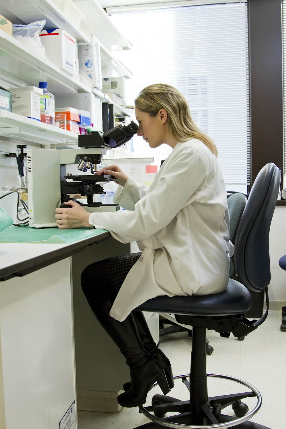
Laboratory Techniques
Advanced molecular biology methods like RNA sequencing and PCR are essential for analyzing gene expression patterns.
Imaging Technology
High-resolution microscopy allows scientists to visualize the regeneration process at the cellular level.
The Future of Mending Broken Hearts
The journey from a zebrafish's aquarium to a human hospital is long, but the path is now illuminated. By understanding the precise genetic language that commands regeneration, we are taking the first steps toward a future where we can "talk" to our own cells .
The research rooted in the tiny, transparent zebrafish heart is providing the foundational dictionary for that future conversation—a conversation that could one day silence the scar and let the heart speak the language of healing once more .
The Promise of Regenerative Medicine
What we learn from zebrafish today could transform how we treat heart disease, spinal cord injuries, and other conditions tomorrow.
