UMI Barcoding in Stem Cell scRNA-seq: A Complete Guide to Quantitative Analysis and Heterogeneity Resolution
Single-cell RNA sequencing (scRNA-seq) with Unique Molecular Identifiers (UMIs) has become an indispensable tool for dissecting the complex heterogeneity of stem cell populations, tracing lineage commitment, and understanding the molecular...
UMI Barcoding in Stem Cell scRNA-seq: A Complete Guide to Quantitative Analysis and Heterogeneity Resolution
Abstract
Single-cell RNA sequencing (scRNA-seq) with Unique Molecular Identifiers (UMIs) has become an indispensable tool for dissecting the complex heterogeneity of stem cell populations, tracing lineage commitment, and understanding the molecular basis of self-renewal and differentiation. This article provides a comprehensive resource for researchers and drug development professionals, covering the foundational principles of UMI barcoding, from its role in correcting amplification bias to its statistical advantages over read counts. It details practical methodological applications for studying stem cell dynamics, explores common troubleshooting and optimization strategies for data quality control, and offers a comparative validation of analysis workflows and emerging multiplexing technologies. By synthesizing established knowledge with the latest methodological advances, this guide aims to empower robust and quantitative single-cell transcriptomic studies in stem cell biology.
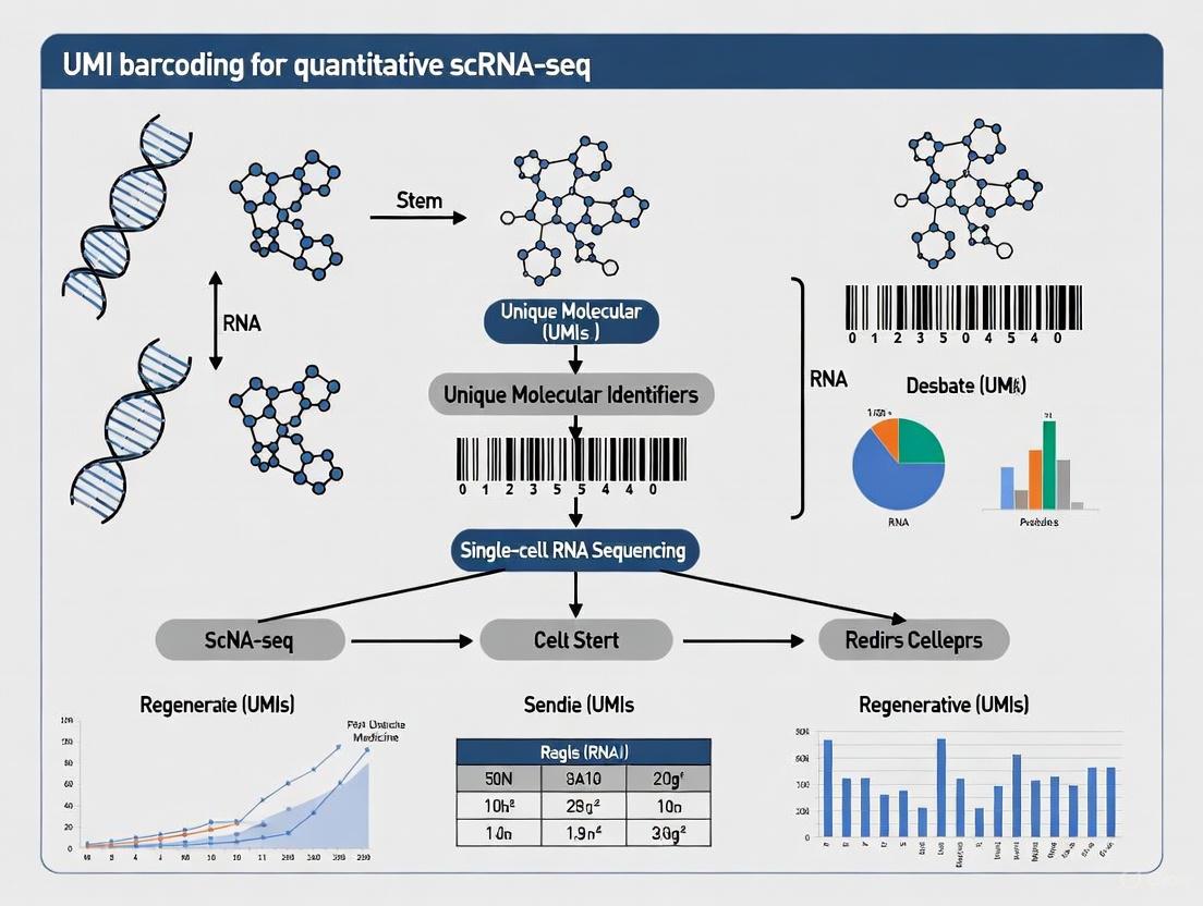
Demystifying UMI Barcoding: The Foundation for Quantitative scRNA-seq in Stem Cell Biology
In single-cell RNA sequencing (scRNA-seq), the ultimate goal is to accurately quantify the absolute number of RNA transcript molecules within individual cells [1]. This quantification is fundamentally challenged by the polymerase chain reaction (PCR) amplification step, an essential process in library preparation that ensures sufficient material for sequencing [2] [1]. Amplification bias occurs because certain sequences are preferentially amplified over others during PCR, leading to overrepresentation of particular transcripts in the final sequencing library that does not reflect their original biological abundance [3] [1]. In stem cell studies, where understanding subtle differences in heterogeneous populations is crucial, this bias can distort the true transcriptome landscape, leading to inaccurate biological interpretations.
Unique Molecular Identifiers (UMIs) are short, random oligonucleotide sequences that provide an elegant solution to this problem [4] [3]. Incorporated into each mRNA molecule during the initial library preparation steps—before any amplification occurs—UMIs uniquely tag each original transcript [1]. All PCR-amplified copies derived from the same original molecule will carry the identical UMI sequence. During bioinformatic analysis, reads sharing the same UMI and mapping to the same genomic locus are identified as PCR duplicates and collapsed into a single digital count [3] [5]. This process effectively removes the amplification bias, enabling researchers to count the number of original molecules directly, thus transforming analog, biased read counts into accurate, digital transcript counts [1].
Table 1: Core Components of the UMI Digital Counting Principle
| Component | Function | Impact on Quantification |
|---|---|---|
| UMI Tagging | Labels each original mRNA molecule with a unique random barcode before PCR amplification [1]. | Enables tracing of molecule ancestry through amplification process. |
| PCR Amplification | Generates sufficient copies of tagged molecules for sequencing [2]. | Introduces quantitative bias that UMI correction is designed to remove. |
| Computational Deduplication | Collapses reads sharing UMI and alignment coordinates into a single count [3] [5]. | Converts analog read counts into digital molecular counts, eliminating amplification noise. |
The following diagram illustrates the core workflow of how UMIs enable digital counting by correcting for PCR amplification bias.
Key Technological Protocols and Reagent Solutions
Experimental Protocol for UMI-Based scRNA-seq
The successful implementation of UMI-based digital counting relies on a meticulously followed experimental protocol. The following steps outline a standard workflow, such as that used in 10x Genomics platforms, which have been recently advanced by GEM-X technology [6].
- Single-Cell Suspension Preparation: A high-viability single-cell or nucleus suspension is prepared from stem cell cultures or tissues. For tissues difficult to dissociate, single-nucleus RNA sequencing (snRNA-seq) is a viable alternative that minimizes artificial stress responses [2].
- Gel Bead and Reagent Loading: The cell suspension is combined with lysis buffer and loaded onto a microfluidic chip, along with Gel Beads and partitioning oil. The GEM-X chips generate twice as many Gel Beads-in-emulsion (GEMs) as previous versions, reducing multiplet rates and improving efficiency [6].
- GEM Generation and Cell Partitioning: Within the Chromium instrument, the mixture is partitioned into nanoliter-scale GEMs. The redesigned GEM-X architecture utilizes oil flow to facilitate GEM formation, enabling faster (6-minute) partitioning and recovery of up to 80% of input cells [6].
- Cell Lysis and Barcoding: Inside each GEM, the single cell is lysed, releasing its RNA. The Gel Bead dissolves, exposing oligonucleotides containing several key functional regions [6]:
- A 10x Barcode unique to each Gel Bead, marking every transcript from the same cell.
- A Unique Molecular Identifier (UMI), a random 12-base sequence that uniquely tags each individual mRNA molecule.
- A Poly(dT) sequence for capturing the poly-adenylated tail of mRNA.
- Reverse Transcription: The primed RNA undergoes reverse transcription within the GEM, creating barcoded cDNA where all copies from a single original molecule share the same UMI [2] [6].
- Library Preparation and Sequencing: The barcoded cDNA is purified, amplified via PCR, and prepared into a sequencing library. The library is then sequenced on a high-throughput platform [2].
Essential Research Reagent Solutions
The UMI scRNA-seq workflow depends on several critical reagents and solutions, each playing a vital role in the digital counting process.
Table 2: Key Reagents for UMI scRNA-seq Experiments
| Reagent / Solution | Critical Function | Technical Note |
|---|---|---|
| Gel Beads | Microbeads coated with barcoded oligos (10x Barcode, UMI, Poly(dT)) for mRNA capture and tagging [6]. | GEM-X technology uses optimized beads for increased sensitivity, detecting up to 98% more genes [6]. |
| Partitioning Oil & Microfluidic Chips | Creates nanoliter-scale reaction vessels (GEMs) for single-cell isolation and barcoding [6]. | GEM-X chip architecture improves GEM generation, halves multiplet rates (to 0.4%), and increases throughput to 20,000 cells per channel [6]. |
| Reverse Transcription (RT) Reagents | Enzymes and buffers to convert captured mRNA into stable, barcoded cDNA [2]. | Must have high efficiency to maximize transcript capture, a key factor for detecting lowly expressed genes in stem cells. |
| UMI Adapters/Oligos | Short, random nucleotide sequences (e.g., 10nt = ~1 million unique combinations) that label individual molecules [1]. | Can be incorporated via RT primers or template-switching oligos. Must have sufficient complexity to avoid UMI saturation [1]. |
Computational Analysis and Error Correction for UMI Data
Bioinformatic Processing of UMI Counts
Following sequencing, raw data must be processed to generate an accurate cell-by-gene digital expression matrix. A standard pipeline involves the following key steps, with UMI error correction being particularly critical.
- Demultiplexing and Alignment: Sequencing reads are demultiplexed using the 10x Barcode to assign reads to their cell of origin and then aligned to a reference genome [5].
- UMI Deduplication (Collapsing): For each cell, reads that align to the same genomic coordinate and share the same UMI sequence are identified and collapsed into a single count, representing one original molecule [3] [5].
- UMI Error Correction: This step is crucial for accurate quantification. PCR amplification and sequencing can introduce errors (substitutions, insertions, deletions) into the UMI sequences themselves, creating artifactual UMIs that inflate transcript counts [4] [3]. Multiple computational strategies exist to correct these errors:
- Network-Based Methods (e.g., UMI-tools): These tools model sequencing errors in UMIs by grouping similar UMIs at the same genomic locus into networks. Methods like "directional" use read counts and edit distances to resolve which UMIs are likely errors derived from a more abundant parent UMI [3].
- Homotrimeric Nucleotide Blocks: A recent advanced method synthesizes UMIs using blocks of three identical nucleotides (homotrimers). Errors can be corrected via a 'majority vote' within each block, significantly improving counting accuracy over traditional monomeric UMIs, especially with increasing PCR cycles [4].
The diagram below illustrates the logical process of UMI deduplication and error correction.
Quantitative Impact of PCR Errors and Correction
The necessity of robust UMI error correction is underscored by empirical data showing how PCR amplification directly introduces inaccuracies in transcript counting.
Table 3: Impact of PCR Cycles and Error Correction on UMI Accuracy
| Experimental Condition | Finding | Implication for scRNA-seq |
|---|---|---|
| Increasing PCR Cycles | A controlled experiment showed a substantial increase in errors within common molecular identifiers (CMIs) as PCR cycles increased from 20 to 25 to 35 [4]. | Protocols should use the minimum number of PCR cycles necessary to maintain library complexity and avoid inflating UMI counts. |
| Homotrimer vs. Monomer UMI Correction | After 25 PCR cycles, homotrimer UMI correction achieved ~96-100% accuracy, outperforming monomer-based tools (UMI-tools, TRUmiCount) which left a significant error rate [4]. | Advanced UMI designs and correction algorithms are critical for absolute molecular counting, especially in sensitive applications. |
| Effect on Differential Expression | In a splicing perturbation experiment, 7.8% of differentially expressed genes were discordant between monomer UMI-tools and homotrimer correction, with homotrimer results yielding more biologically relevant gene ontology terms [4]. | Inaccurate UMI correction can lead to false positives/negatives in downstream analysis, potentially misleading biological conclusions. |
Application in Stem Cell Research
Within the context of stem cell studies, UMI-based scRNA-seq has become an indispensable tool for dissecting cellular heterogeneity, defining differentiation trajectories, and identifying rare subpopulations [7].
- Resolving MSC Heterogeneity and Defining Markers: Mesenchymal Stem/Stromal Cells (MSCs) are known to be highly heterogeneous. SCS technology, empowered by accurate UMI counting, has been pivotal in moving beyond the classical surface marker definitions (e.g., CD105, CD73, CD90) to reveal distinct transcriptional subpopulations within MSC cultures from bone marrow, adipose tissue, and umbilical cord [7]. This allows for a more precise functional characterization of MSC subtypes.
- Mapping Differentiation Trajectories: Understanding the multi-lineage differentiation potential of MSCs into adipocytes, osteocytes, and chondrocytes is a central research focus. UMI-based scRNA-seq enables researchers to study the transcriptome dynamics of individual cells during differentiation, revealing the sequence of transcriptional changes and identifying key regulatory pathways and transient cell states that are masked in bulk analyses [7].
- Characterizing Immunomodulatory Functions: The immunomodulatory capacity of MSCs is a key mechanism in their therapeutic application. By analyzing the interaction between MSCs and immune cells at a single-cell resolution, researchers can identify the specific MSC subclusters that express critical regulatory molecules and unravel the complex cellular crosstalk that underlies their immunomodulatory function [7].
The application of advanced UMI technologies like GEM-X, which offers increased sensitivity and cell recovery, is particularly beneficial in stem cell research. It improves the detection of rare transcripts and lowly expressed regulatory genes, and enhances the capture of rare stem cell subpopulations or cells from precious samples like small tissue biopsies, thereby empowering deeper insights into stem cell biology [6].
Single-cell RNA sequencing (scRNA-seq) has transformed our ability to dissect cellular heterogeneity, a crucial feature in stem cell studies where populations are often diverse and dynamic. The quantitative accuracy of scRNA-seq, however, hinges on advanced molecular barcoding strategies that enable researchers to trace each sequenced transcript back to its cell of origin while controlling for technical artifacts. These barcoding systems are particularly vital for stem cell research, where accurately quantifying expression differences between rare stem cell sub-populations can reveal critical insights into differentiation pathways and regulatory mechanisms.
Barcodes in scRNA-seq are short nucleotide sequences that serve as unique labels during library preparation [8]. The core barcode ecosystem comprises three principal components: Cell Barcodes that tag all transcripts from an individual cell, Unique Molecular Identifiers (UMIs) that label individual mRNA molecules, and Sample Barcodes that enable multiplexing of multiple libraries. Together, this tripartite system transforms complex sequencing data into quantitatively accurate, cell-resolved transcriptomes by providing information about cellular origin, molecular identity, and experimental sample [8] [9]. Understanding the distinct functions, applications, and implementation of each barcode type is foundational to designing robust scRNA-seq experiments in stem cell research.
Core Barcode Components: Definitions and Distinctive Functions
Cell Barcodes
Cell Barcodes are short nucleotide sequences (~16 base pairs) used to "label" all sequences that originate from a single cell source [8]. During single-cell isolation—whether through droplet-based systems (e.g., 10x Genomics, inDrops) or well-based methods—each cell is co-encapsulated with a bead containing a unique cell barcode sequence. During reverse transcription, this barcode is incorporated into all cDNA molecules derived from that specific cell [8] [10]. Following sequencing and bioinformatic processing, sequences sharing the same cell barcode are grouped together as having originated from the same cell, enabling the reconstruction of individual cell transcriptomes from a pooled library.
The primary function of cell barcodes is to enable multiplexing at the cellular level, allowing thousands of cells to be sequenced simultaneously in a single run while maintaining the ability to deconvolute the data back to individual cells [11]. In droplet-based systems, the theoretical diversity of cell barcode libraries is immense—reaching up to 147,456 unique barcodes in some platforms—ensuring a very low probability of two cells receiving the same barcode [11]. However, a key technical consideration is the occurrence of multiplets or doublets, where two or more cells are coincidentally encapsulated together and receive the same cell barcode, potentially leading to misinterpretation of cellular identities [12]. The empirical "technical doublet" rate is often determined by mixing cells from two different species and monitoring barcode purity [11].
Unique Molecular Identifiers (UMIs)
Unique Molecular Identifiers (UMIs) are short, random nucleotide sequences (typically 4-10 base pairs) that serve as molecular barcodes for quality control and quantitative accuracy [8] [9]. Unlike cell barcodes, which are identical for all transcripts from the same cell, each individual mRNA molecule is tagged with a unique UMI during the reverse transcription process [8]. This molecular-level labeling enables bioinformatic correction of amplification biases that inevitably occur during library preparation.
The core function of UMIs is to distinguish between biological duplicates (multiple transcripts from the same original mRNA molecule) and technical duplicates (multiple reads generated through PCR amplification of the same cDNA fragment) [8] [10]. During data processing, reads sharing the same cell barcode, gene assignment, and UMI are collapsed into a single count, representing one original mRNA molecule [8]. This UMI collapsing process mitigates the effects of PCR amplification bias, where some molecules are amplified more efficiently than others, and provides a more accurate quantitative representation of the true molecular count in the original sample [9]. This is especially crucial in stem cell studies where detecting subtle expression differences in key regulatory genes can have significant biological implications.
UMIs also enhance variant detection sensitivity by helping distinguish true biological variants from errors introduced during amplification or sequencing [8] [9]. Since each original molecule is uniquely tagged, sequencing errors can be identified and filtered out, enabling more reliable detection of rare variants and improving the overall quality of quantitative gene expression data [13].
Sample Barcodes (Indexes)
Sample Barcodes (also known as sample indexes) are sequences used to multiplex multiple libraries during sequencing runs [9]. Unlike cell barcodes and UMIs, which operate at the cellular and molecular levels respectively, sample barcodes are added during library preparation and are identical for all sequences derived from the same library. After sequencing, these barcodes enable bioinformatic demultiplexing, where pooled sequences are sorted computationally into their original sample groups.
The primary function of sample barcodes is cost efficiency and experimental design flexibility, allowing researchers to sequence multiple samples simultaneously on the same flow cell while maintaining sample identity [9]. With the advent of unique dual indexes (UDIs), where each sample receives a unique combination of two barcodes, the potential for index hopping (misassignment of reads to wrong samples) is significantly reduced, further enhancing data integrity [9].
Table 1: Comparative Overview of Barcode Types in scRNA-seq
| Feature | Cell Barcodes | UMIs | Sample Barcodes |
|---|---|---|---|
| Primary Function | Demultiplex cells | Quantify molecules | Demultiplex samples |
| Sequence Length | ~16 bp [8] | 4-10 bp [8] | Varies (typically 6-10 bp) |
| Scope of Application | Individual cell | Individual mRNA molecule | Entire library/sample |
| Key Applications | Single-cell resolution, cell tracking [8] | PCR duplicate removal, quantitative analysis [8] [9] | Multiplexing, cost reduction [9] |
| Added During | Cell isolation/encapsulation | Reverse transcription | Library preparation |
| Bioinformatic Processing | Cell calling, doublet detection [12] | UMI collapsing, error correction [8] | Demultiplexing |
Barcoding Technologies and Experimental Workflows
Single-cell RNA sequencing technologies have evolved substantially, with current platforms predominantly utilizing droplet-based or well-based approaches for cell barcoding [14]. Droplet-based systems (e.g., 10x Genomics, inDrops) employ microfluidics to co-encapsulate individual cells with barcoded beads in nanoliter-scale droplets, achieving high throughput of thousands to millions of cells [11] [14]. Well-based methods (e.g., CEL-Seq2, SMART-Seq) distribute cells into multiwell plates containing unique barcodes, offering greater flexibility but lower throughput [10] [14].
The inDrop platform exemplifies a droplet-based approach, encapsulating cells into droplets with lysis buffer, reverse transcription reagents, and barcoded oligonucleotide primers [11]. Each barcoded hydrogel microsphere carries covalently coupled, photo-releasable primers encoding one of thousands of barcodes. Similarly, the CEL-Seq2 protocol employs a paired-end sequencing approach where Read1 contains the barcoding information (cell barcode and UMI) followed by a polyT tail, while Read2 contains the actual transcript sequence [10].
Detailed Protocol: CEL-Seq2 Barcoding Workflow
The following diagram illustrates the core experimental workflow for barcode incorporation in scRNA-seq protocols like CEL-Seq2:
The wet-lab workflow begins with single-cell isolation, where a cell suspension is partitioned into individual compartments [14]. For droplet-based methods, this occurs through microfluidic encapsulation; for well-based methods, through fluorescence-activated cell sorting (FACS) or limiting dilution. Next, cell lysis releases mRNA, which is captured by barcoded primers containing three functional elements: the cell barcode, a UMI, and a poly-dT sequence that binds to the mRNA poly-A tail [8] [10]. During reverse transcription, these primers generate barcoded cDNA. The cDNA is then amplified, sample barcodes are added during library preparation, and the pooled libraries are sequenced [10]. The subsequent bioinformatic processing involves demultiplexing samples by sample barcodes, grouping reads by cell barcodes, and collapsing duplicate reads by UMIs to generate accurate quantitative expression matrices [8] [10].
Bioinformatic Processing and Data Analysis
Barcode Extraction and Processing Pipeline
Following sequencing, bioinformatic processing of barcodes involves multiple critical steps to transform raw sequencing data into a quantitative gene expression matrix. The first step is demultiplexing, where sequences are assigned to their original samples based on sample barcodes [9]. Next, barcode extraction occurs, where cell barcodes and UMIs are identified from the sequencing reads—typically from Read1 in paired-end protocols like CEL-Seq2 [10].
A crucial quality control step is barcode validation, where cell barcodes are filtered against a whitelist of known valid barcodes to exclude those with sequencing errors [10]. For UMI processing, error correction is performed to account for sequencing errors, typically by clustering similar UMIs (within a certain Hamming distance) and collapsing them [12]. The final and most critical step is UMI deduplication, where reads sharing the same cell barcode, gene assignment, and UMI are collapsed into a single count, representing one original mRNA molecule [8] [10]. This process effectively removes PCR duplicates, providing a digital count of transcript molecules per gene per cell.
Statistical Considerations for UMI Count Data
Unique statistical properties distinguish UMI-count data from read-count data in scRNA-seq analysis. UMI counts follow a negative binomial distribution rather than requiring more complex zero-inflated models [13]. Research has demonstrated that while read-count measurements often necessitate zero-inflated negative binomial models to account for excess zeros, UMI counts are adequately modeled by a standard negative binomial distribution, with a significant proportion of genes even following a Poisson distribution [13]. This statistical simplicity reflects the reduced technical noise in UMI-based protocols and has important implications for differential expression analysis in stem cell studies.
For differential expression analysis of UMI count data, methods based on the negative binomial model with independent dispersions (NBID) have shown superior performance in controlling false discovery rates while maintaining good power [13]. This is particularly relevant in stem cell research where accurately detecting subtle expression changes in key regulatory genes can have significant biological implications.
Table 2: Quantitative Comparison of Read-Count vs. UMI-Count Data Characteristics
| Characteristic | Read-Count Data | UMI-Count Data |
|---|---|---|
| Amplification Bias | High sensitivity to amplification biases [13] | Reduced impact of amplification biases [13] |
| Statistical Distribution | Often requires zero-inflated models [13] | Better fit to negative binomial distribution [13] |
| Percentage of Genes Following Poisson | 2.6% (range: 1.0-4.1%) [13] | 80.2% (range: 65.7-95.1%) [13] |
| Goodness of Fit to Negative Binomial | 14.2% reject NB model (range: 1.1-35.3%) [13] | 0.1% reject NB model (range: 0-0.4%) [13] |
| Recommended DE Analysis Method | Zero-inflated negative binomial models [13] | Negative binomial with independent dispersions (NBID) [13] |
Advanced Applications in Stem Cell Research
Resolving Stem Cell Heterogeneity
The integration of barcoding technologies has dramatically advanced stem cell research by enabling the resolution of cellular heterogeneity within seemingly homogeneous populations. In a landmark study profiling mouse embryonic stem cells, droplet-based barcoding of thousands of cells revealed population structure and the heterogeneous onset of differentiation after leukemia inhibitory factor (LIF) withdrawal [11]. The high-throughput nature of barcoded scRNA-seq allowed researchers to identify rare sub-populations expressing markers of distinct lineages that would be difficult to detect when profiling only a few hundred cells [11].
Barcoding technologies have further enabled the investigation of correlation structures in gene expression across entire stem cell populations, revealing how key pluripotency factors fluctuate in a coordinated manner [11]. During differentiation, dramatic changes in these correlation structures occur, resulting from asynchronous inactivation of pluripotency factors and the emergence of novel cell states [11]. Such insights would be impossible without the quantitative accuracy provided by UMI-based counting and the cellular resolution enabled by cell barcoding.
Lineage Tracing and Synthetic Barcoding
Beyond transcriptome quantification, synthetic DNA barcodes have emerged as powerful tools for lineage tracing in stem cell biology. Recent approaches use heritable synthetic DNA barcodes to reconstruct cell lineage relationships alongside transcriptomic profiling [12]. These methods enable researchers to answer fundamental questions about stem cell fate decisions, clonal dynamics, and developmental trajectories.
An innovative application of synthetic barcodes is the identification of "ground-truth singlets" in scRNA-seq datasets [12]. The "singletCode" framework leverages the fact that each synthetically barcoded cell possesses a unique DNA sequence before scRNA-seq processing, enabling definitive identification of true single cells and accurate simulation of doublets for benchmarking computational methods [12]. This approach is particularly valuable in stem cell research where cell aggregation or similar transcriptional states can challenge conventional doublet detection methods.
Table 3: Research Reagent Solutions for scRNA-seq Barcoding
| Reagent/Resource | Function | Example Applications |
|---|---|---|
| Barcoded Beads | Delivery of cell barcodes and UMIs to individual cells | 10x Genomics GemCode, inDrop Barcoded Hydrogel Microspheres [11] |
| Barcoded Primers | Reverse transcription primers containing barcodes | CEL-Seq2 barcoded primers [10] |
| Sample Indexing Kits | Multiplexing samples in sequencing runs | Illumina Indexing Kits [9] |
| Barcode Whitelists | Quality control of cell barcodes | 10x Genomics barcode whitelists [10] |
| UMI-Tools | Bioinformatic processing of UMI data | UMI extraction, error correction, deduplication [10] |
| Synthetic Barcode Libraries | Lineage tracing and singlet identification | FateMap, ClonMapper, SPLINTR, LARRY [12] |
The tripartite barcoding system—comprising cell barcodes, UMIs, and sample barcodes—forms the technological foundation of quantitative single-cell RNA sequencing. Each component addresses distinct challenges in single-cell analysis: cell barcodes enable multiplexing at cellular resolution, UMIs provide molecular-level quantification by correcting for amplification biases, and sample barcodes allow efficient library multiplexing. For stem cell researchers, understanding these core components and their integrated function is crucial for designing robust experiments, interpreting data accurately, and advancing our understanding of stem cell biology at single-cell resolution. As barcoding technologies continue to evolve, particularly with the integration of synthetic barcodes for lineage tracing, they will undoubtedly unlock new dimensions in the study of stem cell heterogeneity, fate decisions, and regulatory mechanisms.
In single-cell RNA sequencing (scRNA-seq) studies of stem cells, Unique Molecular Identifiers (UMIs) have transitioned from a technical refinement to an essential component for quantitative accuracy. UMIs are short, random oligonucleotide barcodes that tag individual mRNA molecules before PCR amplification, enabling precise molecule counting and distinguishing biological signal from technical artifacts [3] [9]. In stem cell research—where resolving subtle heterogeneity, identifying rare transitional states, and accurately tracing lineages are paramount—UMIs provide the mathematical foundation for distinguishing true biological variation from PCR amplification bias and sequencing errors [15]. Without UMI incorporation, attempts to quantify gene expression across heterogeneous stem cell populations remain semi-quantitative at best, as PCR duplicates artificially inflate counts for highly expressed genes and obscure true transcript diversity [16]. This technical note details the essential methodologies and applications that make UMIs non-negotiable for advanced stem cell research, providing structured protocols and analytical frameworks for leveraging their full potential.
Resolving Cellular Heterogeneity in Complex Stem Cell Populations
The Problem of PCR Amplification Bias
Stem cell populations, even those derived from clonal origins, demonstrate remarkable transcriptional heterogeneity that can reflect differential potency, metabolic states, or early lineage priming. Conventional scRNA-seq without UMIs struggles to accurately resolve this heterogeneity because PCR amplification during library preparation generates duplicate reads from original mRNA molecules [3]. These duplicates do not represent distinct biological molecules but rather technical artifacts that skew expression estimates. In studies of glioblastoma stem cells (GSCs), for instance, this amplification bias can obscure critical differences between stem-like states and more differentiated populations, potentially masking therapeutically relevant subpopulations [17].
UMI-Based Error Correction and Deduplication
UMIs solve this fundamental problem by providing a unique tag for each original molecule prior to amplification. Through UMI deduplication bioinformatics processes, reads sharing both genomic coordinates and identical UMIs are identified as technical replicates deriving from a single molecule, enabling accurate quantification of original transcript numbers [3] [18]. Advanced tools like UMI-tools and UMI-nea implement network-based clustering methods that account for sequencing errors in UMI sequences themselves—a common issue that can otherwise create artifactual UMIs and inflate diversity estimates [3] [18]. These tools model sequencing errors and strategically group similar UMIs that likely originated from the same source molecule, significantly improving quantification accuracy [3].
Diagram 1: UMI-integrated scRNA-seq workflow for stem cell studies. The process begins with a heterogeneous stem cell population, incorporates UMIs during reverse transcription, and culminates in accurate transcript quantification after computational deduplication.
Application in Identifying Stem Cell States
The power of UMI-based resolution is particularly evident in studies of cellular plasticity. Research on glioblastoma stem cells has demonstrated that cells expressing stem cell-associated surface markers (CD133, CD15, CD44, A2B5) do not represent fixed hierarchical entities but rather plastic states that most cancer cells can adopt in response to microenvironmental cues [17]. Without UMIs to provide accurate single-cell quantification, the dynamic nature of these states and their rapid interconversion would be difficult to capture with confidence. UMI-enabled scRNA-seq revealed that all GBM subpopulations—regardless of surface marker expression—retained stem cell properties and tumorigenic potential, fundamentally challenging hierarchical stem cell models [17].
Detecting Rare Stem Cell Populations with Confidence
Technical Limitations in Rare Cell Detection
The reliable identification of rare stem cell populations—such as quiescent stem cells, transitional intermediates, or therapy-resistant precursors—represents a significant challenge in stem cell biology. These populations often constitute less than 1% of total cells yet possess critical functions in tissue regeneration, cancer recurrence, and developmental processes. Conventional sequencing approaches struggle to distinguish true biological rare populations from technical artifacts caused by sequencing errors and PCR amplification bias, especially when analyzing low-input samples [19].
Enhanced Sensitivity with Molecular Barcoding
Dual-molecular barcode sequencing technologies significantly enhance sensitivity for detecting rare variants and low-abundance transcripts. In a study of tumor and cell-free DNA, molecular barcode sequencing enabled detection of variants with allele fractions as low as 0.17%—a sensitivity level unattainable with conventional non-UMI approaches [19]. This precision is equally valuable in stem cell research for identifying rare subpopulations defined by unique transcriptional signatures. The UMI-based approach allows researchers to set statistically rigorous thresholds for rare population identification, distinguishing true biological signals from technical noise with high confidence [19] [18].
Experimental Design Considerations
For optimal detection of rare stem cell populations, specific experimental design considerations are essential:
- UMI Length and Complexity: Longer UMIs (12-18bp for short-read sequencing) reduce the probability of "UMI collisions" where distinct molecules receive identical barcodes [18]
- Sequencing Depth: Increased sequencing depth compensates for low cellular abundance while UMI deduplication prevents artificial inflation from PCR duplicates
- Cell Number: Capturing sufficient cell numbers ensures adequate sampling of rare populations while maintaining single-cell resolution
- Bioinformatic Processing: Tools like UMI-nea that use Levenshtein distance (accounting for insertions/deletions) rather than just Hamming distance (substitutions only) provide more accurate error correction, especially valuable in long-read sequencing applications [18]
Lineage Tracing and Trajectory Reconstruction
Uncovering Developmental Hierarchies
Stem cell differentiation follows complex trajectories with branching points that define lineage commitment. UMI-enhanced scRNA-seq enables powerful computational reconstruction of these developmental pathways through pseudotime analysis [20]. By accurately quantifying transcriptomes without PCR distortion, UMIs provide the clean data necessary for algorithms to order cells along differentiation trajectories, identify branch points, and uncover genes driving fate decisions [20]. This approach has been successfully applied to diverse systems, from hematopoietic stem cell differentiation to the branching lineages in colonic epithelium, where absorptive and secretory cells diverge from common progenitors [21].
RNA Velocity and Beyond
Recent methodological advances like RNA velocity leverage UMI-based quantification to predict future cell states from single-cell snapshots [20]. By comparing the ratio of unspliced to spliced mRNAs—a measurement requiring accurate quantification of both forms—RNA velocity infers the direction and pace of cellular state transitions. For stem cell biologists, this enables not just observation of current states but prediction of developmental futures, identifying which stem cells are poised to differentiate and along which lineages [20]. When combined with UMI-based lineage barcoding that permanently marks cells and their progeny, these approaches provide a comprehensive view of stem cell lineage relationships in developing systems [20].
Diagram 2: UMI-enabled lineage trajectory reconstruction in stem cell differentiation. Accurate transcriptome quantification allows mapping of differentiation pathways and prediction of lineage commitment through RNA velocity analysis.
Essential Protocols for UMI Implementation in Stem Cell Studies
Wet-Lab Protocol: UMI Integration in scRNA-seq Library Preparation
Materials Required:
- Commercially available UMI-containing scRNA-seq kits (e.g., 10x Genomics, Parse Biosciences)
- Stem cell population of interest in single-cell suspension
- Laboratory equipment: microcentrifuge, thermal cycler, magnetic separator
Step-by-Step Procedure:
Cell Viability Assessment: Confirm >90% viability using trypan blue exclusion or similar method.
Single-Cell Partitioning and Lysis:
- Load cells following manufacturer's recommendations (targeting 5,000-10,000 cells)
- Ensure proper lysis to release RNA while maintaining cell integrity
Reverse Transcription with UMI Barcoding:
- Perform reverse transcription immediately after lysis
- UMI incorporation occurs automatically in commercial systems via barcoded beads
cDNA Amplification and Library Construction:
- Amplify with limited PCR cycles (typically 12-16) to minimize bias
- Incorporate platform-specific sequencing adapters
Quality Control and Sequencing:
- Assess library quality (Bioanalyzer/Fragment Analyzer)
- Sequence with sufficient depth (≥50,000 reads/cell recommended for heterogeneous stem cell populations)
Critical Considerations:
- For stem cell populations with extreme size variation (e.g., hematopoietic vs. mesenchymal stem cells), optimize cell loading concentration
- Include extraction controls to monitor background RNA contamination
- For rare populations (<1%), consider targeted enrichment approaches
Computational Protocol: UMI Deduplication and Analysis
Software Requirements:
- UMI processing tools (UMI-tools, UMI-nea, Calib)
- Single-cell analysis suite (Seurat, Scanpy, Monocle)
- Computing resources (minimum 16GB RAM for datasets <10,000 cells)
Processing Pipeline:
FASTQ Preprocessing:
- Extract UMIs from read sequences
- Trim adapter sequences and low-quality bases
Read Alignment:
- Align to reference genome using Spliced Transcripts Alignment to a Reference (STAR) or similar
- Generate gene count matrix incorporating UMIs
UMI Deduplication:
- For each cell, group reads by genomic coordinates
- Cluster UMIs using edit distance threshold (typically 1-2 bases)
- Apply directional network-based methods to resolve complex UMI groups [3]
Downstream Analysis:
- Normalize UMI counts across cells
- Perform dimensionality reduction and clustering
- Conduct differential expression and trajectory analysis
Troubleshooting Notes:
- High UMI error rates may indicate poor library quality or insufficient UMI complexity
- Adjust clustering parameters for different UMI lengths and sequencing depths
- Validate rare population findings with orthogonal methods when possible
Research Reagent Solutions for UMI-Enhanced Stem Cell Studies
Table 1: Essential Research Reagents and Platforms for UMI-Based Stem Cell Research
| Reagent/Platform | Function | Key Features for Stem Cell Applications |
|---|---|---|
| Twist UMI Adapter System | Ligation-based UMI incorporation | Compatible with low-input samples; enables detection of rare variants in heterogeneous populations [22] |
| 10x Genomics Single Cell Gene Expression | Droplet-based scRNA-seq with UMIs | High cell throughput ideal for capturing rare stem cell subpopulations; integrated workflow |
| Illumina UMI Adaptors | Sample preparation for UMI sequencing | Reduces false-positive variant calls; increases sensitivity for low-frequency transcripts [9] |
| QIAGEN UMI-nea Bioinformatics Tool | Computational UMI deduplication | Levenshtein distance accounting for indels; robust performance across sequencing platforms [18] |
| UMI-tools | Network-based UMI grouping | Directional method resolves complex UMI networks; improves quantification accuracy [3] |
The integration of UMIs into stem cell scRNA-seq workflows represents a fundamental advancement that transforms qualitative observations into quantitative measurements. By eliminating PCR amplification bias and enabling precise molecular counting, UMIs provide the technical foundation necessary to resolve stem cell heterogeneity, identify rare populations with statistical confidence, and accurately reconstruct lineage trajectories. As stem cell research increasingly focuses on dynamic processes, rare transitional states, and therapeutic applications, the implementation of UMI-based methodologies becomes not merely advantageous but essential. The protocols and frameworks outlined herein provide a pathway for researchers to leverage these powerful tools, ensuring that technical limitations do not constrain biological discovery in the complex landscape of stem cell biology.
Unique Molecular Identifier (UMI) barcoding has revolutionized quantitative single-cell RNA sequencing (scRNA-seq) in stem cell studies by enabling accurate transcript counting. This technology mitigates amplification bias by tagging individual mRNA molecules, allowing bioinformatic removal of PCR duplicates. A critical challenge in analyzing the resulting UMI count data involves selecting appropriate statistical models that account for its characteristic high proportion of zeros without introducing unnecessary complexity. The fundamental question addressed in this Application Note is whether negative binomial (NB) models provide superior fit for UMI count data compared to zero-inflated negative binomial (ZINB) models, particularly within the context of stem cell research where accurately identifying subtle expression differences is paramount.
The distinction between UMI counts and read counts is essential for proper model selection. While read counts from full-length scRNA-seq protocols often show characteristics requiring zero-inflated modeling, evidence increasingly suggests that UMI counts follow a different statistical distribution. Understanding this distinction helps researchers avoid model misspecification, which can lead to reduced statistical power, false positives in differential expression analysis, and inaccurate biological interpretations in stem cell differentiation studies.
Theoretical Foundation: Distribution Properties of UMI-Count Data
The Multinomial Sampling Foundation of UMI Counts
UMI-count data originates from a fundamentally different generative process than read-count data. When a cell containing ti total mRNA transcripts is processed through UMI-based scRNA-seq protocols, the resulting UMI count ni is substantially lower (ni ≪ ti) due to technical losses during capture, reverse transcription, and library preparation. The critical insight is that which molecules successfully become UMIs is essentially a random sampling process [23]. This sampling process can be effectively modeled using the multinomial distribution, which naturally accounts for the zeros observed in scRNA-seq data without requiring special zero-inflation parameters.
The multinomial model for UMI counts posits that the observed count for gene j in cell i, denoted xij, arises from sampling a fixed number of molecules (ni) across all genes according to probability parameters pij that reflect true relative expression levels. Under this model, the abundance of zeros is adequately explained by low capture efficiency and biological variation in true expression levels—no separate zero-generating mechanism is required. Empirical evidence from negative control datasets supports this theoretical foundation, demonstrating that UMI counts follow a discrete distribution with no zero inflation [23].
Comparative Analysis of Statistical Models
Table 1: Comparison of Statistical Models for scRNA-seq Data
| Model | Key Parameters | Assumed Zero Mechanism | Suitability for UMI Data | Computational Complexity |
|---|---|---|---|---|
| Poisson | Mean (λ) | Sampling variation | Poor (underestimates variance) | Low |
| Negative Binomial | Mean (μ), Dispersion (θ) | Sampling variation + biological noise | Excellent | Moderate |
| Zero-Inflated Negative Binomial (ZINB) | Mean (μ), Dispersion (θ), Zero-inflation (π) | Sampling variation + technical dropouts | Overparameterized for UMI data | High |
| Hurdle Models | Separate parameters for zero vs. non-zero | Distinct processes for zero and positive counts | Unnecessary for UMI data | High |
The negative binomial model effectively captures the mean-variance relationship observed in UMI count data through its dispersion parameter, which accounts for overdispersion beyond Poisson sampling variance. This overdispersion arises from both biological heterogeneity (e.g., stochastic expression bursts) and technical noise. Extensive model comparisons using likelihood ratio tests on real UMI datasets reveal that the ZINB model does not provide significantly better fit than the NB model for the vast majority of genes, indicating that the additional zero-inflation parameter is unnecessary [24]. In one comprehensive evaluation, exactly 0% of genes tested across multiple UMI-based protocols showed preference for ZINB over NB at a false discovery rate of 0.05 [24].
Experimental Evidence: Empirical Support for Negative Binomial Models
Systematic Model Comparisons
Several rigorous studies have directly compared the performance of negative binomial and zero-inflated models for UMI count data. In a landmark analysis, researchers examined four UMI-based scRNA-seq protocols (CEL-Seq2/C1, Drop-Seq, MARS-Seq, and SCRB-Seq) using a backward selection strategy on three nested models: Poisson, NB, and ZINB [24]. The results were striking—while read-count data from the same protocols showed 9.4–34.5% of genes preferring ZINB over NB, exactly 0% of genes measured with UMI counts preferred ZINB over NB. Furthermore, a substantial proportion of genes (39.4–84.0%) were adequately modeled by the simple Poisson distribution for UMI counts, suggesting relatively modest overdispersion [24].
These findings challenge the prevailing assumption that scRNA-seq data universally requires complex zero-inflated models. The evidence strongly indicates that UMI counting substantially simplifies the statistical properties of scRNA-seq data, making NB models sufficient for most genes. This has important implications for stem cell researchers, as NB models offer greater numerical stability and computational efficiency compared to ZINB models, which frequently encounter convergence issues during optimization [25].
Table 2: Sources of Zero Counts in scRNA-seq Experiments
| Source Type | Specific Mechanisms | Relevance to UMI Data |
|---|---|---|
| Biological | Stochastic transcription bursts, Phased gene expression, Transcript degradation | Affects all technologies |
| Technical | Inefficient reverse transcription, Low mRNA capture efficiency, Cell dissociation effects | Affects all technologies |
| Protocol-specific | PCR amplification bias (read counts), Molecular sampling (UMI counts) | Technology-dependent |
| Cell quality | Cell death, Cytoplasmic RNA leakage, Poor cell viability | Affects all technologies |
Understanding the sources of zeros in scRNA-seq data helps explain why NB models suffice for UMI counts. The "dropout" phenomenon, often cited to justify zero-inflated models, may be less relevant to UMI data than previously assumed. While UMI-based scRNA-seq can have high dropout rates, the pattern differs from read-count data. For UMI counts, zeros primarily result from a combination of low actual expression and the fundamental sampling nature of the measurement process, rather than a distinct technical failure mechanism that randomly sets counts to zero irrespective of true expression levels [26] [27].
Experimental evidence shows that even strongly expressed genes can occasionally show zeros in some cells with UMI protocols, but these zeros are consistent with NB sampling variation rather than requiring a separate zero-generating process. This distinction is crucial for stem cell researchers investigating heterogeneous populations, where accurate modeling of zero counts affects the identification of rare subpopulations and transitional states.
Practical Implementation Protocols
Differential Expression Analysis Workflow for UMI Data
Detailed Protocol for Negative Binomial-Based Differential Expression Analysis
Protocol: NBID (Negative Binomial with Independent Dispersions) Algorithm
Purpose: To accurately identify differentially expressed genes in UMI-count scRNA-seq data from stem cell populations using a negative binomial framework.
Reagents and Software Requirements:
- R statistical environment (version 4.0 or higher)
- NBID package (available from St. Jude Children's Research Hospital)
- UMI-count matrix from scRNA-seq experiment
- Cell metadata including experimental conditions
Procedure:
Input Data Preparation (Duration: 10-15 minutes)
- Format raw UMI count matrix with genes as rows and cells as columns
- Prepare sample metadata table with cell-type annotations and experimental conditions
- For stem cell studies, include relevant covariates (differentiation stage, cell cycle status)
Model Initialization (Duration: 2-5 minutes)
Parameter Estimation (Duration: 15-60 minutes, depending on dataset size)
- Estimate gene-specific dispersions using conditional maximum likelihood
- Fit negative binomial generalized linear models for each gene
- Incorporate batch effects as random effects when applicable [28]
Hypothesis Testing (Duration: 5-15 minutes)
- Perform likelihood ratio tests between experimental conditions
- Apply false discovery rate correction for multiple testing
- Filter results based on fold-change and adjusted p-value thresholds
Result Interpretation (Duration: 30+ minutes)
- Identify significantly differentially expressed genes
- Perform pathway enrichment analysis on results
- Validate key findings using independent methods
Troubleshooting Tips:
- For convergence issues, reduce the number of covariates in the model
- For unstable dispersion estimates, use trended dispersion approaches
- When analyzing rare stem cell populations, consider mixed models to account for subject-level effects [28]
Advanced Analytical Frameworks
Mixed Model Extensions for Complex Experimental Designs
Stem cell research often involves multi-subject designs where cells are collected from multiple donors or experimental replicates. In such cases, advanced negative binomial mixed models (NBMMs) account for hierarchical data structures. The NEBULA algorithm efficiently decomposes total overdispersion into subject-level and cell-level components, addressing both technical and biological sources of variation [28].
For stem cell researchers investigating disease mechanisms or treatment responses across multiple patient-derived induced pluripotent stem cell lines, NBMMs provide crucial advantages. They properly control false positive rates when testing subject-level variables (e.g., genotype, treatment condition) by accounting for the non-independence of cells from the same subject. Simulation studies demonstrate that NBMMs maintain appropriate type I error rates while achieving better power compared to models that ignore the hierarchical structure [28].
Feature Selection and Dimension Reduction for UMI Data
Based on the multinomial foundation of UMI counts, feature selection using deviance statistics outperforms traditional highly variable gene selection methods. The deviance effectively measures each gene's contribution to total heterogeneity while accounting for the mean-variance relationship of count data. Similarly, generalized principal component analysis (GLM-PCA) applied directly to raw UMI counts provides superior dimension reduction compared to PCA on log-normalized data, which can be distorted by the high proportion of zeros [23].
Table 3: Recommended Computational Tools for UMI-Count Analysis
| Tool Name | Primary Function | Model Foundation | Applicable to Stem Cell Research |
|---|---|---|---|
| NBID | Differential expression | Negative binomial | Yes - heterogeneous populations |
| NEBULA | Multi-subject analysis | Negative binomial mixed model | Yes - patient-derived lines |
| SwarnSeq | Differential expression | Zero-inflated negative binomial | Limited advantage for UMI data |
| scMMST | Batch effect correction | Mixed models | Yes - multi-batch experiments |
| TensorZINB | Large-scale analysis | ZINB with deep learning | Overparameterized for UMI data |
Application to Stem Cell Research
Case Study: Identifying Stem Cell Subpopulations
In a practical application to stem cell biology, researchers applied NB-based differential expression analysis to identify marker genes defining subpopulations in rhabdomyosarcoma cells [29]. The NBID algorithm successfully identified genes separating subpopulations with distinct expression patterns, suggesting novel mechanisms of solid tumor progression. This demonstrates the utility of NB models for uncovering biologically meaningful heterogeneity in stem cell systems.
For stem cell researchers investigating differentiation processes, NB models provide sensitive detection of expression changes in transitional states, where cell-to-cell heterogeneity is high but zero-inflation is minimal in UMI data. The numerical stability of NB estimation ensures reliable results even for genes with moderate to low expression, which often include key regulators of stem cell fate decisions.
Recommendations for Experimental Design
Based on the statistical properties of UMI-count data, we recommend:
- Protocol Selection: Prioritize UMI-based scRNA-seq protocols over read-count methods for quantitative expression analysis
- Sample Size Considerations: Include sufficient biological replicates (subjects/stem cell lines) rather than maximizing cell numbers per subject
- Sequencing Depth: Aim for 20,000-50,000 UMIs per cell to adequately capture mid-to-low abundance transcripts
- Quality Control: Monitor technical metrics but recognize that zeros are expected biological features rather than necessarily indicating failed measurements
The statistical foundations and empirical evidence consistently demonstrate that negative binomial models provide superior fit for UMI-count scRNA-seq data compared to zero-inflated alternatives in most stem cell research contexts. The multinomial sampling process underlying UMI counting naturally produces zeros consistent with NB distributions without requiring additional zero-inflation parameters. By adopting appropriately parameterized NB models, stem cell researchers can achieve more numerically stable, computationally efficient, and biologically accurate analysis of single-cell transcriptomes, ultimately advancing our understanding of stem cell biology and its therapeutic applications.
From Theory to Bench: Implementing UMI scRNA-seq to Decipher Stem Cell Fate and Function
Single-cell RNA sequencing (scRNA-seq) has revolutionized biological research by enabling the dissection of cellular heterogeneity at unprecedented resolution. For stem cell studies, where cellular plasticity and diverse differentiation trajectories are fundamental, scRNA-seq provides unparalleled insights into molecular networks and cellular states [14]. The incorporation of unique molecular identifiers (UMIs) has been particularly transformative for quantitative scRNA-seq, as they mitigate amplification bias and enable precise molecular counting of transcripts [13] [30]. This technical advance is crucial for accurately capturing the subtle expression differences that define stem cell heterogeneity, identify rare subpopulations, and trace developmental lineages.
The journey from a complex biological sample to a sequencing library ready for interpretation is a multistep process where each stage critically influences the final data quality. This application note provides a comprehensive experimental workflow breakdown, detailing best practices from single-cell isolation through cDNA synthesis and library preparation, with particular emphasis on their application within stem cell research utilizing UMI barcoding for quantitative analysis.
Single-Cell Isolation: The Foundation of scRNA-seq
Isolation Strategies and Their Applications
The initial step of single-cell isolation is arguably the most critical, as it determines the representativeness and viability of the input material. The choice of method involves trade-offs between throughput, viability, and compatibility with downstream applications.
Table 1: Comparison of Single-Cell Isolation Methods for scRNA-seq
| Method | Throughput | Principle | Key Advantages | Key Limitations | Ideal for Stem Cell Studies |
|---|---|---|---|---|---|
| Droplet-Based (e.g., 10x Genomics) | High (Thousands to millions of cells) | Microfluidics to encapsulate single cells in oil droplets [31] | High throughput, commercial scalability, early barcoding | Limited capture efficiency (2-50%), specialized equipment required, higher multiplet rates [14] | Profiling large, heterogeneous populations (e.g., organoids) |
| Plate-Based (e.g., SMART-Seq) | Low to Medium (96-384 wells) | FACS or manual deposition of single cells into multi-well plates [32] | High sensitivity, full-length transcript coverage, flexible input | Lower throughput, higher reagent costs, requires pre-amplification [14] | Deep characterization of predefined stem cell subsets |
| Combinatorial Barcoding (e.g., Parse Biosciences) | Very High (Thousands to millions of cells) | Cells act as reaction chambers; barcodes are added over multiple rounds of splitting and pooling [31] | Scalability, does not require specialized equipment, low multiplet rates [31] | Protocol complexity, longer hands-on time, compatible with fixed cells | Large-scale perturbation screens or time-course experiments |
| Laser Capture Microdissection | Low | Direct microscopic visualization and isolation of cells from tissue context [14] | Preserves spatial context, precise selection | Very low throughput, technically challenging, potential RNA degradation | Studying stem cells in their anatomical niche (e.g., intestinal crypts) |
Practical Considerations for Stem Cell Samples
Successful isolation of stem cells requires careful handling to preserve viability and minimize transcriptional stress. For solid tissues, enzymatic digestion must be optimized to dissociate the extracellular matrix without damaging cell surface markers critical for stem cell identity.
- Enzyme Selection: A blend of collagenase (targets collagen), dispase (targets fibronectin and collagen IV), and hyaluronidase (targets hyaluronan) is often effective for breaking down the extracellular matrix [33]. For sensitive epitopes, trypsin alternatives like Accutase or TrypLE are recommended as they are less aggressive on cell surface proteins [33].
- Viability Preservation: The dissociation process can activate stress responses, including the induction of immediate early genes, which can confound transcriptomic analysis [14]. This is particularly relevant for neural stem cells. Rapid processing and maintaining samples on ice can mitigate this. Using nuclei instead of intact cells (single-nucleus RNA-seq) is a valuable alternative for particularly sensitive cell types or frozen samples [14] [34].
- Quality Control: The resulting cell suspension must be assessed for viability (typically >80% via trypan blue or propidium iodide exclusion), concentration, and single-cell efficiency. Cell clumps (doublets or multiplets) must be minimized as they can be misidentified as novel cell types during analysis [31]. Adding DNase I during preparation can reduce stickiness caused by genomic DNA release [31].
Cell Lysis and RNA Capture: Preserving Molecular Integrity
Once single cells are isolated, they are lysed to release RNA. Lysis must be immediate and thorough to inhibit RNases and maximize RNA recovery. Common lysis buffers contain guanidine thiocyanate (a potent denaturant) and RNase inhibitors [32]. Following lysis, mRNA is captured using oligo(dT) primers that hybridize to the poly-A tail of mature mRNAs. This step enriches for messenger RNA and depletes ribosomal RNA. In UMI-based protocols, the capture oligonucleotides are conjugated with cell barcodes (to label all transcripts from a single cell) and UMIs (to label individual mRNA molecules) [13] [35]. These barcodes are essential for the quantitative nature of the protocol, as they allow bioinformatic demultiplexing of cells and correction for amplification bias.
cDNA Synthesis: Converting RNA to a Stable Amplifiable Library
Reverse Transcription and Template Switching
The minute quantity of RNA from a single cell (∼10–50 pg) must be converted to a more stable and amplifiable complementary DNA (cDNA) library. This is achieved through reverse transcription (RT), primed by the barcoded oligo(dT) primers. The reverse transcriptase enzyme copies the RNA template into first-strand cDNA. Many advanced protocols (e.g., SMART-Seq) employ reverse transcriptases with terminal transferase activity. Upon reaching the 5' end of the mRNA, this enzyme adds a few non-templated nucleotides (typically deoxycytosines), creating an overhang [32]. A specially designed "template-switch" oligonucleotide (TSO) with riboguanosines at its 3' end then base-pairs with this overhang, allowing the reverse transcriptase to continue replication, effectively adding a universal primer sequence to the 5' end of the cDNA [32]. This mechanism ensures that full-length transcripts are captured with common adapter sequences on both ends, which is crucial for efficient downstream amplification and library construction.
Optimizing Reverse Transcription
The choice of reverse transcriptase significantly impacts cDNA yield, length, and representation, especially for challenging RNA with secondary structures.
Table 2: Reverse Transcriptase Attributes for cDNA Synthesis
| Attribute | AMV Reverse Transcriptase | MMLV Reverse Transcriptase | Engineered MMLV (e.g., SuperScript IV) |
|---|---|---|---|
| RNase H Activity | High | Medium | Low/None [36] |
| Reaction Temperature | Up to 42°C | Up to 37°C | Up to 55°C [36] |
| Typical Reaction Time | 60 minutes | 60 minutes | 10 minutes [36] |
| Optimal Target Length | ≤5 kb | ≤7 kb | Up to 14 kb [36] |
| Relative Yield with Suboptimal RNA | Medium | Low | High [36] |
For stem cell applications, where transcripts of key regulatory genes can be long and complex, using an engineered MMLV reverse transcriptase (RNase H–, thermostable) is advantageous. The higher reaction temperature (50–55°C) helps denature GC-rich regions and secondary structures, leading to increased yield, better representation of complex transcripts, and higher sensitivity [36].
cDNA Amplification and Library Preparation for Sequencing
The synthesized cDNA is amplified by PCR using primers targeting the universal sequences added during reverse transcription and template switching [32]. Following amplification, the cDNA is converted into a sequencing-ready library. The Nextera XT system (Illumina), which uses a Tn5 transposase for simultaneous fragmentation and adapter tagging ("tagmentation"), is a common and efficient method [32]. This step appends sequencing adapters, including sample-specific indices (i.e., i7 and i5 indexes), enabling multiplexing of multiple libraries in a single sequencing run. Final library quality is assessed using fragment analyzers or bioanalyzers to confirm a distribution of fragment sizes, typically between 300–400 bp to 9–10 kb for pre-amplified cDNA, and a sharper peak around 400–500 bp for the final sequencing library [31] [32].
The Scientist's Toolkit: Essential Reagents and Solutions
Table 3: Key Research Reagent Solutions for scRNA-seq Workflows
| Reagent / Solution | Function | Application Notes |
|---|---|---|
| Collagenase/Dispase Blend | Enzymatic digestion of extracellular matrix components (collagen, fibronectin) [33] | Critical for liberating stem cells from solid tissues; concentration and time must be optimized to maintain viability. |
| DNase I | Degrades free DNA released by dying cells [33] | Reduces cell clumping and stickiness in suspension, lowering multiplet rates [31]. |
| Agencourt RNAClean XP SPRI Beads | Solid-phase reversible immobilization (SPRI) for RNA and cDNA cleanup and size selection [32] | Used for purifying RNA after lysis and cDNA after amplification; removes enzymes, salts, and short fragments. |
| SMARTer Ultra Low Input RNA Kit | All-in-one system for reverse transcription and cDNA amplification via template-switching [32] | Ideal for plate-based protocols; ensures high sensitivity for low-input RNA. |
| Nextera XT DNA Library Prep Kit | Prepares sequencing libraries via tagmentation [32] | Enables fast, efficient, and multiplexed library construction from amplified cDNA. |
| 10x Genomics Chromium Single Cell Gene Expression Kits | Integrated reagent kit for droplet-based scRNA-seq [14] | Provides a complete, commercialized workflow from cells to libraries, including all barcodes and enzymes. |
| InvITrogen ezDNase Enzyme | Thermolabile, double-strand-specific DNase for gDNA removal [36] | Efficiently removes contaminating genomic DNA from RNA samples without requiring a separate inactivation step that can damage RNA. |
The following diagram summarizes the complete experimental workflow for UMI-based scRNA-seq, from sample preparation to sequencing.
A rigorous and optimized wet-lab workflow is the foundation for any successful scRNA-seq study. For stem cell research, where questions often revolve around subtle transitions and rare cell states, the quantitative accuracy afforded by UMI barcoding is indispensable. By carefully executing each step—from gentle cell isolation to efficient cDNA synthesis and library preparation—researchers can generate high-quality data that truly reflects the underlying biology. This detailed protocol provides a roadmap for leveraging scRNA-seq to unlock the dynamic transcriptional landscapes of stem cells, fueling discoveries in development, disease, and regenerative medicine.
For stem cell researchers, selecting the appropriate single-cell RNA sequencing (scRNA-seq) platform is crucial for accurately capturing cellular heterogeneity and dynamic transitions. The table below summarizes the core characteristics of three major technology approaches to guide your experimental design.
| Feature | 10x Genomics (3' v3.1) | Parse Biosciences (Evercode) | Traditional Plate-Based (e.g., CEL-Seq2) |
|---|---|---|---|
| Core Technology | Droplet-based microfluidics [37] [38] | Split-pool combinatorial barcoding (SPLiT-seq) [37] [31] [39] | Multi-well plate-based isolation |
| Multiplexing Capacity | Limited per run; requires cell hashing [39] | High (up to 96-384 samples in a single run) [37] [39] | Inherently low; limited by plate well number |
| Cell Throughput | High (80K-960K cells per kit) [38] | Very High (up to 1 million cells per experiment) [37] [39] | Low (typically hundreds to thousands of cells) |
| Cell Recovery/Capture Efficiency | ~53-56.5% [37] [39] | ~27-54.4% [37] [39] | Highly variable; can be high with careful handling |
| Genes Detected per Cell | ~1,900-2,000 (median) [37] | ~2,300-2,800 (median); nearly twice in some studies [37] [39] | Variable; often lower sensitivity |
| Key Strength | Standardized protocol, low technical variability [39] | High multiplexing, superior gene detection, no custom equipment [37] [39] | Low equipment cost, well-established protocols |
| Key Limitation | Lower gene detection sensitivity, higher multiplet rates [37] [31] | Lower cell capture efficiency, higher inter-sample variability [37] [39] | Low throughput, high hands-on time, limited scalability |
| Ideal for Stem Cell Applications | Profiling large, complex populations (e.g., organoids); immune profiling in differentiation [38] | Large-scale longitudinal studies, rare cell type identification, piloting sequencing depth [37] [31] | Small-scale, targeted studies with limited cell numbers |
Performance Analysis for Stem Cell Research
Sensitivity and Accuracy in Gene Detection
Quantitative data from benchmark studies is essential for evaluating a platform's ability to resolve subtle transcriptional differences, a key requirement in stem cell biology.
Table 2: Performance Metrics from Benchmarking Studies
| Metric | 10x Genomics | Parse Biosciences | Notes & Implications for Stem Cell Research |
|---|---|---|---|
| Median Genes per Cell | 1,884 - 1,984 [37] | 2,283 - 2,319 [37] | Parse's higher sensitivity is critical for identifying rare cell states, lowly expressed transcription factors, and subtle heterogeneity within stem cell populations. |
| Cell Capture Efficiency | 53% - 56.5% [37] [39] | 27% - 54.4% [37] [39] | 10x offers more predictable cell recovery, advantageous for precious or low-input stem cell samples. Parse's efficiency is sample-dependent [39]. |
| Multiplet Rate | Low double-digit percentage [31] | Low single-digit percentage [31] | Parse's lower multiplet rate reduces data artifacts, providing a more accurate picture of cell identities, which is vital for lineage tracing. |
| Technical Variability | Lower; high reproducibility between replicates [39] | Higher inter-sample variability observed [39] | 10x provides more precise data, beneficial for quantifying expression changes during differentiation or in response to perturbations. |
| Transcriptome Coverage | 3'-biased [37] | Whole-transcriptome (via oligo-dT + random hexamers) [37] | Parse's method reduces 3' bias, offering a more uniform view of the transcriptome, which can be valuable for isoform-level analyses. |
Technical and Practical Considerations
Library Efficiency and Sequencing: 10x Genomics demonstrates a higher fraction of valid reads (~98% vs. ~85% for Parse), meaning less sequencing capacity is wasted on background noise [37]. Parse's unique sub-library structure allows researchers to pilot sequencing depth with one sub-library to determine the optimal saturation point for cost-effective sequencing of the entire experiment [31].
Compatibility with Complex Samples: Stem cell-derived samples can be challenging. Droplet-based methods like 10x are sensitive to ambient RNA released from dying cells, which can lead to misattribution of transcripts [31] [39]. Parse's wash steps during the split-pool process reduce this issue, making it potentially more robust for samples with varying viability [31]. For fixed samples, 10x Genomics' Flex assay is specifically designed to preserve biology and is compatible with FFPE tissues and fixed whole blood [38].
Experimental Protocols and Workflows
10x Genomics Chromium (3' Gene Expression)
The 10x workflow is designed for high-throughput cell partitioning and barcoding via proprietary microfluidics chips [38].
Key Protocol Steps:
- Sample Preparation: Create a high-viability (>90%) single-cell suspension in PBS with 0.04% BSA, targeting 1,000-1,600 cells/μL [40].
- GEM Generation: On a Chromium chip, single cells, barcoded Gel Beads, and RT reagents are co-partitioned into nanoliter-scale Gel Beads-in-emulsion (GEMs). Cell lysis and reverse transcription occur within each GEM, labeling all cDNA from a single cell with the same cellular barcode and each mRNA molecule with a unique UMI [38].
- Library Prep: GEMs are broken, and cDNA is purified and amplified. Following fragmentation, end-repair, and adapter ligation, libraries are enriched for final sequencing-ready products [40] [38].
Parse Biosciences Evercode (SPLiT-seq)
This protocol uses the cell itself as a reaction vessel through fixation and permeabilization, eliminating the need for specialized partitioning equipment [31].
Key Protocol Steps:
- Cell Fixation: Cells are fixed and permeabilized, stabilizing the transcriptome and allowing for workflow flexibility [31] [39].
- Combinatorial Barcoding: Fixed cells are distributed to a 96-well plate for the first round of in-situ reverse transcription, where well-specific barcodes are added. Cells are then pooled, split into a new plate, and a second barcode is added. This split-pool process is repeated, typically four times, to assign each cell a unique combination of barcodes [37] [31].
- Library Preparation: Cells are pooled and lysed. cDNA is fragmented, and a final barcode is added via adapter ligation to create sublibraries, which are then amplified and ready for sequencing [31].
Plate-Based Methods (CEL-Seq2)
As a representative plate-based method, CEL-Seq2 provides a reference for lower-throughput, more accessible approaches.
Key Protocol Steps:
- Cell Sorting: Single cells are manually or robotically sorted into individual wells of a 96- or 384-well plate containing lysis buffer.
- In-Well Reverse Transcription: mRNA from each cell is reverse-transcribed, and the second strand is synthesized with a template-switching oligonucleotide (TSO) to incorporate universal priming sites.
- Pooling and Amplification: cDNA from all wells is pooled and then amplified by in vitro transcription (IVT), a hallmark of CEL-Seq2. The resulting amplified RNA is fragmented and converted into a sequencing library.
The following diagram illustrates the core technological and workflow differences between these three major platforms.
The Scientist's Toolkit: Essential Research Reagent Solutions
This table outlines key materials and reagents required for implementing these scRNA-seq protocols in a stem cell research setting.
Table 3: Essential Research Reagents and Materials
| Item | Function / Description | Platform Relevance |
|---|---|---|
| Viability Stain (e.g., DAPI, Propidium Iodide) | Distinguishes live/dead cells for assessing suspension quality and FACS sorting. | Universal - Critical for all platforms to ensure high-quality input [40]. |
| Dissociation Enzymes (e.g., Collagenase, Trypsin) | Breaks down extracellular matrix to create single-cell suspensions from tissues or organoids. | Universal - Required for sample preparation [41]. |
| RNase Inhibitor | Protects RNA integrity during dissociation and library preparation. | Universal - Essential for preserving transcriptome fidelity [40]. |
| Barcoded Gel Beads | Microparticles containing cell barcode and UMI oligonucleotides for transcript capture. | 10x Genomics - Core consumable for droplet-based partitioning [38]. |
| Fixation/Permeabilization Kit | Reagents to cross-link and permeabilize cells for in-situ barcoding. | Parse Biosciences - Enables the SPLiT-seq workflow [39]. |
| Evercode Barcoded Plates | Pre-plated oligonucleotides for combinatorial barcoding rounds. | Parse Biosciences - Core consumable for the split-pool process. |
| Template Switching Oligo (TSO) | Enables template switching during RT for full-length cDNA synthesis. | Plate-Based (CEL-Seq2) & 10x (5' kit) - Key component of the reaction [38]. |
| SPRIselect Beads | Magnetic beads for size selection and cleanup of cDNA and final libraries. | Universal - Used in purification steps across all protocols [31]. |
| Unique Dual Indexes (UDIs) | Sample-specific barcodes for multiplexing libraries during sequencing. | Universal - Allows pooling of multiple libraries on one sequencing run [40]. |
The choice between 10x Genomics, Parse Biosciences, and plate-based methods hinges on the specific goals and constraints of the stem cell research project.
Choose 10x Genomics when your study requires high cell throughput from a limited number of samples, demands high technical reproducibility with low variability, and leverages standardized, widely supported protocols. It is ideal for large-scale atlases of organoids or differentiating cultures [39] [38].
Choose Parse Biosciences for large-scale studies involving many samples or conditions, such as detailed time-course experiments of stem cell differentiation or drug screens. Its superior gene detection sensitivity is paramount for identifying rare stem cell subtypes or transient progenitor states, and its scalability offers a lower cost per cell in highly multiplexed designs [37] [39].
Consider Plate-Based Methods like CEL-Seq2 primarily for pilot studies with very limited cell numbers, or in laboratories where equipment budgets are constrained and the research questions can be answered with lower-throughput, targeted profiling.
Ultimately, the integration of UMI barcoding across these platforms provides the quantitative accuracy needed to resolve the dynamic transcriptional landscape of stem cells, from pluripotency through lineage commitment.
Single-cell RNA sequencing (scRNA-seq) with Unique Molecular Identifiers (UMIs) has revolutionized our ability to trace developmental pathways by providing precise quantitative transcriptome data. UMI counting enables accurate molecular quantification by effectively mitigating PCR amplification bias, allowing researchers to track subtle transcriptional changes as cells transition through developmental states [13] [42]. This technical advancement has proven particularly powerful for reconstructing lineage trajectories in both embryonic stem cell models and increasingly complex organoid systems. By combining UMI-based scRNA-seq with innovative barcoding strategies, researchers can now systematically explore how combinatorial signaling cues drive cell fate decisions, map clonal relationships across developmental stages, and identify molecular vulnerabilities in disease models [43] [44] [45].
The fundamental challenge in developmental biology has been understanding how cellular heterogeneity emerges from uniform progenitor populations. Traditional bulk RNA sequencing approaches obscure this heterogeneity by averaging gene expression across cell populations [42]. scRNA-seq technologies overcome this limitation by capturing transcriptomes from individual cells, but early methods suffered from technical artifacts introduced during cDNA amplification. The incorporation of UMIs - random 4-12 bp sequences added during reverse transcription - has transformed the quantitative potential of scRNA-seq by enabling researchers to distinguish original mRNA molecules from PCR duplicates [13] [42].
When applied to developmental systems, UMI-counting provides the precision required to order cells along pseudotemporal trajectories, reconstruct branching lineage decisions, and identify rare transitional states that would otherwise be masked in population averages. The statistical properties of UMI counts make them particularly suitable for modeling gene expression in single cells, with studies demonstrating that UMI-based data follows a negative binomial distribution that can be modeled without zero-inflation parameters required for read count data [13]. This mathematical robustness underpins the reliability of trajectory inference algorithms that leverage UMI-count data to reconstruct developmental pathways.
UMI-Count Modeling: Statistical Foundation for Trajectory Analysis
Comparative Analysis of UMI vs. Read Count Models
The quantitative advantages of UMI counting become evident when comparing their statistical distribution to traditional read counts. A comprehensive analysis of multiple scRNA-seq datasets revealed fundamental differences in their statistical properties, with profound implications for differential expression analysis and trajectory inference [13].
Table 1: Statistical Model Comparison for UMI and Read Counts
| Quantification Scheme | Preferred Statistical Model | Zero-Inflation Requirement | Goodness of Fit (NB Model) |
|---|---|---|---|
| UMI Counts | Negative Binomial or Poisson | Not required | >99.9% of genes adequate fit |
| Read Counts | Zero-Inflated Negative Binomial | Required for significant fraction | ~85.8% of genes adequate fit |
This analysis demonstrated that while read count measurements frequently require complex zero-inflated models (34.5% of genes in MARS-Seq data), UMI counts are effectively modeled by simpler negative binomial or even Poisson distributions [13]. The practical implication for developmental studies is that UMI-based data provides more reliable detection of differentially expressed genes along trajectories and at branch points, which is crucial for identifying key regulators of cell fate decisions.
Experimental Implications for Lineage Reconstruction
The statistical advantages of UMI counting translate into practical benefits for trajectory inference:
- Reduced technical noise enables more accurate identification of branching events in lineage trajectories
- Improved detection of low-abundance transcripts facilitates identification of rare transitional states
- Enhanced quantification precision allows more reliable ordering of cells along pseudotemporal axes
These properties make UMI-based scRNA-seq particularly valuable for studying developmental processes where cells undergo rapid transcriptional changes and where distinguishing true biological zeros (genes not expressed) from technical dropouts is essential for accurate trajectory reconstruction [13].
Advanced Applications in Embryonic and Organoid Models
Multiplexed Perturbation Screening with barRNA-seq
The barRNA-seq approach represents a powerful application of UMI technology for systematically investigating combinatorial signaling in embryonic stem cell differentiation. This method enables simultaneous manipulation and tracking of up to seven developmental pathways in a single highly-multiplexed experiment [43].
Table 2: barRNA-seq Experimental Configuration for Germ Layer Specification
| Component | Specification | Function in Experimental Design |
|---|---|---|
| Barcodelets | ~100 nt RNA molecules with 8-11 nt condition-specific barcodes | Tag individual cells based on treatments received |
| Pathways Manipulated | Wnt, RA, Tgfβ, Bmp, Fgf, Shh, Notch | Combinatorial modulation of developmental signaling |
| Labeling Strategy | 2-5 distinct barcodelet species per condition | Theoretical disambiguation of hundreds of thousands of populations |
| Library Preparation | Separation of short (<500 bp) and long (>500 bp) cDNA pools | Prevents barcodelet reads from swamping transcriptome reads |
In practice, epiblast-stage mESCs are divided into treatment groups comprising every combination of activation or inhibition of key developmental signaling pathways. Each population is transfected with a unique barcodelet combination, then pooled for droplet-based scRNA-seq. This approach allowed identification of 32 distinct treatment conditions from 10 possible barcodelet species, with 68.2% of cells confidently assigned to specific treatment combinations at a 1% false positive rate [43].
Single-Cell Lineage Tracing with SISBAR
For mapping clonal relationships across developmental stages, Single-Cell Split Barcoding (SISBAR) enables coupling of clonal tracking with transcriptomic profiling. Applied to human neural differentiation, this approach revealed previously uncharacterized converging and diverging trajectories [44].
Key findings from SISBAR analysis include:
- Transcriptome-defined cell types can arise from distinct lineages that leave molecular imprints on their progenies
- Multipotent progenitor cell types represent collective results of distinct clonal fates rather than similar individual progenitors
- Ventral midbrain progenitor clusters serve as common clonal origins for multiple neuronal and non-neuronal cell types
This methodology demonstrated that a multipotent progenitor cell type consists of cells with distinct clonal fates, each with distinct molecular signatures that could be identified through UMI-enhanced scRNA-seq [44].
Cerebral Organoid Screening with CHOOSE System
The CRISPR-human organoids–single-cell RNA sequencing (CHOOSE) system combines inducible CRISPR-Cas9 with UMI-based single-cell transcriptomics for pooled loss-of-function screening in mosaic cerebral organoids [45]. This approach enables systematic functional analysis of neurodevelopmental disorder genes during human brain development.
Table 3: CHOOSE System Experimental Parameters
| Parameter | Specification | Utility in Organoid Screening |
|---|---|---|
| Genetic Perturbation | 36 high-risk ASD genes with verified dual sgRNA pairs | Ensures efficient generation of loss-of-function alleles |
| Barcoding Strategy | Unique Clone Barcodes (1.4×10^7 combinations) | Labels individual lentiviral integration events for clonal tracking |
| Cell Type Diversity | Dorsal/ventral progenitors, excitatory neurons, interneurons, glia | Captures comprehensive neural lineage relationships |
| Perturbation Rate | ~21.8% mutant cells (GFP+/dTomato+) by day 120 | Maintains mosaic tissue environment while enabling phenotypic detection |
Application of CHOOSE to ASD risk genes revealed that perturbation of the BAF chromatin remodeling complex subunit ARID1B affects the fate transition of progenitors to oligodendrocyte and interneuron precursor cells, a phenotype confirmed in patient-specific iPSC-derived organoids [45].
Experimental Protocols
barRNA-seq Protocol for Multiplexed Perturbation Screening
Day 1: Cell Preparation and Barcodelet Transfection
- Culture epiblast-stage mouse ESCs in appropriate maintenance medium.
- Split cells into 32 treatment groups in separate culture vessels.
- Prepare activator/inhibitor combinations for five signaling pathways (Wnt, RA, Tgfβ, Bmp, Fgf) using predetermined active concentrations [43].
- Transfect each population with a unique combination of 5 of 10 possible barcodelet species using TransIT-mRNA transfection reagent.
- Incubate for 4-6 hours before pooling all treatment groups.
Day 2-4: Differentiation and Sample Preparation
- Culture pooled cells under differentiation conditions for 48-72 hours.
- Harvest cells and prepare single-cell suspension for 10X Chromium.
- Confirm cell viability >80% before loading.
Library Preparation and Sequencing
- Load cells onto 10X Chromium Chip following manufacturer's instructions.
- Perform reverse transcription within GEMs to barcode cDNA.
- Break emulsions and purify cDNA.
- Separate cDNA into short (<500 bp) and long (>500 bp) pools using SPRIselect beads.
- Prepare barcodelet-specific library from short cDNA pool and transcriptome library from long cDNA pool.
- Sequence with sufficient depth: >4×10^8 transcriptome reads and >1×10^7 barcodelet reads per experiment.
Data Analysis
- Assign cells to treatment conditions by identifying valid barcodelet combinations.
- Set threshold on summed barcode count fraction to control false positive rate at 1%.
- Perform integrated analysis of treatment conditions and transcriptomic states.
Cerebral Organoid Morphological Selection Protocol
Organoid Differentiation and Classification
- Induce cerebral organoid differentiation from hiPSCs using established protocols [46].
- After 5-6 weeks of induction, morphologically classify organoids into seven categories:
- Variant 1: Rosette-like concentric layered structures throughout
- Variant 2: Low transparency with no clear internal structures
- Variant 3: Balloon-like cystic structures
- Variant 4: Fibrous epithelial-like structures
- Variant 5: Visible pigmentation
- Variant 6: Transparent with cyst-like internal structures
- Variant 7: Transparent periphery without clear internal structures
Validation and Selection
- For each morphological variant, perform scRNA-seq to establish cellular composition reference.
- Confirm cell type identities through marker gene expression:
- Variant 1: Cortical tissue/glutamatergic neurons (SLC17A7, EMX1, NEUROD6)
- Variant 2: GABAergic neurons (GAD2, DLX1, DLX2, DLX5, DLX6)
- Variant 3/4: CNS fibroblasts (COL1A1)
- Variant 5: Melanocytes (TYR)
- Variant 7: Choroid plexus (TTR)
- Use non-destructive morphological assessment to select organoids with desired cellular composition for downstream experiments.
Computational Analysis of Lineage Trajectories
Trajectory Inference Methods
Multiple computational approaches have been developed to reconstruct lineage trajectories from UMI-based scRNA-seq data. The TSCAN algorithm employs a cluster-based minimum spanning tree (MST) approach, which identifies discrete cell states then constructs the most parsimonious trajectories connecting them [47]. Alternatively, Slingshot fits principal curves that pass through the high-dimensional expression space, ordering cells based on their projection onto these curves [47]. For more complex trajectory topologies, STREAM (Single-cell Trajectories Reconstruction, Exploration And Mapping) uses elastic principal graphs to model branching processes and provides specialized visualization through stream plots [48].
A critical advantage of STREAM is its explicit mapping function, which enables projection of new cells onto previously reconstructed reference trajectories without recomputing the entire structure. This is particularly valuable for comparing perturbation conditions or different timepoints while maintaining a consistent trajectory framework [48].
Pseudotime Estimation and Branch Point Analysis
The foundation of trajectory analysis is pseudotime estimation, which assigns each cell a numerical value representing its progression along a developmental continuum [47]. In branched trajectories, cells typically have multiple pseudotime values representing their progression along different lineage paths. The detection of branch points relies on identifying genes with divergent expression patterns between emerging lineages, with UMI counts providing the quantitative precision necessary to distinguish these patterns from technical noise [48].
The Scientist's Toolkit: Essential Research Reagents and Platforms
Table 4: Key Research Reagent Solutions for UMI-Based Lineage Tracing
| Reagent/Platform | Function | Application Note |
|---|---|---|
| 10X Genomics Chromium GEM-X | Microfluidic partitioning with improved sensitivity | Enables detection of 98% more genes compared to previous generation; 80% cell recovery efficiency [6] |
| Barcodelet Systems | Multiplexed condition tracking | RNA barcodelets (~100 nt) with poly-A tails enable labeling of 32-384 distinct populations in single experiments [43] |
| Unique Molecular Identifiers (UMIs) | Molecular counting and PCR duplicate removal | 4-12 bp random sequences incorporated during reverse transcription; enable accurate transcript quantification [13] [42] |
| SISBAR Barcodes | Clonal tracking across developmental stages | Viral barcoding strategy enabling association of single-cell transcriptomes with clonal origins across stages [44] |
| CHOOSE System | Pooled CRISPR screening in organoids | Combines inducible Cas9, dual sgRNAs, and unique clone barcodes for lineage-aware perturbation screening [45] |
Signaling Pathway Diagrams
Developmental Signaling Pathways in Lineage Specification
Organoid Model Development and Analysis Workflow
UMI-enhanced scRNA-seq technologies have fundamentally transformed our approach to mapping developmental trajectories in both embryonic and organoid models. The quantitative precision offered by UMI counting provides the statistical foundation for reliable identification of branching points, rare transitional states, and molecular drivers of cell fate decisions. When combined with innovative barcoding strategies for multiplexed perturbation screening and lineage tracing, these approaches enable systematic deconstruction of developmental pathways at unprecedented resolution. As organoid models continue to increase in complexity and physiological relevance, UMI-based methods will play an increasingly crucial role in validating their fidelity to in vivo development and establishing their utility for disease modeling and therapeutic development.
Identifying Rare Stem Cell Subpopulations and Characterizing Transcriptional Bursting in Pluripotent States
Within the seemingly homogeneous population of pluripotent stem cells lies a rich heterogeneity driven by stochastic gene expression, a phenomenon that is crucial for understanding cell fate decisions, regenerative medicine, and the fundamental principles of developmental biology. This application note provides a detailed protocol for leveraging Unique Molecular Identifier (UMI)-based single-cell RNA sequencing (scRNA-seq) to dissect this complexity. We frame this within a broader research thesis focused on UMI barcoding for quantitative scRNA-seq in stem cell studies, detailing a comprehensive workflow from experimental design through computational analysis to biological interpretation. The protocols herein are designed to enable researchers to identify rare stem cell subpopulations and quantitatively characterize their transcriptional bursting dynamics—the fundamental process where gene expression occurs in stochastic, episodic bursts. By integrating wet-lab techniques with advanced computational models, we provide a roadmap for moving beyond static snapshots of gene expression to a dynamic understanding of the regulatory kinetics that define pluripotent states.
Background and Significance
UMI Barcoding for Quantitative scRNA-seq
The miniscule starting material in scRNA-seq protocols necessitates cDNA amplification, which inevitably introduces substantial technical bias and noise [24]. UMI barcoding has emerged as a powerful solution to this problem. In this approach, individual mRNA transcripts are tagged with random barcodes before amplification [24]. This allows bioinformaticians to accurately quantify transcript counts by counting unique barcodes rather than sequencing reads, effectively mitigating amplification bias and providing a more digital, quantitative measure of gene expression [24]. The statistical characteristics of UMI-count data are distinct from those of read-count data; while read counts often require complex zero-inflated models to account for technical noise and "dropout" events, UMI counts typically fit simpler negative binomial distributions, making them more amenable to robust differential expression analysis and kinetic parameter inference [24].
Transcriptional Bursting in Gene Regulation
Gene transcription is not a continuous, clock-like process but rather occurs in irregular, stochastic bursts [49]. This "transcriptional bursting" creates significant heterogeneity in mRNA and protein levels between genetically identical cells, potentially driving cellular phenotypic diversity [50]. The phenomenon is nearly universal across species and is commonly described using a two-state model where genes randomly switch between transcriptionally active ("ON") and inactive ("OFF") states [49] [51]. The key kinetic parameters of this process are burst frequency (how often a gene switches to the ON state) and burst size (the number of RNA molecules produced during an ON episode) [50]. Evidence suggests that these parameters are encoded by different regulatory elements: enhancers primarily control burst frequency, while core promoter elements affect burst size [51]. In stem cell biology, understanding how these bursting parameters vary across subpopulations and pluripotency states provides critical insights into the molecular mechanisms underlying cell fate decisions and the maintenance of pluripotent states.
Experimental Design and Workflow
The comprehensive workflow for identifying rare stem cell subpopulations and characterizing their transcriptional bursting kinetics involves both wet-lab and computational phases, integrating sample preparation, single-cell library construction with UMI barcoding, sequencing, and sophisticated data analysis.
Critical Experimental Considerations
- Sample Origin and Heterogeneity: For studies of pluripotent states, carefully consider the source of stem cells (e.g., embryonic stem cells, induced pluripotent stem cells) and their culture conditions, as these significantly impact population heterogeneity [52].
- Cell Throughput and Multiplexing: When studying rare subpopulations, ensure sufficient cell numbers are sequenced to achieve statistical power. For nested case-control designs within larger cohorts, sample multiplexing approaches are recommended [52].
- Replicate Strategy: Include biological replicates to account for donor-to-donor variability and technical replicates to assess protocol consistency. The number of individuals in each experimental group should be carefully considered to control for possible covariates [52].
Wet-Lab Protocols
Single-Cell Isolation and UMI Barcoding
Principle: Isolate viable single cells from stem cell cultures and barcode individual transcripts with UMIs before amplification to enable accurate transcript counting.
Materials:
- Stem Cell Culture: Maintained under appropriate pluripotency conditions
- Cell Dissociation Reagent: Such as accutase for gentle dissociation [53]
- Viability Stain: Propidium iodide or similar for assessing cell integrity
- Single-Cell Platform: 10x Genomics Chromium, Singleron systems, or similar [52]
- UMI Barcoding Reagents: Platform-specific master mix and barcoded beads
- RNAse Inhibitors: To preserve RNA integrity during processing
Procedure:
- Cell Harvesting: Harvest stem cells at ~80% confluence using accutase treatment for 5 minutes at 37°C [53].
- Cell Quality Control: Centrifuge suspension at 200 × g for 5 minutes, resuspend in appropriate buffer, and assess viability and cell count using an automated cell counter [53].
- Single-Cell Partitioning: Load cells following manufacturer's instructions for your specific platform (e.g., 10x Genomics Chromium) to achieve targeted cell recovery.
- UMI Barcoding and Reverse Transcription: Perform cell lysis, barcoded oligo-dT primer binding, and reverse transcription within partitions.
- Library Construction: Pool barcoded cDNA, amplify, and add sequencing adapters following platform-specific protocols.
- Sequencing: Perform paired-end sequencing on Illumina platforms with sufficient depth (typically 50,000 reads/cell).
Troubleshooting:
- Low Cell Viability: Optimize dissociation protocol; use gentle pipetting; include viability-preserving buffers.
- High Doublet Rate: Titrate cell loading concentration to achieve optimal target recovery rate.
- Low RNA Quality: Work quickly on ice; use fresh RNAse inhibitors; minimize processing time.
Counterflow Centrifugal Elutriation for Subpopulation Enrichment
Principle: Separate stem cell subpopulations by size and density using counterflow centrifugal elutriation (CCE) prior to scRNA-seq, enabling targeted analysis of rare populations.
Materials:
- Elutriation System: Beckman J6-MC with JE-5.0 rotor and standard 5ml elutriation chamber [53]
- Digital Flow Controller: For precise pump speed regulation [53]
- Collection Tubes: Sterile containers for fraction collection
- Cell Viability Analyzer: Such as Vi-CELL Series Analyzer [53]
Procedure:
- System Setup: Sterilize elutriation system and establish baseline conditions (1,600 rpm at 24°C) with PBS buffer [53].
- Cell Preparation: Harvest approximately 4×10⁸ exponentially growing stem cells, resuspend in PBS, and load into elutriation chamber [53].
- Fraction Collection: Collect 100ml aliquots at progressively increasing pump speeds:
Table 1: CCE Fractionation Parameters
| Fraction | Flow Rate (ml/min) | Average Cell Diameter (μm) | Cell Viability (%) |
|---|---|---|---|
| 1 | 0.8 | 11.1 ± 1.3 | 65.0 ± 15.3 |
| 2 | 1.2 | 12.4 ± 1.1 | 88.3 ± 0.7 |
| 3 | 1.5 | 14.0 ± 1.9 | 94.5 ± 3.9 |
| 4 | 2.0 | 14.3 ± 1.0 | 86.9 ± 9.4 |
| 5 | 2.8 | 15.4 ± 1.1 | 80.7 ± 10.8 |
| 6 | 2.8 (without centrifugation) | 19.1 ± 3.1 | 75.1 ± 9.4 |
Data adapted from [53]
- Fraction Analysis: Examine each fraction for cell size distribution, viability, and count.
- Downstream Processing: Proceed with scRNA-seq library preparation on enriched fractions of interest.
Validation:
- Flow Cytometry: Confirm subpopulation identity using stem cell markers (CD44, CD73, CD90, CD105) [53].
- Functional Assays: Assess proliferative capacity and differentiation potential of enriched fractions.
Computational Analysis Pipeline
Raw Data Processing and Quality Control
Principle: Process raw sequencing data into UMI count matrices while implementing rigorous quality control to remove technical artifacts.
Software Tools: Cell Ranger [52], CeleScope [52], or UMI-tools [52] [54]
Procedure:
- Demultiplexing: Assign reads to specific cells based on cellular barcodes.
- UMI Counting: Extract UMIs and collapse PCR duplicates using UMI-tools [54] or similar.
- Quality Control Metrics: Calculate and apply thresholds for:
- Total UMI count per cell (count depth)
- Number of detected genes per cell
- Fraction of mitochondrial reads per cell [52]
QC Threshold Guidelines:
- Low-quality cells: Low UMI counts and few detected genes
- Dying cells: High mitochondrial read fraction
- Doublets: Exceptionally high UMI counts and gene detection [52]
Rare Subpopulation Identification
Principle: Utilize dimensionality reduction and clustering to identify distinct cell states, including rare subpopulations.
Procedure:
- Normalization: Normalize UMI counts using methods accounting for sequencing depth variation.
- Feature Selection: Identify the 500-5,000 most variable genes for downstream analysis [21].
- Dimensionality Reduction:
- Apply Principal Component Analysis (PCA) for linear dimension reduction
- Use UMAP or t-SNE for visualization in 2D/3D [21]
- Clustering: Apply graph-based clustering (e.g., Louvain algorithm) to identify distinct cell groups [21].
- Cluster Annotation: Identify marker genes for each cluster and annotate cell types using reference databases.
Considerations for Rare Populations:
- Adjust clustering resolution parameters to avoid over-fragmentation
- Validate rare population markers using orthogonal methods
- Ensure sufficient sequencing depth to capture rare cell types
Transcriptional Bursting Parameter Inference
Principle: Infer transcriptional burst kinetics (burst frequency and size) from UMI count distributions using stochastic models of gene expression.
Theoretical Framework: The two-state model of gene expression provides the foundation for inferring burst parameters [50] [51]:
Computational Implementation:
- Model Selection: Apply negative binomial models to UMI count data, as they provide a good approximation without requiring zero-inflation parameters [24].
- Parameter Inference: Use moment estimation or likelihood-based methods to infer burst parameters (kon, koff, k_syn) from the observed UMI count distributions [50] [51].
- Allele-Specific Analysis: Where possible, leverage natural genetic variation or engineered alleles to obtain more accurate burst parameter estimates [51].
Software Tools: Custom scripts implementing the two-state model inference [51], SCALE [50], or Poisson-beta models [50].
Expected Results and Data Interpretation
Quantitative Analysis of Bursting Parameters
Application of the above protocols should yield quantitative measurements of transcriptional burst kinetics across different stem cell subpopulations. The table below summarizes expected bursting parameters for different gene categories:
Table 2: Expected Transcriptional Bursting Parameters by Gene Category
| Gene Category | Burst Frequency | Burst Size | Representative Genes | Regulatory Mechanism |
|---|---|---|---|---|
| Pluripotency Factors | Intermediate | Large | OCT4, SOX2, NANOG | Enhancer-controlled frequency [51] |
| Housekeeping Genes | High | Small | GAPDH, ACTB | Promoter-controlled size [51] |
| Developmental Regulators | Low | Large | TBXT, HOX genes | Dual control [51] |
| Stress Response | Variable | Variable | HSP genes | Environmentally responsive |
Biological Interpretation Framework
- Burst Frequency vs. Size Modulation: Determine whether changes in gene expression between subpopulations are driven primarily by changes in how often genes burst (frequency) or how many transcripts are produced per burst (size) [51].
- Regulatory Element Mapping: Associate differences in burst frequency with enhancer activity and differences in burst size with promoter sequence features [51].
- Cell Fate Correlations: Correlate bursting parameters with functional stem cell properties such as differentiation potential, cell cycle state, and spatial position within colonies.
Research Reagent Solutions
Table 3: Essential Research Reagents for scRNA-seq in Stem Cell Studies
| Reagent Category | Specific Product | Function in Protocol | Key Considerations |
|---|---|---|---|
| Single-Cell Platform | 10x Genomics Chromium | Partitioning cells & barcoding | Optimize cell loading concentration |
| UMI Reagents | Chemically modified nucleotides | Molecular barcoding | Ensure random incorporation |
| Cell Viability Assay | Propidium iodide/Flow cytometry | Quality control pre-sequencing | >80% viability recommended |
| Stem Cell Markers | CD44, CD73, CD90, CD105 antibodies [53] | Subpopulation validation | Expression levels vary by subpopulation |
| cDNA Synthesis Kit | SMARTScribe Reverse Transcriptase | cDNA generation with UMI | High efficiency crucial |
| Sequencing Kit | Illumina sequencing reagents | Final library sequencing | Adjust read depth for complexity |
Troubleshooting and Optimization
Common Technical Challenges
- High Doublet Rates: Solution: Optimize cell loading concentration; use doublet detection algorithms (e.g., singletCode [55]) in analysis.
- Low UMI Recovery: Solution: Check reverse transcription efficiency; optimize template switching reactions.
- Poor Cluster Separation: Solution: Adjust feature selection parameters; try alternative normalization methods.
- Ambient RNA Contamination: Solution: Implement background correction algorithms; improve cell viability.
Validation Strategies
- Orthogonal Validation: Confirm rare subpopulation identity using flow cytometry with stem cell markers [53].
- Technical Replication: Assess reproducibility across independent sample preparations.
- Functional Validation: Isolate identified subpopulations and test their functional properties (e.g., differentiation potential).
The integrated experimental and computational framework presented here enables comprehensive characterization of stem cell heterogeneity at unprecedented resolution. By combining UMI-based quantitative scRNA-seq with advanced analysis of transcriptional bursting kinetics, researchers can move beyond cataloging cellular diversity to understanding the dynamic regulatory processes that underlie pluripotency and cell fate decisions. The protocols outlined provide a practical roadmap for implementing these approaches, with particular attention to the challenges of working with rare subpopulations. As single-cell technologies continue to evolve, the integration of transcriptional bursting analysis with other single-cell modalities promises to further illuminate the molecular mechanisms controlling stem cell identity and function.
Single-cell RNA sequencing (scRNA-seq) has transformed our ability to profile cellular heterogeneity, but it cannot establish long-term dynamic relationships between cells and their progeny. The integration of DNA barcoding for clonal tracking with scRNA-seq enables researchers to simultaneously interrogate cell lineage relationships and transcriptional states. This integrative multi-omics approach provides unprecedented resolution for understanding cellular dynamics in development, stem cell biology, and disease pathogenesis. This Application Note details experimental protocols and analytical frameworks for combining these powerful technologies, with particular emphasis on applications in stem cell research.
Single-Cell RNA Sequencing (scRNA-seq)
Single-cell RNA sequencing analyzes transcriptomes at single-cell resolution, enabling the identification of differential gene expression, new cell-specific markers, and previously unrecognized cell types [56]. In cancer research and stem cell biology, scRNA-seq reveals cellular heterogeneity and monitors developmental progress by characterizing transcriptomic profiles of individual cells [56]. The technology can identify rare cell populations that may play crucial roles in tissue regeneration, disease progression, or therapy resistance—populations that are often obscured in bulk sequencing approaches [56].
The fundamental workflow of scRNA-seq consists of four critical steps: (1) isolation of single cells, (2) reverse transcription, (3) cDNA amplification, and (4) sequencing library construction [56]. Cell isolation methods include fluorescence-activated cell sorting (FACS), microfluidic technologies, and laser capture microdissection, with each approach offering distinct advantages for specific applications [56] [14].
DNA Barcoding for Lineage Tracing
DNA lineage barcoding utilizes unique DNA sequences to prospectively label individual cells by inserting heritable barcodes into the genome of host cells [57]. These barcodes are inherited by offspring cells through cell division, enabling precise long-term lineage tracking [57]. The number of potential barcodes increases exponentially with the length and multiplicity of the random nucleotide sequence, providing a virtually unlimited array of unique labels [57].
This approach represents a paradigm shift from earlier lineage tracing methods that relied on fluorescent protein reporting, which was limited by the number of spectrally distinct fluorophores available [57]. When combined with scRNA-seq, DNA barcoding enables researchers to correlate lineage relationships with transcriptional profiles, uncovering the molecular mechanisms underlying cell fate decisions [57].
Experimental Design and Workflow
Integrated Experimental Framework
The successful integration of DNA barcoding with scRNA-seq requires careful experimental design across multiple stages:
Initial Planning:
- Define research question and appropriate model system
- Determine optimal barcode delivery method
- Establish single-cell capture strategy
- Plan sequencing depth and replicate numbers
Barcode Design and Delivery: DNA barcodes can be introduced into cells via several systems, each with distinct characteristics [57]:
Table 1: DNA Barcode Delivery Systems
| Delivery System | Mechanism | Advantages | Limitations |
|---|---|---|---|
| Lentiviral/Retroviral | Integration of exogenous DNA into host genome | High efficiency, stable inheritance | Potential for insertional mutagenesis |
| Transposon-Based | DNA transposition into genome | Simpler design, reduced size constraints | Potentially lower integration efficiency |
| CRISPR-Based | Targeted integration via homology-directed repair | Precise genomic location | Technical complexity, lower throughput |
For stem cell studies, barcode delivery should occur at the earliest relevant progenitor stage to ensure comprehensive labeling of all lineages of interest. The multiplicity of infection (MOI) must be optimized to ensure each cell receives a unique barcode while maintaining cell viability [57].
Single-Cell Partitioning and Library Preparation: Modern high-throughput approaches typically use droplet-based microfluidics (e.g., 10X Genomics, inDrops, Drop-seq) to encapsulate single cells in nanoliter droplets with barcoded beads [56] [58]. Each bead contains oligonucleotides with:
- Cell Barcode (CB): A 16bp sequence that identifies each individual cell [8] [59]
- Unique Molecular Identifier (UMI): A 8-10bp random sequence that labels individual mRNA molecules [8]
- Poly(dT) sequence: For mRNA capture via hybridization to the poly-A tail [8]
The use of UMIs is particularly important for accurate transcript quantification, as they enable correction for PCR amplification biases by distinguishing biological duplicates from technical duplicates [8] [59].
The diagram below illustrates the integrated workflow for combining DNA barcoding with scRNA-seq:
Critical Technical Considerations
Cell Capture Efficiency: Different single-cell isolation methods offer varying capture efficiencies. Drop-seq, inDrops, and 10X Genomics capture approximately 2-4%, 75%, and 50% of input cells, respectively [14]. The choice of method should align with research goals, weighing throughput against sensitivity.
Amplification Bias: The minimal RNA content in single cells requires substantial amplification before sequencing. UMIs are essential for correcting the resulting amplification biases, enabling accurate quantification of transcript abundance [8] [59].
Multiplexing Capability: Incorporating sample-specific barcodes allows pooling of multiple samples for sequencing, reducing costs and batch effects [60]. Methods such as cell hashing or natural genetic variation (e.g., demuxlet) can distinguish samples from different sources [60].
Doublet Rate: In droplet-based systems, the rate of multiple cells occupying a single droplet (doublets) must be monitored and controlled. Empirical doublet rates can be determined by mixing cells from different species or using genetic polymorphisms [14].
Research Reagent Solutions
Table 2: Essential Research Reagents for Integrated ScRNA-seq and DNA Barcoding
| Reagent Category | Specific Examples | Function | Technical Notes |
|---|---|---|---|
| Barcode Delivery Systems | Lentiviral vectors, PiggyBac transposon, Sleeping Beauty transposon | Heritable labeling of progenitor cells and their progeny | Optimize MOI for single-copy integration; include purification markers |
| Single-Cell Isolation Platforms | 10X Genomics Chromium, BD Rhapsody, Fluidigm C1 | Partitioning single cells with barcoded beads | Consider cell throughput, capture efficiency, and cost per cell |
| Library Preparation Kits | Smart-seq2, Smart-seq3, 10X 3' Gene Expression | cDNA synthesis, amplification, and library construction | Smart-seq3 offers full-length coverage with 5' UMIs for improved quantification [60] |
| UMI Design | 8-10nt random nucleotides | Unique labeling of mRNA molecules for quantification | Position within read structure varies by protocol [8] [59] |
| Cell Barcode Design | 16nt sequence | Labeling all mRNAs from a single cell | Whitelisting required to distinguish true cells from background [59] |
| Analysis Tools | UMI-tools, Seurat, Monocle, STAR aligner | Processing sequencing data, demultiplexing, clustering, trajectory inference | UMI-tools corrects PCR errors and counts unique molecules [59] |
Applications in Stem Cell Research
Mapping Developmental Hierarchies
The combination of DNA barcoding and scRNA-seq has proven particularly powerful for reconstructing developmental lineages. A landmark study investigating yolk sac hematopoiesis utilized in vivo barcoding to demonstrate that blood and endothelial lineages emerge through three distinct precursors with dual-lineage outcomes: the haemangioblast, the mesenchymoangioblast, and a previously undescribed cell type termed the haematomesoblast [61]. This application revealed the complex ancestral relationships governing early hematopoietic development, demonstrating how multi-optic approaches can uncover novel biological mechanisms.
In this study, researchers combined single-cell transcriptomics with in vivo cellular barcoding to unravel the relationships between haematopoietic, endothelial, and mesenchymal lineages in the yolk sac between E5.5 and E7.5 in mouse embryos [61]. The integrated analysis revealed that mesodermal derivatives are produced by three distinct precursors with dual-lineage outcomes, challenging previous models of hematopoietic development [61].
Characterizing Clonal Dynamics
In cancer and stem cell biology, integrated lineage tracing enables researchers to track the behavior of individual clones over time, correlating clonal kinetics with transcriptional programs. A study of CAR-T cells in patients undergoing immunotherapy demonstrated how TCRB sequencing and scRNA-seq can reveal distinct patterns of clonal kinetics following infusion [62]. Researchers observed that while CAR-T cells in infusion products were highly polyclonal, clonal diversity decreased after infusion, with certain clones expanding while others diminished [62].
Through single-cell transcriptional profiling, the study further revealed that clones expanding after infusion primarily originated from clusters with higher expression of cytotoxicity and proliferation genes, providing insights into the molecular programs associated with persistent anti-tumor activity [62].
The following diagram illustrates the analytical workflow for processing integrated lineage barcoding and transcriptomic data:
Detailed Protocols
Protocol 1: Lentiviral Barcoding and scRNA-seq of Stem Cell Populations
Materials:
- Lentiviral barcode library (complexity >10⁶)
- Polybrene (4-8μg/mL)
- Stem cell culture medium with growth factors
- 10X Genomics Chromium Controller and Single Cell 3' Reagent Kits
- Bioanalyzer or TapeStation
Procedure:
Barcode Delivery:
- Day 1: Seed stem cells at 30-50% confluence in growth medium
- Day 2: Replace medium with fresh medium containing polybrene
- Add lentiviral barcode library at MOI of 0.3-0.5 to ensure single integrations
- Centrifuge at 1000 × g for 60 minutes (optional, enhances infection)
- Incubate at 37°C for 6-24 hours
Selection and Expansion:
- Day 3: Replace with fresh medium without virus
- Day 5: Begin antibiotic selection if using resistance markers
- Expand barcoded cells for 7-14 days to establish stable integration
- Verify barcode representation by bulk sequencing
Single-Cell Suspension Preparation:
- Harvest cells using appropriate dissociation reagent
- Filter through 40μm flow cytometry strainer
- Count and assess viability (>90% recommended)
- Adjust concentration to 700-1200 cells/μL targeting 10,000 cells
Single-Cell Library Preparation:
- Load cells onto 10X Genomics Chromium Chip
- Perform GEM generation and barcoding per manufacturer's protocol
- Conduct reverse transcription, cDNA amplification, and library construction
- Assess library quality using Bioanalyzer High Sensitivity DNA kit
Sequencing:
- Pool libraries appropriately based on cell number
- Sequence on Illumina platform with recommended read length:
- Read 1: 28 cycles (10X cell barcode + UMI)
- Read 2: 90 cycles (transcript)
- Index 1: 8 cycles (sample index)
Protocol 2: Integrated Analysis of Lineage Barcodes and Transcriptomes
Computational Requirements:
- Linux-based system with ≥16GB RAM
- Python 3.7+ with scanpy, scprep packages
- R 4.0+ with Seurat, Monocle3 packages
- UMI-tools, STAR aligner
Analysis Steps:
Preprocessing and Quality Control:
Read Alignment and Quantification:
Lineage Barcode Extraction:
- Extract lineage barcodes from cDNA sequences or separate genomic DNA
- Collapse PCR duplicates using UMI information
- Construct barcode count matrix for each cell
Integrated Data Analysis in R:
Troubleshooting and Optimization
Table 3: Common Technical Challenges and Solutions
| Problem | Potential Causes | Solutions |
|---|---|---|
| Low cell viability after sorting | Harsh dissociation, delayed processing | Optimize dissociation protocol; process within 30 minutes of sorting |
| High doublet rate | Cell concentration too high, inadequate mixing | Adjust cell concentration; implement doublet detection algorithms |
| Low barcode diversity | MOI too high, insufficient library complexity | Titrate viral concentration; use higher complexity barcode library |
| Batch effects | Different processing times, reagent lots | Implement sample multiplexing; include control reference samples |
| Low sequencing saturation | Insufficient sequencing depth, poor RT efficiency | Increase read depth; optimize reverse transcription conditions |
Future Perspectives
The integration of scRNA-seq with DNA barcoding continues to evolve with emerging technologies. Recent advances include:
- Multiomic enhancements: Simultaneous measurement of transcriptome, chromatin accessibility, and protein expression alongside lineage tracing [63]
- Spatial context preservation: Integration with spatial transcriptomics to maintain tissue architecture information
- Higher throughput methods: Novel combinatorial indexing approaches that scale to millions of cells [60]
- CRISPR-based recording: Engineered systems that record molecular events in cellular DNA over time
These technological advances, combined with increasingly sophisticated computational methods, promise to further enhance our ability to decipher the complex relationships between cellular lineage and identity in development, regeneration, and disease.
Optimizing Your scRNA-seq Data: A Troubleshooting Guide for UMI Quality Control and Analysis
This application note presents a novel machine-learning framework designed to overcome the challenge of arbitrary UMI threshold selection in scRNA-seq data analysis. The method systematically identifies the lowest possible UMI threshold that maintains high cell classification accuracy, enabling researchers to rescue valuable cellular data that would otherwise be lost during standard quality control procedures. In a breast cancer case study, this approach reduced the minimum UMI threshold from 1,500 to 450, resulting in a 49% increase in recovered cells while maintaining a classification accuracy exceeding 90% [64] [30]. The protocols and methodologies outlined herein are specifically framed within stem cell research applications, where preserving rare progenitor and differentiating cell populations is paramount for accurate lineage reconstruction.
Single-cell RNA sequencing has revolutionized our ability to dissect cellular heterogeneity in complex biological systems, including stem cell populations and their differentiation trajectories. A critical technical aspect of droplet-based scRNA-seq platforms is the use of unique molecular identifiers (UMIs) to quantify transcript abundance while mitigating amplification bias [13]. During standard quality control (QC) pipelines, cells are filtered based on UMI counts, gene detection levels, and mitochondrial content to remove low-quality cells [64] [30].
However, the selection of UMI thresholds remains largely arbitrary in the literature, with values ranging from 100 to 2,500 UMIs without clear justification [64]. This practice creates a fundamental trade-off: while stringent thresholds remove technical artifacts, they inevitably discard biologically relevant cells, particularly quiescent stem cells, rare progenitors, and low-expression cell populations critical for understanding differentiation hierarchies. This framework addresses this problem by replacing arbitrary cutoffs with a data-driven, systematic approach for UMI threshold optimization.
Experimental Framework and Workflow
The machine learning framework for UMI threshold optimization consists of a sequential workflow that integrates gold standard annotation, systematic downsampling, and classifier validation.
Diagram: Machine Learning Framework for UMI Threshold Optimization
Phase 1: Creation of Gold Standard Reference Annotations
Objective: Establish high-confidence cell type labels through integrated computational and expert-led validation [64] [30].
Protocol Steps:
- Stringent Initial QC: Process raw scRNA-seq data through conventional stringent filters
Cell Type Annotation:
- Perform unsupervised clustering (Seurat v4.1.1) using 2,000 highly variable genes and 10 principal components [64]
- Generate preliminary labels with reference-based classification (SingleR) using the Human Primary Cell Atlas [64] [30]
- Validate lineage assignments with canonical marker genes:
- For cancer studies: Implement InferCNV on epithelial cells using stromal/immune cells as reference to identify malignant cells based on copy number alterations [30]
Quality Assessment:
- Verify cluster coherence through Uniform Manifold Approximation and Projection (UMAP) visualization
- Confirm marker gene expression specificity across annotated clusters
Phase 2: Machine Learning Classifier Training
Objective: Develop predictive models capable of accurately classifying cell lineages and subtypes [64].
Protocol Steps:
- Data Partitioning: Randomly split gold-standard dataset into training (50%) and test (50%) sets [64]
Classifier Implementation:
- Utilize established classification algorithms (SingleR and SingleCellNet mentioned in framework) [64]
- Train separate models for:
- Major lineage classification (epithelial, stromal, immune)
- Subtype classification (T-cell subsets, neutrophil states, etc.)
- Implement appropriate feature selection and cross-validation
Model Validation:
- Assess baseline performance on high-quality test set cells
- Establish accuracy benchmarks before downsampling
Phase 3: Systematic UMI Threshold Determination
Objective: Identify the minimum UMI threshold that maintains classification accuracy >0.9 [64] [30].
Protocol Steps:
- Read Depth Simulation:
- Apply Poisson downsampling model to test set cells across a range of target UMI thresholds (e.g., 200-1,500 UMIs) [64]
- Generate count matrices simulating lower sequencing depth
Accuracy Assessment:
- Apply trained classifiers to downsampled test sets
- Calculate prediction accuracy by comparing with known gold standard labels
- Compute accuracy metrics at each UMI threshold
Optimal Threshold Selection:
- Identify the lowest UMI threshold that maintains >0.9 classification accuracy [64]
- Validate threshold robustness through bootstrap resampling
Phase 4: Application to Full Dataset
Objective: Rescue additional cells using the optimized UMI threshold.
Protocol Steps:
- Apply Optimal Threshold: Filter full dataset using determined optimal UMI cutoff
- Cell Classification: Assign lineage and subtype labels to rescued cells using trained classifiers
- Downstream Analysis: Incorporate additional cells into subsequent biological analyses
Performance Metrics and Validation
Table 1: Quantitative Performance of ML Framework in Case Study
| Metric | Original Threshold | Optimized Threshold | Change |
|---|---|---|---|
| Minimum UMI Threshold | 1,500 | 450 | -70% |
| Total Cells Recovered | 176,644 | 263,202 | +49% |
| Classification Accuracy | >0.95 | >0.90 | -5.3% |
| Cell Lineage Accuracy | N/A | >0.90 | Maintained high |
| Cell Subtype Accuracy | N/A | >0.85 | Slight decrease |
Note: Performance data based on FELINE breast cancer dataset as reported in [64] [30]
Validation Across Biological Contexts
The framework has been successfully validated across multiple biological contexts:
- Rare Cell Populations: Applied to low-expression cells and neutrophil subtypes with maintained accuracy [64]
- External Datasets: Validated on two independent external datasets demonstrating framework generalizability [64]
- Stem Cell Applications: Particularly valuable for preserving rare stem and progenitor cell populations that typically exhibit lower transcript counts
The Scientist's Toolkit: Essential Research Reagents and Computational Tools
Table 2: Key Research Reagent Solutions for Implementation
| Category | Specific Tool/Reagent | Function in Protocol |
|---|---|---|
| scRNA-seq Platform | 10X Chromium Platform | High-throughput single-cell partitioning and barcoding [64] |
| Reference Databases | Human Primary Cell Atlas (HPCA) | Reference for cell type annotation [64] [30] |
| Computational Tools | Seurat (v4.1.1) | scRNA-seq data processing, normalization, and clustering [64] [30] |
| Classification Algorithms | SingleR, SingleCellNet | Cell type classification using reference datasets [64] |
| Copy Number Inference | InferCNV | Identification of malignant cells via copy number alterations [30] |
| Programming Environment | R/Bioconductor | Primary computational environment for framework implementation [64] |
Technical Considerations and Optimization Guidelines
Statistical Foundations of UMI Count Modeling
The framework leverages key statistical properties of UMI-count data:
- UMI counts follow a negative binomial distribution, enabling reliable modeling without zero-inflation components required for read-count data [13]
- This distributional characteristic ensures that standard classification algorithms perform robustly even at lower UMI thresholds [13]
Critical Experimental Parameters
- Minimum Cell Requirements: Sufficient cells (>1,000 recommended) in gold standard set to train accurate classifiers
- Cell Type Heterogeneity: Framework performs best when all major cell populations are represented in training data
- Sequencing Depth: Original sequencing depth should be sufficient to capture biological signal after downsampling
- Marker Gene Validation: Essential for verifying gold standard labels, particularly for rare populations
Diagram: UMI Count Distribution and Threshold Impact
Adaptation to Stem Cell Research Applications
For stem cell studies, particular considerations enhance framework utility:
- Pluripotency Marker Integration: Include core pluripotency factors (OCT4, SOX2, NANOG) in gold standard validation
- Lineage Tracing: Combine with DNA barcoding methods to track clonal relationships alongside transcriptomic states
- Differentiation Time Series: Apply framework across differentiation timepoints to capture transitional states with variable UMI content
- Rare Population Preservation: Specifically optimize for tissue stem cells typically characterized by lower transcriptional activity
This machine learning framework provides a systematic, data-driven approach to replace arbitrary UMI thresholds in scRNA-seq analysis. By implementing this protocol, researchers can:
- Rescue biologically relevant cells typically lost during standard QC (49% increase demonstrated)
- Maintain high classification accuracy (>0.9) for downstream analysis
- Preserve rare and low-expression cell populations critical for stem cell biology
- Improve reproducibility by eliminating subjective threshold selection
The methodology is particularly valuable for stem cell research applications where comprehending cellular heterogeneity and preserving rare progenitor populations is essential for accurate lineage reconstruction and differentiation modeling. Future developments may integrate multimodal data, incorporate long-read scRNA-seq technologies [65] [66], and adapt to emerging single-cell sequencing platforms.
In single-cell RNA sequencing (scRNA-seq) research, particularly in stem cell studies utilizing UMI barcoding for quantitative assessment, quality control (QC) presents a critical analytical challenge. The fundamental dilemma lies in distinguishing true low-quality cells—those with compromised membranes or technical artifacts—from biologically relevant populations such as quiescent, small, or metabolically distinct stem cells. This distinction is paramount because overzealous filtering can remove rare but biologically critical stem cell populations, while insufficient QC allows technical artifacts to distort downstream analysis, including clustering, differential expression, and cell trajectory inference [67] [68].
The integration of unique molecular identifiers (UMIs) in droplet-based technologies has revolutionized quantitative scRNA-seq by minimizing amplification bias and enabling precise molecular counting [30]. However, this technological advancement does not eliminate the core QC challenge: cells with low UMI counts may represent either dying cells with leaked cytoplasmic RNA or genuine biological states such as quiescence, small cell size, or unique metabolic profiles [30] [68]. Similarly, elevated mitochondrial percentages can indicate either cellular stress or naturally high respiratory activity, a particular concern in studying metabolically active stem cell populations [67]. This application note establishes a framework for navigating these pitfalls within the context of stem cell research, providing structured protocols for making informed, biologically-grounded QC decisions.
Critical QC Metrics and Their Biological Interpretations
Standard QC Metrics and Threshold Guidelines
Quality control in scRNA-seq analysis typically relies on three primary metrics, each with distinct biological and technical interpretations that must be carefully considered in stem cell studies [67] [68].
Table 1: Standard QC Metrics and Their Interpretations in Stem Cell Research
| QC Metric | Technical/Artifact Interpretation | Biological Interpretation in Stem Cells | Common Initial Thresholds |
|---|---|---|---|
| UMI Counts per Cell | Empty droplets (very low counts); Doublets/multiplets (very high counts) [67] | Small cell size; Quiescent state; Distinct stem cell subpopulation [68] | >200-500 genes (Seurat/Scanpy default) [67] |
| Genes Detected per Cell | Low-quality/dying cell (few genes detected) [68] | Quiescent cell population; Specific cell cycle stage [68] | >200 genes (Seurat/Scanpy default) [67] |
| Mitochondrial Gene Percentage | Dying cell with broken membrane (cytoplasmic RNA loss) [67] [69] | High respiratory activity; Metabolic state; Stem cell differentiation status [67] | <5-20% (protocol/tissue dependent) [67] [70] |
The default thresholds applied in common analysis pipelines like Seurat and Scanpy (filtering cells that express <200 genes, have >5% mitochondrial counts, or where genes are detected in <3 cells) provide a starting point but require careful validation for each stem cell dataset [67]. The critical insight is that these metrics exist on a biological continuum, where the same quantitative value may indicate either a technical artifact or a legitimate biological state.
Advanced and Emerging QC Measures
Beyond standard metrics, advanced QC approaches provide additional layers of quality assessment. SkewC represents an emerging methodology that identifies poor-quality cells based on skewed gene body coverage profiles, which can reveal technical artifacts that standard metrics might miss [71]. This tool is particularly valuable as it functions independently of the scRNA-seq protocol used. Additionally, specialized tools have been developed to address specific technical artifacts: DoubletFinder and Scrublet systematically identify and remove doublets/multiplets, while SoupX and CellBender computationally remove ambient RNA contamination that can blur true biological signals [67] [72]. These methods are especially crucial in heterogeneous stem cell populations where doublets can create false intermediate states.
A Protocol for Differentiating True Low-Quality Cells from Biological Outliers in Stem Cell Research
Stage 1: Data Acquisition and Initial Processing
Research Reagent Solutions and Computational Tools
Table 2: Essential Toolkit for scRNA-seq QC in Stem Cell Studies
| Tool/Resource Category | Specific Examples | Primary Function |
|---|---|---|
| Raw Data Processing | Cell Ranger, zUMIs, Bioinformatics the ExperT SYstem [30] [68] | Demultiplexing, genome alignment, UMI counting to generate count matrices |
| Quality Control & Filtering | Seurat, Scanpy, Scater [72] [68] [70] | Calculation of QC metrics, visualization, and initial filtering |
| Doublet Detection | DoubletFinder, Scrublet, scDblFinder [67] [72] | Identification and removal of multiplets using artificial doublet generation |
| Ambient RNA Removal | SoupX, CellBender, DecontX [67] [72] | Computational removal of cell-free RNA background contamination |
| Cell Type Classification | SingleR, InferCNV [30] | Cell type annotation and identification of putative cancer cells |
Experimental Workflow Protocol:
- Sample Preparation and Sequencing: Process stem cell samples through an appropriate droplet-based scRNA-seq platform (e.g., 10X Genomics) that incorporates UMIs for quantitative molecular counting [30]. Ensure proper cell viability and concentration optimization during sample loading to minimize technical artifacts.
- Raw Data Processing: Utilize pipelines such as Cell Ranger or an equivalent bioinformatic system to align reads to a reference genome (e.g., GRChg38) and generate a UMI count matrix where rows represent genes and columns represent cellular barcodes [30] [68].
- Initial Metric Calculation: Using a framework like Seurat or Scanpy, calculate the three primary QC metrics for each barcode: total UMI counts, number of genes detected, and the percentage of counts originating from mitochondrial genes [68] [70].
Stage 2: Multivariate QC Assessment and Threshold Optimization
Visual Inspection and Threshold Determination Protocol:
- Generate Diagnostic Plots: Create violin plots, histograms, or scatter plots to visualize the distributions of all three QC metrics simultaneously [67] [70]. This multivariate visualization is crucial for identifying outlier populations that may represent technical artifacts.
- Identify "Elbow" Points: Examine the distribution of UMI counts per cell to identify the "elbow" point—the inflection where the distribution drastically reduces—which often provides a more objective threshold than arbitrary cutoffs [67].
- Implement Flexible, Sample-Specific Thresholds: Rather than applying universal thresholds, determine QC parameters separately for different samples if their distributions of QC covariates differ significantly [67]. This is particularly important when comparing stem cells from different tissue origins or culture conditions.
- Apply MAD-Based Filtering: For a more automated approach, consider using median absolute deviation (MAD) as implemented in sc-best-practices.org, which identifies outliers based on robust statistical measures rather than fixed thresholds [67].
The following decision framework visualizes the critical process of distinguishing true low-quality cells from biologically relevant populations:
Stage 3: Iterative Validation and Machine Learning Approaches
Validation and Refinement Protocol:
- Initial Relaxed Filtering: Begin with more permissive QC thresholds to retain a broader population of cells, including potential biological outliers [67]. This conservative approach prevents premature exclusion of rare stem cell populations.
- Downstream Analysis Assessment: Proceed with preliminary clustering and visualization (e.g., UMAP/t-SNE) and examine whether cells filtered by QC metrics form distinct clusters or are intermingled with high-quality populations [67] [72]. Cells removed by stringent QC that cluster separately from main populations may represent legitimate biological states.
- Marker Gene Validation: Investigate the expression of known stem cell markers, quiescence markers, and mitochondrial genes across clusters. Biologically relevant populations with naturally high mitochondrial content should express appropriate cell-type markers, whereas low-quality cells typically show random or stress-related gene expression patterns [69].
- Iterative Threshold Adjustment: Based on clustering and marker expression results, revisit and adjust QC thresholds as needed. This iterative process ensures that biologically relevant populations are preserved while true technical artifacts are removed [67].
Machine Learning Framework for Optimal Thresholding:
For large-scale stem cell studies, implement a systematic machine learning approach to determine optimal UMI thresholds as demonstrated in recent methodologies [30]:
- Apply stringent initial QC to create a gold-standard dataset of high-confidence cells with validated cell type labels using marker genes and, where applicable, copy number variation analysis [30].
- Train cell lineage and subtype classifiers on this high-quality subset.
- Systematically downsample UMI counts using a Poisson model to assess the minimum threshold at which classification accuracy remains >0.9.
- Apply this optimized threshold to recover additional viable cells—this approach has been shown to increase recovered cell counts by up to 49% while maintaining classification accuracy [30].
Application to Stem Cell Research: Special Considerations and Best Practices
The following workflow diagram integrates these QC considerations into a comprehensive analytical pipeline for stem cell scRNA-seq studies:
Stem-Cell Specific QC Recommendations
When working with stem cell populations, several specific considerations should guide QC decisions:
Quiescent Stem Cells: Populations with naturally low transcriptional activity (e.g., hematopoietic stem cells, satellite cells) may exhibit low UMI and gene counts as their biological characteristic rather than a quality issue [68]. Validate these populations using known quiescence markers and functional assays when possible.
Metabolically Distinct Populations: Stem cells often display unique metabolic profiles, including variations in mitochondrial activity. Elevated mitochondrial percentages may reflect genuine metabolic states rather than cell death, particularly in primed versus naive pluripotent states [67].
Differentiation Continuums: During stem cell differentiation, transitional states may exhibit mixed QC characteristics. Apply sample-specific thresholds when processing cells from different differentiation time points or conditions [67].
Small Stem Cell Populations: Certain stem cell types (e.g., primordial germ cells, certain progenitor populations) are naturally small in size, resulting in lower RNA content. Be particularly cautious when filtering these populations based solely on UMI thresholds [68].
Validation Techniques for Questionable Populations
When encountering cell populations with ambiguous QC metrics, employ these validation strategies:
- Cross-modality Correlation: Where available, correlate transcriptional profiles with protein expression data (e.g., CITE-seq) or spatial positioning to confirm biological validity [73].
- Pseudotime Analysis: Utilize trajectory inference to determine whether questionable cells occupy logical positions within differentiation continuums.
- Functional Enrichment: Perform Gene Ontology analysis on genes expressed in borderline populations—legitimate stem cells typically show enrichment for relevant biological processes rather than stress responses [69].
- Comparison to Published References: Compare gene expression profiles to established stem cell signatures in public databases.
Robust quality control in scRNA-seq analysis of stem cells requires a nuanced approach that balances technical stringency with biological insight. By implementing the protocols and decision frameworks outlined in this application note—including flexible thresholding, iterative validation, and machine learning optimization—researchers can significantly improve their ability to distinguish true technical artifacts from biologically relevant quiescent, small, or metabolically distinct stem cell populations. This approach ensures that critical biological signals are preserved throughout the analytical pipeline, ultimately leading to more accurate and meaningful insights into stem cell biology and therapeutic development.
Single-cell RNA sequencing (scRNA-seq) has revolutionized stem cell studies by enabling the dissection of cellular heterogeneity, tracing lineage development, and identifying rare subpopulations [35] [74]. The accurate interpretation of scRNA-seq data, however, hinges on rigorous quality control (QC) practices that distinguish technical artifacts from genuine biological signals. For research utilizing unique molecular identifiers (UMIs)—short nucleotide sequences that uniquely tag individual mRNA molecules to correct for amplification biases—understanding the interplay between three fundamental QC metrics is essential: UMI counts, gene detection, and mitochondrial content [9] [75]. These metrics provide complementary insights into cell integrity, library quality, and cellular physiological state. In the context of stem cell research, where cellular states are often transient and metabolically dynamic, appropriate interpretation and thresholding of these metrics are critical for avoiding the elimination of rare but biologically relevant stem cell populations or the retention of compromised cells that can obscure true biological variation.
Decoding the Core QC Metrics
UMI Counts: Quantifying Transcriptional Capacity
Unique Molecular Identifiers (UMIs) are short random oligonucleotide barcodes incorporated during library preparation to label individual mRNA molecules before PCR amplification [9]. The primary function of UMIs is to enable bioinformatics tools to collapse PCR duplicates, thereby distinguishing biologically meaningful transcript counts from amplification artifacts [9] [75]. The total UMI count per cell (also known as library size or count depth) serves as a fundamental metric of transcriptional capacity and overall cell quality.
UMI-count data demonstrates distinct statistical properties compared to conventional read-count data. Empirical analyses reveal that UMI counts generally follow a unimodal distribution and can be effectively modeled by simpler statistical distributions like the Poisson or Negative Binomial, whereas read counts often require more complex zero-inflated models due to higher technical noise [24] [13]. This statistical characteristic makes UMI counts more reliable for quantitative gene expression analysis in stem cell studies.
In practice, cells with abnormally low UMI counts typically indicate:
- Damaged or dying cells with degraded RNA
- Incomplete cell lysis resulting in poor RNA capture
- Technical failures during reverse transcription or library preparation
- Empty droplets in droplet-based protocols
Conversely, cells with exceptionally high UMI counts may indicate:
- Multiplets (doublets or triplets) where two or more cells were captured together
- Cell clumps or aggregates
- Over-amplification artifacts during library preparation
Gene Detection: Assessing Transcriptome Complexity
The number of detected genes per cell (where detection typically means at least one UMI-counted transcript) reflects transcriptome complexity. This metric complements UMI counts by providing information about the diversity of expressed genes rather than simply the total transcriptional output.
In stem cell research, monitoring gene detection patterns is particularly valuable because:
- Pluripotent stem cells often exhibit rich transcriptomes with high gene diversity
- Differentiating cells may show dynamic changes in transcriptome complexity during lineage specification
- Specialized cell types typically express a more restricted subset of genes relevant to their function
The relationship between UMI counts and gene detection follows a generally positive correlation, but the specific ratio provides additional quality insights. Abnormally high gene counts relative to UMI counts may indicate multiplets, while low gene counts relative to UMI counts might suggest dominance of a few highly expressed genes, potentially indicating stressed or dying cells.
Mitochondrial Content: Monitoring Cellular Stress and State
The mitochondrial proportion (mtDNA%) represents the percentage of RNA transcripts derived from mitochondrial genes relative to total transcripts. This metric serves as a sensitive indicator of cellular stress and metabolic state, as mitochondrial gene expression increases during apoptosis and various stress responses [76] [77].
In stem cell biology, mitochondrial content takes on additional significance because:
- Metabolically active stem cells may naturally exhibit higher mitochondrial content during specific states
- Pluripotent stem cells undergo metabolic reprogramming during differentiation
- Cell dissociation protocols can induce stress that elevates mitochondrial RNA
Recent evidence challenges the universal application of standardized mitochondrial thresholds, particularly in specialized contexts like cancer and stem cell biology [78]. Malignant cells—and potentially certain stem cell populations—naturally exhibit higher baseline mitochondrial gene expression without a corresponding increase in dissociation-induced stress markers [78]. This underscores the importance of context-specific threshold determination rather than relying exclusively on conventional cutoffs.
Table 1: Interpretation of QC Metrics in scRNA-seq Data
| QC Metric | What It Measures | Low Value Indicates | High Value Indicates | Stem Cell Considerations |
|---|---|---|---|---|
| UMI Counts | Transcriptional capacity & cDNA conversion efficiency | Damaged cell, poor RNA capture, empty droplet | Multiplet (doublet/triplet), cell clump | Pluripotent states may have higher counts; varies with differentiation |
| Gene Detection | Transcriptome complexity & diversity | Technically compromised cell, low viability | Multiplets, over-amplification | Dynamic during differentiation; useful for identifying transitional states |
| Mitochondrial Content | Cellular stress & metabolic state | Healthy cell with intact membrane | Apoptosis, dissociation stress, metabolic activity | Metabolic reprogramming in stem cells may cause natural variation |
Statistical Foundations and Threshold Determination
Statistical Properties of UMI Count Data
The statistical characterization of UMI count distributions provides a foundation for establishing appropriate QC thresholds. Comparative analyses of scRNA-seq protocols reveal that UMI counts generally follow simpler statistical distributions compared to read counts. A comprehensive model comparison study evaluated three candidate distributions—Poisson, Negative Binomial (NB), and Zero-Inflated Negative Binomial (ZINB)—for their ability to fit both UMI and read count data [24] [13].
The findings demonstrated striking differences between these quantification schemes. For UMI counts, a large proportion of genes (39.4–84.0% across platforms) were adequately modeled by the simple Poisson distribution, and no genes significantly preferred the ZINB model over the NB model at a false discovery rate (FDR) of 0.05 [24]. In contrast, read-count measurements showed a sharp drop in Poisson model adequacy (2.4–9.5%), with significant percentages of genes (9.4–34.5%) requiring the more complex ZINB model [24].
Goodness-of-fit tests further confirmed that UMI counts are well-approximated by the Negative Binomial model, with only 0.1% (range: 0–0.4%) of genes rejecting the NB model for UMI counts at FDR 0.05, compared to 14.2% (range: 1.1–35.3%) for read counts from the same datasets [13]. This statistical foundation supports the use of NB-based models for differential expression analysis of UMI-count data and informs threshold-setting practices for QC metrics.
Context-Dependent Thresholding for Mitochondrial Content
The determination of appropriate thresholds for mitochondrial content requires consideration of biological context, species differences, and experimental conditions. Systematic analysis of 5,530,106 cells from 1,349 datasets revealed significant differences in mitochondrial proportions between human and mouse tissues, with human tissues generally exhibiting higher mtDNA% [76].
Table 2: Mitochondrial Content Variation Across Biological Contexts
| Context Factor | Impact on mtDNA% | Recommended Approach | Rationale |
|---|---|---|---|
| Species | Human tissues show significantly higher mtDNA% than mouse | Use species-specific references | Biological differences in mitochondrial gene regulation |
| Tissue Type | High-energy tissues (e.g., heart) naturally have higher mtDNA% | Establish tissue-specific thresholds | Metabolic requirements drive mitochondrial abundance |
| Cell Type | Malignant cells show elevated mtDNA% without stress | Avoid uniform filtering across cell types | Cancer cells undergo metabolic reprogramming |
| Protocol | Dissociation methods can induce stress-related mtDNA increase | Optimize protocols to minimize stress | Technical artifacts can confound biological signals |
For mouse tissues, the conventional 5% threshold generally performs well for distinguishing healthy from low-quality cells. However, in human tissues, this threshold fails to accurately discriminate between healthy and low-quality cells in 29.5% (13 of 44) of tissues analyzed [76]. This evidence strongly supports adopting context-aware, rather than universal, thresholds for mitochondrial content filtering.
Experimental Protocols for QC Implementation
Standardized QC Workflow for scRNA-seq Data
A robust QC workflow incorporates multiple metrics to comprehensively assess cell quality. The following protocol outlines a standardized approach for QC implementation in stem cell scRNA-seq studies:
Step 1: Raw Data Processing and UMI Counting
- Process raw FASTQ files using established pipelines (Cell Ranger, kallisto bustools, salmon alevin, or scPipe) [77] [75]
- Generate UMI-count matrices with cell barcodes and gene annotations
- Perform initial cell calling based on UMI count distributions
Step 2: Multi-Metric QC Assessment
- Calculate three core metrics for each cell:
- Total UMI counts (library size)
- Number of detected genes
- Mitochondrial proportion (mtDNA%)
- Visualize distributions using violin plots, scatter plots, or histograms
Step 3: Threshold Determination and Application
- For UMI counts: Remove cells in the extreme lower tail of distribution (typically <500-1000 UMIs, protocol-dependent)
- For gene detection: Filter cells with unusually low or high gene counts relative to UMI counts
- For mitochondrial content: Apply context-aware thresholds (see Section 4.2)
Step 4: Doublet Detection and Removal
- Identify potential multiplets using specialized tools (DoubletFinder, scDblFinder)
- Remove doublets based on UMI/gene count anomalies and computational predictions
Step 5: Data Verification
- Confirm that QC filtering preserves expected cell types and biological heterogeneity
- Verify that technical artifacts have been reduced without introducing bias
Context-Aware Mitochondrial Thresholding Protocol
Based on emerging evidence, particularly from cancer biology [78], the following adaptive approach for mitochondrial thresholding is recommended for stem cell studies:
Option A: Data-Driven Threshold Identification
- Calculate median absolute deviation (MAD) of mtDNA% across all cells
- Flag cells as outliers if mtDNA% > median + 3×MAD
- Visually inspect outlier cells for additional quality issues
- Consider biological context before filtering
Option B: Experimental Determination
- Process control samples with known quality status
- Establish mtDNA% distributions for healthy vs. compromised cells
- Set thresholds that maximize separation between groups
- Validate thresholds with independent metrics (e.g., stress gene signatures)
Option C: Population-Aware Filtering
- Perform initial clustering without mtDNA filtering
- Calculate cluster-specific mtDNA% distributions
- Apply cluster-specific thresholds when justified biologically
- Retain high mtDNA% populations if they show coherent biological signatures
QC Workflow for scRNA-seq Data
The Scientist's Toolkit: Essential Reagents and Computational Tools
Successful implementation of scRNA-seq QC requires both wet-lab reagents and computational resources. The following table summarizes key solutions for generating high-quality UMI-count scRNA-seq data:
Table 3: Essential Research Reagent Solutions for UMI-based scRNA-seq
| Category | Specific Examples | Function | QC Relevance |
|---|---|---|---|
| Library Prep Kits | 10x Genomics Chromium, Singleron protocols | Single-cell partitioning, barcoding, UMI incorporation | Determines initial data quality and UMI efficiency |
| UMI Design | Various UMI configurations (8-12 bp randomers) | Unique molecular tagging for PCR duplicate removal | Enables accurate transcript counting and reduces noise |
| Cell Viability Assays | Fluorescent dyes (propidium iodide, calcein AM) | Assess membrane integrity before library prep | Predicts mitochondrial content and overall data quality |
| mRNA Capture Beads | Poly(dT)-conjugated magnetic beads | mRNA selection with UMI/barcode incorporation | Affects gene detection sensitivity and 3' bias |
| Reverse Transcriptase | SmartScribe, SuperScript IV | cDNA synthesis with template switching | Impacts UMI incorporation efficiency and library complexity |
| Bioinformatic Pipelines | Cell Ranger, Optimus, salmon alevin, kallisto bustools | Raw data processing, UMI counting, QC metric generation | Standardized processing enables cross-study QC comparisons |
Advanced Considerations for Stem Cell Applications
Stem cell biology presents unique challenges for QC metric interpretation that require specialized approaches:
Metabolic Heterogeneity: Pluripotent and differentiating stem cells exhibit dynamic metabolic states, with mitochondrial content fluctuating during metabolic reprogramming. Conventional mitochondrial thresholds may inadvertently eliminate metabolically distinct but biologically relevant subpopulations.
Rare Population Preservation: Stem cell hierarchies often contain rare transitional states with potentially unusual QC metric profiles. Overly stringent filtering may eliminate these biologically significant populations.
Differentiation Time Series: During differentiation experiments, global changes in transcriptional activity (UMI counts) and transcriptome complexity (gene detection) are expected biological phenomena rather than technical artifacts.
Protocol-Specific Optimization: Stem cell dissociation protocols vary in their stress induction. Enzymatic dissociation can artificially elevate mitochondrial content, potentially necessitating protocol-specific QC thresholds.
Troubleshooting Framework for QC Metric Anomalies
The interpretation of UMI counts, gene detection, and mitochondrial content represents a critical foundation for rigorous scRNA-seq analysis in stem cell research. Rather than applying universal thresholds, researchers should adopt a context-aware approach that considers biological expectations, technical parameters, and species-specific patterns. The statistical properties of UMI count data support the use of simpler models for downstream analysis, while emerging evidence challenges conventional practices in mitochondrial content filtering, particularly for dynamic cellular systems like stem cells. By implementing the structured QC framework, experimental protocols, and troubleshooting strategies outlined in this application note, researchers can enhance the reliability and biological relevance of their single-cell stem cell studies while preserving rare but important cellular states that might otherwise be lost to overly stringent filtering practices.
Single-cell RNA sequencing (scRNA-seq) has revolutionized stem cell research by enabling the dissection of cellular heterogeneity within seemingly uniform populations. However, the accurate quantification of gene expression using UMI barcoding in sensitive stem cell samples is critically compromised by two major sources of technical noise: ambient RNA contamination and doublets. Ambient RNA contamination arises from freely floating transcripts originating from ruptured or dying cells during sample preparation, which are subsequently captured in droplets containing other cells [79] [80]. This contamination leads to the erroneous detection of genes that are not actually expressed in the encapsulated cell, potentially obscuring true biological signals and leading to misinterpretation of cell identities [81]. In droplet-based systems, doublets occur when two cells are inadvertently encapsulated together, generating hybrid expression profiles that can be mistaken for novel cell types or transitional states [82] [83]. For stem cell researchers, these technical artifacts are particularly problematic as they can confound the identification of rare progenitor populations, obscure subtle lineage commitment signatures, and compromise the quantitative accuracy essential for tracking transcriptional dynamics during differentiation.
Origins and Impact of Ambient RNA
Ambient RNA contamination stems primarily from mRNA molecules released into the cell suspension from cells that undergo stress, apoptosis, or mechanical rupture during tissue dissociation and single-cell suspension preparation [79] [80]. In the droplet-based scRNA-seq workflow, these extracellular transcripts can be co-encapsated with intact cells, reverse-transcribed, and sequenced alongside endogenous transcripts. The resulting contamination manifests as a "background" expression profile that blurs distinctions between cell populations [79]. Highly expressed cell type-specific genes from abundant cell types become particularly problematic when they appear as low-level contamination in other cell populations. In stem cell cultures, where multiple differentiation states may coexist, this contamination can lead to misclassification of cell states and false detection of multilineage primed cells [80]. Experimental evidence demonstrates that contamination levels can vary substantially from cell to cell (0.43–45.09% in human/mouse mixture experiments), highlighting the need for individual cell-level correction approaches rather than global normalization methods [79].
Formation and Consequences of Doublets
Doublets form primarily due to statistical limitations in droplet microfluidic systems, where the random partitioning of cells into droplets follows a Poisson distribution [82] [83]. While modern systems maintain multiplet rates below 5% under optimal loading conditions, this still translates to thousands of compromised cells in large-scale experiments. Doublets pose a particular challenge in stem cell research because they can create the illusion of intermediate cellular states or novel cell populations that don't actually exist biologically [82]. For instance, a doublet formed from a pluripotent stem cell and a differentiating progeny may exhibit a hybrid expression profile that resembles a putative progenitor population. The consequences include inaccurate trajectory inference in differentiation time courses, inflated estimates of cellular heterogeneity, and potential misidentification of rare transitional states that are actually technical artifacts [82].
Computational Tools for Decontamination and Doublet Detection
Ambient RNA Removal Algorithms
Several computational methods have been developed specifically to address ambient RNA contamination, each employing distinct statistical frameworks and assumptions:
DecontX: A Bayesian method that models observed gene expression in each cell as a mixture of counts from two multinomial distributions: (1) a native transcript distribution from the cell's actual population, and (2) a contaminating transcript distribution from all other cell populations [79]. The method uses variational inference to deconvolute the gene-by-cell count matrix into native and contamination components, requiring cell population labels as input [79] [81]. Validation studies using species-mixing experiments demonstrate DecontX's high accuracy in estimating contamination levels (R = 0.99 with observed contamination) [79].
SoupX: This method operates by first estimating the ambient RNA expression profile from empty droplets (containing no cell) and then subtracting this profile from each cell's expression matrix based on an estimated or user-defined contamination fraction [80] [81]. SoupX provides both automated estimation and manual specification of contamination levels using known marker genes that should not be expressed in particular cell types, offering flexibility for researchers with prior biological knowledge [80].
CellBender: A deep learning approach that implements a deep generative model to distinguish cell-containing from cell-free droplets without supervision, simultaneously learning the background noise profile and retrieving a noise-free quantification [80] [81]. This end-to-end framework performs both cell calling and ambient RNA removal, potentially offering a more integrated solution, though with higher computational costs compared to other methods [80].
Table 1: Comparison of Computational Tools for Ambient RNA Removal
| Tool | Statistical Approach | Input Requirements | Advantages | Limitations |
|---|---|---|---|---|
| DecontX | Bayesian mixture model | Cell population labels | High accuracy in species-mixing experiments; Individual cell contamination estimates | Requires preliminary clustering |
| SoupX | Background profile estimation | Empty droplet matrix | Flexible contamination fraction estimation; Biological prior incorporation | Performance depends on empty droplet quality |
| CellBender | Deep generative model | Raw count matrix | End-to-end cell calling and decontamination | High computational demand; GPU recommended |
Doublet Detection Methods
Doublet detection algorithms employ various strategies to identify hybrid expression profiles resulting from multiple cells:
Scrublet: This method simulates artificial doublets by combining random pairs of observed single-cell profiles and uses a k-nearest neighbor classifier to identify real cells that resemble these simulated doublets in a reduced-dimensional space [79] [81]. Each cell receives a doublet score representing its similarity to simulated doublets, enabling threshold-based classification.
DoubletFinder: This approach operates on pre-clustered data and calculates a doublet score based on the local density of artificial doublet neighbors compared to real cell neighbors [79] [81]. It assumes that real doublets will be located in regions of phenotypic space between genuine cell populations.
scDblFinder: A comprehensive method that combines multiple doublet detection strategies, including simulated doublet density and co-expression analysis of mutually exclusive gene pairs [82]. It employs an iterative classification scheme that improves detection accuracy, particularly for complex datasets with multiple cell types.
findDoubletClusters: This cluster-based approach identifies clusters with expression profiles that lie between two other putative "source" clusters, suggesting they may be composed of doublets rather than genuine biological populations [82]. The method evaluates the number of genes that are differentially expressed in the same direction in the query cluster compared to both source clusters, with fewer unique genes indicating a higher likelihood of being doublets.
Table 2: Comparison of Computational Tools for Doublet Detection
| Tool | Detection Strategy | Clustering Requirement | Advantages | Limitations |
|---|---|---|---|---|
| Scrublet | Artificial doublet simulation | No | Protocol-agnostic; Works on reduced dimensions | May miss heterotypic doublets from similar cells |
| DoubletFinder | Neighborhood comparison | Yes | Effective for identifying inter-cluster doublets | Dependent on clustering quality |
| scDblFinder | Combined approach | No | Integrates multiple evidence sources; High accuracy | More computationally intensive |
| findDoubletClusters | Between-cluster profiling | Yes | Intuitive results interpretation | May miss doublets within homogeneous clusters |
Integrated Experimental and Computational Workflow
The following diagram illustrates a comprehensive workflow integrating both experimental best practices and computational tools to minimize the impact of ambient RNA and doublets in stem cell scRNA-seq studies:
Figure 1: Comprehensive scRNA-seq Quality Control Workflow. This integrated workflow depicts the sequential steps for minimizing technical artifacts in stem cell scRNA-seq experiments, from sample preparation through computational analysis.
Experimental Protocol for Sample Preparation
Cell Viability Optimization
Begin with rigorous assessment and optimization of cell viability, as dead cells are a primary source of ambient RNA:
Material Requirements:
- Viability stain (e.g., Trypan Blue, Acridine Orange/PI)
- Hemocytometer or automated cell counter
- Cell culture reagents for gentle washing
Procedure:
- Harvest stem cells using gentle dissociation reagents suitable for your cell type (e.g., Accutase for pluripotent stem cells)
- Wash cells twice with cold PBS + 0.04% BSA to remove extracellular RNA
- Resuspend cells at approximately 1,000 cells/μL in ice-cold PBS + 0.04% BSA
- Mix 10μL cell suspension with 10μL viability stain and count immediately
- Calculate viability percentage: (Live cells / Total cells) × 100
- CRITICAL: Proceed only if viability exceeds 85%. For lower viability, consider additional purification steps such as:
- Dead cell removal kits (magnetic bead-based)
- FACS sorting based on viability dyes
- Optimization of dissociation protocol to minimize stress
Droplet Generation with Optimized Cell Loading
Proper cell concentration is essential for minimizing doublet rates while maintaining cell capture efficiency:
Material Requirements:
- Single-cell suspension with >85% viability
- Droplet-based scRNA-seq platform (10X Genomics Chromium, etc.)
- Cell counter with high accuracy
Procedure:
- Precisely determine cell concentration using a hemocytometer or automated cell counter
- Prepare dilution series to achieve target concentration of 700-1,000 cells/μL
- CALCULATION: Expected multiplet rate = (Cell concentration × Droplet volume × Fraction of droplets containing cells) × 100
- Target a cell recovery of 2,000-10,000 cells to maintain multiplet rate below 5%
- Load cells according to platform-specific protocols
- Include experimental controls such as:
- Empty wells for ambient RNA profiling
- Species-mixing controls (e.g., human and mouse cells) for contamination assessment
Computational Analysis Protocol
Quality Control and Ambient RNA Removal
Implement a comprehensive computational pipeline following sequencing:
Software Requirements:
- Scanpy or Seurat for single-cell analysis
- DecontX, SoupX, or CellBender for ambient RNA removal
- Scrublet or DoubletFinder for doublet detection
Procedure:
- Process raw sequencing data through standard pipelines (CellRanger, etc.)
- Calculate QC metrics for each cell:
- nUMI: Total UMI counts per cell (filter if <500 or extreme outlier)
- nGene: Number of genes detected per cell (filter if <300 or extreme outlier)
- Percent mitochondrial reads: Filter if >20% for most stem cell types
Apply ambient RNA correction:
Assess decontamination effectiveness:
- Examine expression of known cell type-specific markers in inappropriate clusters
- Verify reduction in cross-species contamination in mixed-species controls
- Check that biological heterogeneity is preserved
Doublet Detection and Validation
Implement complementary doublet detection strategies:
Procedure:
Run multiple doublet detection algorithms:
Compare results across methods and identify consensus doublets
- Validate doublet calls biologically:
- Check for simultaneous expression of mutually exclusive markers
- Examine cells with unusually high UMI counts or gene detection
- Verify that putative doublets localize between clusters in UMAP space
- Remove confirmed doublets before downstream analysis
Quality Assessment and Validation Metrics
Key Performance Indicators
Rigorous quality assessment is essential for validating the success of decontamination and doublet removal:
Table 3: Quality Metrics for Assessing Decontamination and Doublet Removal
| Metric Category | Specific Metrics | Target Values | Interpretation |
|---|---|---|---|
| Sample Quality | Cell viability | >85% | Lower viability increases ambient RNA |
| Cell concentration accuracy | 700-1,000 cells/μL | Optimizes doublet rates | |
| Sequencing Quality | Median UMI counts/cell | >1,000 | Indicates sufficient sequencing depth |
| Median genes detected/cell | >500 | Reflects library complexity | |
| Mitochondrial read percentage | <20% | Indicates cellular stress | |
| Decontamination Efficacy | Cross-species contamination | <1% in mixed species controls | Validates ambient RNA removal |
| Ectopic marker expression | Minimal in inappropriate clusters | Confirms biological fidelity | |
| Doublet Detection | Multiplet rate estimate | <5% | Aligns with expectations |
| Doublet score distribution | Bimodal with clear separation | Indicates effective detection |
Validation Approaches
Implement multiple validation strategies to confirm technical artifact removal:
Species-Mixing Controls: When possible, include a control experiment mixing human and mouse cells in known proportions. After decontamination, the percentage of cross-species transcripts should be substantially reduced (typically to <1%) while preserving genuine species-specific expression patterns [79].
Marker Gene Validation: Examine the expression patterns of well-established, cell type-specific marker genes before and after decontamination. Successful decontamination should reduce the apparent expression of these markers in inappropriate cell types while maintaining strong expression in the correct populations [79].
Doublet Simulation: Artificially generate doublets by combining random cell profiles and verify that detection algorithms correctly identify these simulated doublets. This approach provides a ground truth for assessing method sensitivity and specificity in your specific experimental context [82].
Cluster Stability: Evaluate whether cell clustering results are stable after artifact removal. Effective decontamination should remove spurious intermediate populations while preserving biologically relevant clusters. Similarly, trajectory analysis in stem cell differentiation experiments should show cleaner transitions without anomalous branching points after doublet removal.
The Scientist's Toolkit: Essential Research Reagents and Computational Tools
Table 4: Essential Research Reagents and Computational Tools for Addressing Technical Noise
| Category | Item | Specification/Function | Application Notes |
|---|---|---|---|
| Wet Lab Reagents | Gentle dissociation reagent | Accutase or enzyme-free alternatives | Minimizes cell stress and RNA release |
| Viability stains | Trypan Blue, Acridine Orange/PI, DAPI | Accurate viability assessment pre-loading | |
| Dead cell removal kit | Magnetic bead-based removal | Enhances viability for problematic samples | |
| BSA solution | 0.04-1% in PBS | Reduces nonspecific binding in suspensions | |
| Species-mixing controls | Human and mouse cell lines | Quantifies ambient RNA contamination | |
| Computational Tools | DecontX | Bayesian decontamination | Requires cluster labels; ideal for defined populations |
| SoupX | Background profile subtraction | Effective with empty droplets available | |
| CellBender | Deep learning approach | Integrated cell calling and decontamination | |
| Scrublet | Doublet simulation | Protocol-agnostic; works pre-clustering | |
| DoubletFinder | Neighborhood comparison | Effective for identifying inter-cluster doublets | |
| scDblFinder | Combined approach | High accuracy for complex datasets | |
| Quality Control Software | Scanpy | Python-based ecosystem | Comprehensive QC visualization |
| Seurat | R-based toolkit | Integrated doublet detection modules | |
| FastQC | Sequencing quality control | Identifies technical sequencing issues |
Effective management of ambient RNA contamination and doublets is not merely a technical formality but a fundamental requirement for generating biologically accurate scRNA-seq data in stem cell research. The integrated experimental and computational workflow presented here provides a systematic approach to address these challenges, combining rigorous sample preparation with sophisticated computational correction. By implementing viability optimization, appropriate cell loading concentrations, and validated computational tools like DecontX and Scrublet, researchers can significantly enhance the reliability of their single-cell data. Particularly in stem cell applications where quantitative accuracy is paramount for identifying rare subpopulations and tracing lineage trajectories, these strategies ensure that observed transcriptional heterogeneity reflects biology rather than technical artifacts. As single-cell technologies continue to evolve toward higher throughput and multi-modal integration, maintaining vigilance against these sources of technical noise will remain essential for extracting meaningful biological insights from stem cell systems.
Unique Molecular Identifiers (UMIs) are random nucleotide barcodes pivotal for digital sequencing, enabling the correction of amplification biases and polymerase errors to achieve precise, quantitative genomic data. However, conventional unstructured UMIs with fully randomized sequences are prone to forming non-specific PCR products, compromising assay sensitivity and specificity. This application note explores the transformative potential of structured UMIs—barcodes with predefined nucleotides at specific positions—to mitigate these limitations. Framed within the context of quantitative single-cell RNA sequencing (scRNA-seq) for stem cell research, we summarize recent quantitative evidence, provide detailed protocols for implementation, and visualize key concepts. The data indicate that structured UMIs universally enhance library purity and specificity, offering a path to more reliable clonal tracking and transcriptome quantification in heterogeneous stem cell populations.
In stem cell research, resolving cellular heterogeneity is a fundamental challenge. Quantitative scRNA-seq has emerged as a powerful tool for dissecting this heterogeneity, identifying novel cell states, and tracing lineage trajectories [14] [84]. A cornerstone of quantitative scRNA-seq is the use of UMIs, which tag individual mRNA molecules to control for amplification biases, thereby converting sequencing reads into accurate molecular counts [85] [13].
Despite their utility, traditional unstructured UMIs—typically 8-12 nucleotide fully random sequences—have an inherent flaw: their randomness can promote the formation of unintended secondary structures and non-specific PCR products. These artifacts arise from stable interactions between UMI sequences themselves, with other primers, or with the input DNA [86] [87]. This leads to reduced assay sensitivity, impaired library construction efficiency, and ultimately, compromised data quality. For sensitive applications like tracking single hematopoietic stem cells (HSCs) in vivo or detecting low-frequency variants, these shortcomings are particularly problematic [88].
Recent work has proposed structured UMIs as a solution. By incorporating fixed, predefined nucleotides at specific positions within the UMI sequence, these designs aim to minimize unwanted interactions while maintaining high diversity. This application note synthesizes the latest evidence on structured UMIs, providing a resource for scientists aiming to enhance the precision of their quantitative single-cell assays in stem cell and drug development research.
Quantitative Performance of Structured UMI Designs
A comprehensive study published in 2025 systematically designed and benchmarked 19 different structured UMI designs against an unstructured reference UMI (a conventional 12-nucleotide random sequence) using the SiMSen-Seq protocol [86] [87]. The performance was evaluated using multiple metrics, including assay specificity (measured by quantitative PCR) and library purity (assessed by parallel capillary electrophoresis).
Table 1: Performance Ranking of Select Structured UMI Designs
| UMI Design | Description | Relative Specificity (vs. Reference) | Library Purity (vs. Reference) | Overall Rank |
|---|---|---|---|---|
| Design III | Balanced degenerated nucleotides | 36x higher | +~30 percentage points | 1 |
| Design X | Segmented with adenine | High | +32 percentage points | 2 |
| Design XV | Segmented with A, C, T | High | +~30 percentage points | 3 |
| Design XVII | Segmented with A, C, T | High | High | 4 |
| Unstructured Reference | Fully random 12nt sequence | (Baseline) | 43% (Baseline) | - |
The key findings from this benchmarking are:
- Universal Improvement: All 19 structured UMI designs demonstrated enhanced assay performance compared to the unstructured reference UMI [87].
- Top Performers: Designs III, X, and XV consistently ranked highest, showing significant improvements in specificity and the proportion of specific library products in unpurified samples [86].
- Diversity Considerations: While UMI diversity is crucial to avoid "collisions" (different molecules tagged with the same UMI), the best-performing structured designs achieved their performance without necessarily requiring the highest possible diversity [87].
The following diagram illustrates the core experimental workflow used to generate this quantitative data.
Detailed Experimental Protocol
This section provides a detailed methodology for implementing and evaluating structured UMIs, based on the SiMSen-Seq protocol used in the cited studies.
Reagents and Equipment
Research Reagent Solutions:
- Template DNA: 20 ng genomic DNA per reaction.
- Structured UMI Primers: Resuspended in nuclease-free water to a working concentration of 10 µM. Designs III, X, or XV are recommended based on performance.
- SiMSen-Seq Reagents: Including barcoding PCR mix, adapter PCR mix, and protease-based inactivation buffer.
- Purification Kits: Solid-phase reversible immobilization (SPRI) beads.
- Qubit dsDNA HS Assay Kit or similar for DNA quantification.
- Bioanalyzer High Sensitivity DNA Kit or similar for quality control.
Step-by-Step Procedure
Barcoding PCR
- Prepare the reaction mix on ice:
- Genomic DNA (20 ng)
- Structured UMI forward primer (low concentration, e.g., 50 nM)
- Reverse target-specific primer
- Barcoding PCR master mix
- Run the PCR with the following cycling conditions:
- Initial Denaturation: 95°C for 3 min.
- 15-20 Cycles of:
- Denaturation: 95°C for 30 sec.
- Annealing: 60°C for 30 sec. (Stem-loop is closed, protecting UMI)
- Extension: 72°C for 30 sec.
- Final Extension: 72°C for 5 min.
- Terminate the reaction by adding the provided inactivation buffer containing protease.
- Prepare the reaction mix on ice:
Adapter PCR
- Use 1-2 µL of the barcoding PCR product as template.
- Prepare the reaction mix:
- Barcoded DNA template
- Forward and reverse adapter primers
- Adapter PCR master mix
- Run the PCR with cycling conditions adjusted for adapter primer annealing. During this step, the stem-loop is open, exposing the UMI for sequencing.
Library Purification and QC
- Purify the final library using SPRI beads according to manufacturer's instructions.
- Quantify the purified DNA using the Qubit assay.
- Assess library quality and size distribution using the Bioanalyzer.
Performance Evaluation
Quantitative PCR (qPCR) for Specificity:
- Run the adapter PCR as a quantitative PCR assay using DNA-positive and DNA-negative (water) samples.
- Calculate the difference in cycle of quantification (ΔCq) values. A larger ΔCq indicates higher specificity, as it reflects less background amplification in the negative control.
Parallel Capillary Electrophoresis for Library Purity:
- Analyze the unpurified library using a system like the Fragment Analyzer.
- Library purity is calculated as the percentage of DNA fragments corresponding to the specific library product relative to the total amount of DNA detected.
The Scientist's Toolkit: Essential Reagents for Structured UMI Workflows
Table 2: Key Research Reagents and Their Functions
| Reagent / Material | Function in the Protocol |
|---|---|
| Structured UMI Primers | Contains the structured barcode sequence; labels original DNA molecules during barcoding PCR. |
| Protease Inactivation Buffer | Critically terminates the barcoding PCR to prevent carry-over and generation of non-specific products. |
| SPRI Beads | Purifies PCR products by size-selective binding, removing primers, enzymes, and salts. |
| Adapter Primers with Flow Cell Sequences | Adds sequencing adapters (e.g., P5/P7) to the barcoded products for cluster generation on the sequencer. |
| High-Sensitivity DNA Analysis Kit | Provides precise quality control of final library size distribution and concentration before sequencing. |
Conceptual Framework and Mechanism of Action
The superior performance of structured UMIs can be understood by their ability to reduce unintended molecular interactions. The following diagram contrasts the behavior of unstructured and structured UMIs during the critical library preparation steps.
Application in Stem Cell Research: A Case for Enhanced Clonal Tracking
In stem cell biology, techniques like viral genetic barcoding combined with high-throughput sequencing are used to track the in vivo differentiation of single HSCs, providing a clonal perspective on fate decisions [88]. The accuracy of such digital sequencing is paramount.
- Improved Sensitivity for Rare Clones: Structured UMIs enhance the detection of true, low-abundance molecules by reducing background noise. This is critical for reliably identifying small subpopulations or rare stem cell clones.
- Accurate Quantification in scRNA-seq: For scRNA-seq of heterogeneous stem cell cultures, UMI counting is the gold standard for transcript quantification [13] [89]. Structured UMIs ensure that counts more accurately reflect true cellular RNA abundances, leading to more reliable identification of stem cell markers and differentiation drivers.
- Ground-Truth Validation: Synthetic DNA barcodes can be used to identify true single cells (singlets) in scRNA-seq datasets [55]. Implementing structured UMIs in such barcoding systems would further improve the fidelity of this ground-truth validation, enabling more robust benchmarking of doublet detection algorithms.
Structured UMIs represent a significant advancement over traditional unstructured designs, directly addressing the problem of non-specific PCR products to deliver enhanced assay specificity and library purity. For researchers in stem cell science and drug development, adopting structured UMI designs—particularly top-performing configurations like Design III or X—can substantially improve the reliability of quantitative genomic applications. This includes critical tasks like clonal tracking in vivo, precise transcriptome quantification in scRNA-seq, and the ultrasensitive detection of genetic variants. By integrating these optimized barcodes into existing protocols, the scientific community can push the boundaries of precision in single-cell analysis.
Benchmarking and Validation: Ensuring Accuracy in UMI-Based Stem Cell Profiling
Unique Molecular Identifiers (UMIs) are short, random nucleotide sequences used to tag individual RNA molecules before PCR amplification in single-cell RNA sequencing (scRNA-seq) workflows. This molecular barcoding strategy is critical for accurate transcript quantification because it enables bioinformatic identification and deduplication of PCR-amplified copies, thereby mitigating amplification bias and reducing technical noise [9] [1]. In quantitative scRNA-seq, particularly in stem cell studies where subtle transcriptional differences define cellular states, UMI-based quantification provides a more reliable count of original mRNA molecules than read counts alone, forming a robust foundation for analyzing heterogeneity and identifying rare cell populations [13] [1].
The process of converting raw sequencing data (FASTQ files) into a cell-by-gene count matrix involves multiple critical steps: read alignment/mapping, cell barcode identification and correction, UMI deduplication, and gene assignment [85] [90]. Variations in how these steps are implemented across different preprocessing workflows can influence quantification accuracy and downstream biological interpretations. Several packaged preprocessing workflows have been developed to handle this complex process, creating a need for systematic comparison to guide researchers in selecting appropriate tools for their specific experimental contexts [91] [85].
This application note provides a comparative benchmark of four prominent scRNA-seq preprocessing workflows—Cell Ranger, Optimus, Kallisto Bustools, and Salmon Alevin—focusing on their performance characteristics, quantification properties, and suitability for UMI-based quantitative scRNA-seq in stem cell research.
Workflow Architectures and Methodologies
Systematic benchmarking of scRNA-seq preprocessing workflows requires datasets with known ground truth to objectively evaluate quantification accuracy. The performance of the four featured workflows has been evaluated using datasets of varying biological complexity generated by different platforms, including CEL-Seq2 and 10x Chromium (v2 and v3 chemistry) [91] [85]. These benchmarking approaches typically compare workflows both in terms of their direct quantification properties (read assignment, gene detection) and their impact on downstream analyses like normalization and clustering when combined with various analytical methods.
A key consideration in benchmarking is the use of datasets with available cell type labels that provide a biological ground truth for validating clustering results. This approach enables researchers to assess how workflow-specific quantification differences ultimately affect the ability to resolve biologically meaningful cell states—a particularly important consideration for stem cell studies where distinguishing closely related progenitor populations is often crucial [91].
Workflow Architectures and Technical Approaches
Cell Ranger (10x Genomics) is a comprehensive preprocessing pipeline specifically designed for data from 10x Chromium platforms. It utilizes the STAR aligner for read mapping and employs a complex strategy for UMI deduplication that considers base quality and edit distance. Cell Ranger typically discards multi-mapped reads and uses a predefined allow list of cell barcodes for cell calling [85] [92].
Optimus is the preprocessing workflow developed by the Human Cell Atlas project to uniformly process the millions of human single-cell transcriptomes generated through this international collaboration. Like Cell Ranger, it discards multi-mapped reads and is designed for scalability and consistency across large datasets [85].
Salmon Alevin takes a fundamentally different approach by implementing selective alignment to genome decoys for read mapping. It generates a putative list of highly abundant cell barcodes rather than relying solely on a predefined allow list. For UMI deduplication, Alevin constructs parsimonious UMI graphs and probabilistically assigns ambiguous reads [85].
Kallisto Bustools employs an alignment-free strategy using pseudoalignment for rapid read assignment. It implements a "naive" collapsing strategy for UMI deduplication that its developers found to be effectively simple. The workflow can operate in either standard pseudoalignment mode or with additional constraints to reduce spurious gene assignments [85].
Table 1: Technical Approaches of scRNA-seq Preprocessing Workflows
| Workflow | Mapping Strategy | UMI Deduplication | Cell Calling | Multi-mapped Reads |
|---|---|---|---|---|
| Cell Ranger | STAR alignment (genome) | Quality- and edit-distance aware | Allow list-based | Discarded |
| Optimus | Genome alignment | Not specified | Allow list-based | Discarded |
| Salmon Alevin | Selective alignment (genome+decoys) | Parsimonious UMI graph | Abundance-based filtering | Probabilistic assignment |
| Kallisto Bustools | Pseudoalignment (transcriptome) | "Naive" collapsing | Filtering of low-abundance barcodes | Discarded or constrained |
Experimental Protocol for Workflow Benchmarking
To ensure fair and reproducible comparison of preprocessing workflows, the following experimental protocol outlines key steps for benchmarking:
Input Data Preparation:
- Obtain raw FASTQ files from scRNA-seq experiments, ideally using 10x Chromium or CEL-Seq2 platforms for compatibility with all workflows.
- Include datasets with known cellular composition or synthetic spike-ins to provide ground truth for accuracy assessment.
- Ensure sequencing includes both biological reads (cDNA sequences) and technical reads (cell barcodes and UMIs) [90].
Quality Control Assessment:
- Perform initial quality assessment using FastQC to evaluate read quality scores, base composition, adapter contamination, and other sequencing metrics.
- Use MultiQC to aggregate QC reports across multiple samples when working with large datasets [90].
Workflow Execution:
- Install each preprocessing workflow following developer recommendations (Cell Ranger v7.1.0, Optimus, salmon alevin with selective alignment, kallisto bustools).
- For 10x data, process using Cell Ranger, Optimus, salmon alevin, alevin-fry, scPipe, zUMIs, and kallisto bustools.
- For CEL-Seq2 data, apply celseq2, scruff, scPipe, zUMIs, and kallisto bustools [85].
- Use consistent reference transcriptomes/genomes across all workflows (e.g., GRCh38 for human data).
- Record computational resources (memory, runtime) for efficiency comparisons.
Output Analysis:
- Extract gene count matrices from each workflow for downstream evaluation.
- Compare gene detection rates, cell calling sensitivity, and UMI utilization efficiency.
- Assess downstream impacts using standardized normalization and clustering pipelines.
- Validate against known cell type labels or spike-in controls to measure accuracy [91] [85].
Performance Benchmarking Results
Quantitative Performance Comparison
Systematic benchmarking reveals distinct performance characteristics across the four preprocessing workflows. The comparison metrics include gene detection sensitivity, cell calling accuracy, computational efficiency, and downstream analytical impact.
Table 2: Performance Metrics of scRNA-seq Preprocessing Workflows
| Workflow | Genes Detected | Cell Calling | Computational Efficiency | UMI Handling |
|---|---|---|---|---|
| Cell Ranger | Moderate | High sensitivity | Moderate | Quality- and edit-distance aware |
| Optimus | Moderate | Consistent with Cell Ranger | Moderate | Not specified |
| Salmon Alevin | Variable across datasets | Filtered list based on abundance | High with selective alignment | Parsimonious graph approach |
| Kallisto Bustools | Higher detection (potential false positives) | Detects more cells with low gene content | Very high with pseudoalignment | "Naive" collapsing strategy |
When examining quantification properties directly, preprocessing workflows show variation in their detection and quantification of genes across different datasets [91]. These differences can be attributed to the fundamental architectural variations in their approaches to read assignment and UMI deduplication. For example, Kallisto Bustools has been observed to detect more cells with low gene content, which may represent mapping artifacts in some cases [85].
Impact on Downstream Analysis
Despite variations in direct quantification metrics, the choice of preprocessing workflow appears to have less impact on final biological interpretations when followed by appropriate downstream analysis. Benchmarking studies have demonstrated that after downstream processing with performant normalization and clustering methods, almost all workflow combinations produce clustering results that agree well with known cell type labels that provide biological ground truth [91].
This finding suggests that while preprocessing choices affect initial count matrices, their influence may be mitigated by subsequent analytical steps. However, workflow-specific characteristics can still influence specialized analyses. For example, preprocessing tools have been shown to affect RNA velocity results, indicating that choice of workflow may be particularly important for certain analytical applications [85].
Research Reagent Solutions and Computational Tools
Table 3: Essential Research Reagents and Computational Tools for scRNA-seq Preprocessing
| Item | Function | Example Sources/Platforms |
|---|---|---|
| 10x Chromium Platform | Droplet-based single-cell partitioning | 10x Genomics |
| CEL-Seq2 Reagents | Plate-based scRNA-seq library preparation | Various manufacturers |
| UMI-containing Primers | Molecular barcoding of individual transcripts | Lexogen, Illumina |
| Reference Transcriptomes | Read alignment and quantification | GENCODE, Ensembl |
| Cell Barcode Allow Lists | Cell identification and filtering | 10x Genomics, Parse Biosciences |
| High-Performance Computing | Resource-intensive data processing | Institutional HPC clusters, cloud computing |
Implementation Guidelines and Decision Framework
Workflow Selection for Specific Research Scenarios
Choosing an appropriate preprocessing workflow depends on multiple factors, including experimental platform, sample type, computational resources, and analytical goals. Based on benchmarking results, the following recommendations can guide workflow selection:
For 10x Genomics Data:
- Cell Ranger represents the benchmark standard, providing reliable, platform-optimized processing with high cell calling sensitivity [92].
- Salmon Alevin offers an efficient alternative with selective alignment, particularly suitable for large-scale studies where computational efficiency is prioritized.
- Kallisto Bustools provides the fastest processing time but may require additional filtering to address potentially higher levels of spurious gene assignments [85].
For Studies Requiring Maximum Transcript Detection:
- Salmon Alevin with selective alignment provides a balance of sensitivity and accuracy, with structural constraints helping to reduce false-positive assignments [85].
- Kallisto Bustools detects higher numbers of genes but may include more false positives, necessitating careful quality control.
For Large-Scale Consortia Projects:
- Optimus offers standardized processing optimized for consistency across large datasets, as demonstrated by its adoption in the Human Cell Atlas [85].
Integration with Downstream Analysis
Successful preprocessing requires careful consideration of how count matrices will interface with downstream analytical tools. The following strategies ensure seamless integration:
- Format output matrices to be compatible with standard single-cell analysis ecosystems (Seurat, Scanpy, SingleCellExperiment).
- Perform consistent quality control metrics application across all workflows, including mitochondrial percentage thresholds, UMI/gene counts, and doublet detection.
- For stem cell studies, pay particular attention to workflows that preserve subtle transcriptional differences between closely related progenitor states.
- When integrating datasets processed by different workflows, apply batch correction methods (Harmony, Seurat CCA, scvi-tools) to address technical variations [93] [92].
Diagram 1: scRNA-seq Preprocessing Workflow Architecture
Diagram 2: Workflow Selection Decision Framework
Comprehensive benchmarking of scRNA-seq preprocessing workflows demonstrates that while quantification differences exist between methods, the choice of preprocessing workflow is generally less critical than subsequent analytical steps for determining final biological interpretations [91]. Nevertheless, workflow selection should be guided by experimental platform, study design, and analytical priorities.
For stem cell research applications, where accurate quantification of subtle transcriptional differences is essential for resolving closely related cellular states, workflows that balance sensitivity with precision—such as Salmon Alevin with selective alignment—may offer optimal performance. The implementation of standardized benchmarking protocols and appropriate quality control measures ensures robust and reproducible preprocessing, forming a solid foundation for downstream analyses that explore stem cell heterogeneity, lineage commitment, and developmental trajectories.
As scRNA-seq technologies continue to evolve, with increasing cell throughput and multi-modal assays, preprocessing workflows will likewise advance to address new computational challenges and analytical opportunities. Ongoing benchmarking efforts will remain essential for validating these tools and providing guidance to the research community.
Within stem cell research, understanding transcriptional heterogeneity is crucial for unraveling differentiation trajectories, identifying rare progenitor populations, and evaluating the functional effects of genetic perturbations. Single-cell RNA sequencing (scRNA-seq) powered by Unique Molecular Identifier (UMI) barcoding has become the gold standard for this quantitative exploration. The selection of an appropriate platform significantly influences data quality, experimental design, and ultimately, the biological conclusions. This application note provides a detailed, evidence-based comparison of two leading commercial scRNA-seq platforms—10x Genomics (Chromium) and Parse Biosciences (Evercode)—focusing on the critical performance metrics of sensitivity, library efficiency, and cell recovery, with a specific lens on applications in stem cell studies.
The fundamental difference between the two platforms lies in their core technology for cell barcoding, which directly impacts experimental flexibility, scalability, and cost structure.
10x Genomics Chromium Platform
10x Genomics employs a droplet-based microfluidics system. In this approach, single cells are co-encapsulated with barcoded gel beads in nanoliter-scale water-in-oil emulsion droplets, known as Gel Bead-in-EMulsions (GEMs). Cell lysis and reverse transcription occur within each droplet, where the poly(dT) primers on the beads capture polyadenylated RNA. Each primer contains a cell barcode, a UMI, and the poly(dT) sequence [94] [95]. This process is automated on Chromium X series instruments, which are designed to standardize the most critical step of partitioning and barcoding, reducing hands-on time and technical variability [96] [97].
Parse Biosciences Evercode Platform
Parse Biosciences utilizes a split-pool combinatorial barcoding method. This technology is instrument-free, relying on standard laboratory equipment like multi-well plates and pipettes. The process begins with fixed and permeabilized cells. Barcoding is achieved over multiple rounds of splitting cells into plates with well-specific barcodes and then pooling them. Through several rounds of this process, each cell accrues a unique combination of barcodes that serves as its identifier [98] [37]. This method decouples library preparation from the cell source, allowing for unparalleled scalability and sample multiplexing.
The following diagram illustrates the key procedural differences between these two core technologies:
Comparative Performance Metrics
Independent benchmarking studies, often using complex immune cells like Peripheral Blood Mononuclear Cells (PBMCs) or thymocytes, provide critical quantitative data for platform comparison.
Key Performance Indicators
Table 1: Quantitative Comparison of Platform Performance Metrics
| Performance Metric | 10x Genomics Chromium | Parse Biosciences Evercode | Implications for Stem Cell Research |
|---|---|---|---|
| Gene Detection Sensitivity | ~1,900 median genes/cell (3' v3.1) [37] | ~2,300 median genes/cell (WT v2) [37] | Higher sensitivity can better resolve subtle transcriptional states in heterogeneous stem cell populations. |
| Cell Recovery Efficiency | Up to 80% claimed; ~56% observed in thymocyte study [39] [97] | ~27-54% observed (varies by study) [37] [39] | Higher recovery is critical for precious/limited samples (e.g., primary stem cells, FACS-sorted populations). |
| Library Efficiency (Valid Barcodes) | ~98% [37] | ~85% [37] | Higher library efficiency reduces required sequencing depth, lowering per-sample sequencing costs. |
| Sequencing Saturation | Higher duplicate rate (~50-56%) [37] | Lower duplicate rate (~35-38%) [37] | |
| Hands-on Time | ~3-4 hours (largely automated) [96] | Longer, multi-day protocol (manual) [98] | Instrumentation reduces operator-induced variability, a key factor for core facilities. |
| Sample Multiplexing | Up to 8 samples/chip (on-chip) or 384+/week (Flex) [96] [94] | Up to 96 samples in a single experiment [98] [37] | High multiplexing is ideal for large time-course studies or multi-condition drug screens. |
| Cell Throughput per Run | Up to 80,000 cells/chip (Universal); millions with Flex [96] | Up to 1 million cells per experiment [98] | Very high cell throughput enables the construction of comprehensive atlases. |
Analysis of Platform-Specific Biases
The underlying chemistry of each platform imparts distinct transcriptional biases. A comparative analysis of PBMCs revealed that 10x data, which relies solely on poly(dT) priming, was strongly enriched for exonic reads. In contrast, Parse data, which uses a mix of poly(dT) and random hexamer primers, showed a higher proportion of intronic reads [37]. This suggests that the Parse platform may more effectively capture pre-mRNA and nascent transcripts.
This technical difference has profound implications for study design. The Parse protocol, with its broader coverage, is particularly powerful for research questions involving regulatory non-coding RNAs or for integrating scRNA-seq data with Genome-Wide Association Studies (GWAS), a high percentage of which map to non-coding regulatory regions [99]. In stem cell biology, this can help link genetic variants to specific regulatory mechanisms controlling differentiation or self-renewal in particular cell subtypes.
Detailed Experimental Protocols
To ensure reproducibility, below are condensed protocols derived from the manufacturers' workflows and benchmarking publications.
10x Genomics Chromium Single Cell 3' Reagent Kits Protocol
This protocol is designed for use with the Chromium Controller or Chromium X series instruments.
Table 2: Research Reagent Solutions for 10x Genomics Protocol
| Item | Function | Critical Notes |
|---|---|---|
| Chromium Chip B | Microfluidic chip for generating GEMs. | Single-use; ensures consistent partitioning. |
| Single Cell 3' GEM Beads | Barcoded gel beads containing primers with Cell Barcode, UMI, and poly(dT). | Core reagent for cell barcoding. |
| Partitioning Oil | Creates stable water-in-oil emulsion. | Essential for forming GEMs. |
| RT Reagent Mix | Master mix for reverse transcription within GEMs. | Converts captured RNA to barcoded cDNA. |
| Silane Beads | Cleans up post-amplification cDNA by removing unincorporated primers. | Critical for library quality. |
Procedure:
- Sample Preparation: Prepare a single-cell suspension from your stem cell culture or tissue with >90% viability and a target cell concentration. It is critical to minimize ambient RNA from dead cells.
- Master Mix Assembly: Combine cells, Master Mix, and Partitioning Enzyme in a tube. Load this mixture, along with the GEM Beads and Partitioning Oil, into a Chromium Chip.
- Instrument Run: Place the chip into the Chromium Instrument. The run completes in ~4 minutes, generating up to 80,000 barcoded GEMs.
- Reverse Transcription & cDNA Amplification: Transfer the GEMs to a PCR tube for a thermal cycler run. Reverse transcription occurs inside the droplets, followed by droplet breakage and cDNA amplification via PCR.
- Library Construction: Fragment the amplified cDNA, add adaptors, and index via a second PCR to create sequencing-ready libraries. The entire workflow from cells to libraries can be completed in one day [96] [94] [97].
Parse Biosciences Evercode Whole Transcriptome Kit Protocol
This protocol uses standard laboratory equipment and is divided into stages that can be paused at specified points.
Table 3: Research Reagent Solutions for Parse Biosciences Protocol
| Item | Function | Critical Notes |
|---|---|---|
| Cell Fixation Solution | Preserves cells for delayed processing. | Enables batch experimentation and time-course studies. |
| Permeabilization Buffer | Makes cell membrane permeable to barcoding reagents. | Essential for in-cell reverse transcription. |
| Evercode Barcode Plates | 96-well plates pre-loaded with well-specific barcodes. | Core of the combinatorial indexing system. |
| RT Enzyme Mix | Reverse transcriptase and additives for cDNA synthesis. | Contains template-switching activity. |
| PCR Mix for Library Amp | Amplifies barcoded cDNA for sequencing. | Final step to generate sufficient library material. |
Procedure:
- Cell Fixation and Permeabilization: Fix and permeabilize your stem cell sample. Fixed cells can be stored for weeks or months, allowing for batch processing of samples collected over time.
- Round 1 Barcoding (Sample Multiplexing): Distribute fixed cells into a 96-well Evercode Barcode Plate for the first round of reverse transcription. Each well's barcode acts as a sample tag, enabling computational demultiplexing of up to 96 samples in one experiment.
- Split-Pool Barcoding Rounds 2-4: Pool all cells from Round 1, then redistribute them into new barcode plates for subsequent rounds of barcoding. After each round, cells are pooled and split again. This process assigns each cell a unique combination of four barcodes.
- Library Preparation: After the final barcoding round, the fully barcoded cDNA pool is cleaned up and amplified via PCR to generate the final sequencing library. This multi-step process typically spans several days [98] [37].
The choice between 10x Genomics and Parse Biosciences is not a matter of which platform is universally superior, but which is optimal for a specific research question and experimental design.
- For high-resolution mapping of heterogeneous cultures (e.g., identifying rare transitional states during differentiation), Parse's higher gene detection sensitivity may provide deeper insights [37].
- For precious, low-input stem cell samples (e.g., directly isolated from primary tissues), 10x Genomics' higher cell recovery efficiency helps ensure that every available cell is profiled [97] [39].
- For large-scale, multi-condition experiments (e.g., drug screens, longitudinal studies, or integrating data from multiple patients), Parse's unmatched multiplexing capacity and fixed-sample flexibility offer significant logistical and economic advantages [98] [37].
- For studies requiring rapid turnaround or conducted in core facilities, 10x Genomics' automated, one-day workflow reduces hands-on time and technical variability [96] [97].
In conclusion, both platforms are powerful tools for quantitative scRNA-seq in stem cell research. The decision should be guided by a careful consideration of the specific requirements for sample scale, cellular resolution, and budget.
A fundamental challenge in modern biology lies in confidently linking genetic variation (genotype) to its functional consequences in gene expression and cellular state (phenotype). Over 90% of disease-associated genetic variants identified in genome-wide association studies reside in noncoding regions, making their functional impact particularly difficult to assess [100]. While single-cell RNA sequencing (scRNA-seq) has revolutionized our ability to profile cellular phenotypes, it has traditionally been challenging to correlate these findings with endogenous genetic variation in the same cells. Existing technologies for simultaneous DNA and RNA measurement have been hampered by low throughput, high allelic dropout rates (>96%), or the inability to accurately determine variant zygosity at single-cell resolution [100]. The development of Single-Cell DNA-RNA sequencing (SDR-seq) represents a methodological advance that enables direct, high-throughput linking of precise genotypes to gene expression changes in their endogenous context, providing a powerful platform for validating functional impacts of both coding and noncoding variants [100] [101].
SDR-seq combines targeted genomic DNA (gDNA) and RNA sequencing in thousands of single cells simultaneously, enabling accurate determination of coding and noncoding variant zygosity alongside associated gene expression changes [100]. The method builds upon the Tapestri platform (Mission Bio) through strategic adaptations that enable cDNA capture and barcoding alongside DNA targets [101].
Core Technological Principles
The SDR-seq methodology addresses key limitations in previous multi-omics approaches by featuring:
- High-sensitivity detection of both DNA variants and RNA transcripts in the same cell
- Minimal cross-contamination between gDNA and RNA compartments (<0.16% for gDNA, 0.8-1.6% for RNA) [100]
- Low allelic dropout rates compared to previous droplet-based methods
- Accurate zygosity determination for variants on a single-cell level
- Scalable target panels from tens to hundreds of genomic loci and genes [100]
Detailed Workflow Visualization
The following diagram illustrates the integrated SDR-seq workflow, highlighting the simultaneous processing of DNA and RNA modalities:
SDR-seq Integrated Workflow: This diagram illustrates the complete SDR-seq process from cell preparation to sequencing, highlighting the simultaneous processing of DNA and RNA targets. CS = Capture Sequence; UMI = Unique Molecular Identifier [100] [101].
Quantitative Performance and Validation Data
SDR-seq demonstrates robust performance across multiple metrics, enabling confident genotype-phenotype linking. The table below summarizes key quantitative performance data from validation studies:
Table 1: SDR-seq Performance Metrics Across Experimental Conditions
| Performance Parameter | Proof-of-Principle (28 DNA + 30 RNA targets) | Scaled Panel (480 total targets) | Primary B Cell Lymphoma |
|---|---|---|---|
| Cells Analyzed | ~9,000 cells | Thousands of cells | 2,600-8,400 cells per patient |
| DNA Target Detection | 82% of targets with high coverage | 80% of targets in >80% of cells | Not specified |
| RNA Target Detection | Varying expression levels detected | Minor decrease in larger panels | Tumorigenic expression profiles identified |
| Cross-contamination (gDNA) | <0.16% on average | Not specified | Not specified |
| Cross-contamination (RNA) | 0.8-1.6% on average | Not specified | Not specified |
| Key Application | Method validation in iPSCs | Scalability demonstration | Linking mutational burden to B cell receptor signaling |
Fixation Method Comparison
The SDR-seq protocol has been optimized for fixation conditions, with glyoxal demonstrating advantages over paraformaldehyde (PFA) for RNA target detection [100]:
- Glyoxal fixation: Does not cross-link nucleic acids, providing more sensitive RNA readout
- PFA fixation: Commonly used but can impair gDNA and RNA quality through cross-linking
- RNA detection: Increased UMI coverage and target detection with glyoxal compared to PFA
Panel Scalability Performance
SDR-seq maintains robust performance with expanded target panels, as demonstrated in systematic scaling experiments [100]:
- Detection consistency: 80% of gDNA targets detected in >80% of cells across 120, 240, and 480 target panels
- Minimal performance decrease: Only minor detection reduction for larger panel sizes, predominantly affecting low-coverage targets
- High correlation: Shared target detection and gene expression highly correlated between different panel sizes
Applications in Functional Genomics and Disease Research
Validating Functional Impacts of Genetic Variants
SDR-seq enables systematic study of how genetic variants influence gene expression by directly linking variants to expression changes in the same cells [100] [101]:
- Coding and noncoding variants: Simultaneous assessment of both variant types in endogenous genomic context
- CRISPRi validation: Confident detection of gene expression changes mediated by CRISPR interference
- eQTL characterization: Identification of expression quantitative trait loci effects via prime editing and base editing
- Variant-to-function pipeline: Direct experimental evidence for regulatory mechanisms encoded by genetic variants
Insights into Cancer Biology and Tumor Microenvironment
Application of SDR-seq to primary B cell lymphoma samples demonstrates its utility in cancer research [100] [101]:
- Mutational burden effects: Cells with higher mutational burden showed elevated B cell receptor signaling and tumorigenic gene expression
- Intra-tumor heterogeneity: Resolution of clonal architecture and associated phenotypic differences
- Tumor microenvironment: Uncovering relationships between genetic alterations and cellular phenotypes in complex tissue contexts
Advancing Stem Cell Research
The proof-of-principle experiment in human induced pluripotent stem (iPS) cells highlights applications in stem cell biology [100]:
- Endogenous variant effects: Studying how genetic variation influences pluripotency and differentiation
- Regulatory mechanisms: Dissecting transcriptional regulation in stem cell populations
- Cellular heterogeneity: Resolving expression differences within seemingly homogeneous stem cell populations
Detailed Experimental Protocol
Cell Preparation and Fixation
Cell Dissociation:
- Create single-cell suspension using appropriate dissociation protocol
- Assess cell viability and concentration (>90% viability recommended)
Fixation:
- Option A: Glyoxal fixation (recommended for superior RNA quality)
- Option B: Paraformaldehyde fixation (standard protocol)
- Confirm permeabilization efficiency for downstream access to nucleic acids
In Situ Reverse Transcription:
- Perform RT with custom poly(dT) primers
- Add unique molecular identifiers (UMIs), sample barcodes, and capture sequences to cDNA molecules
- Preserve cell integrity throughout process
Tapestri Platform Steps
Instrument Setup:
- Load fixed cells onto Tapestri machine
- Prepare primer panels for DNA and RNA targets
Droplet Generation and Lysis:
- First droplet: Cell lysis with proteinase K treatment
- Heat-inactivation of enzymes
- Mix with reverse primers for intended gDNA and RNA targets
Barcoding and Amplification:
- Second droplet: Introduce forward primers with CS overhang, PCR reagents, and barcoding beads
- Perform multiplexed PCR to amplify both gDNA and RNA targets
- Cell barcoding through complementary CS overhangs
Library Preparation and Sequencing
Library Separation:
- Break emulsions and recover amplified products
- Separate gDNA and RNA libraries using distinct overhangs on reverse primers
- R2N (Nextera R2) for gDNA targets
- R2 (TruSeq R2) for RNA targets
Sequencing Optimization:
- gDNA libraries: Full-length sequencing to cover variant information with cell barcodes
- RNA libraries: Sequencing focused on transcripts, cell barcodes, sample barcodes, and UMIs
Quality Control:
- Assess library concentration and size distribution
- Verify target coverage and minimal cross-contamination
The Scientist's Toolkit: Essential Research Reagents and Solutions
Table 2: Key Research Reagents and Solutions for SDR-seq Experiments
| Reagent/Solution | Function | Implementation Example |
|---|---|---|
| Mission Bio Tapestri Platform | Microfluidic partitioning and barcoding | Core instrumentation for single-cell processing |
| Custom Poly(dT) Primers | mRNA capture and reverse transcription | Adds UMIs, sample barcodes, and capture sequences during in situ RT |
| Glyoxal Fixative | Cell fixation without nucleic acid cross-linking | Alternative to PFA for superior RNA quality |
| Proteinase K | Cell lysis and protein degradation | Essential for accessing nucleic acids in droplets |
| Barcoding Beads | Single-cell identification | Contains unique cell barcode oligonucleotides with CS overhangs |
| Target-Specific Primers | Amplification of genomic loci and transcripts | Custom panels for DNA variants and RNA targets of interest |
| Capture Sequence (CS) Oligos | Molecular handles for barcoding | Enables linkage between amplicons and cell barcodes |
UMI Barcoding for Quantitative scRNA-seq in Stem Cell Studies
The integration of Unique Molecular Identifiers (UMIs) is critical for accurate transcript quantification in SDR-seq, particularly for stem cell applications where subtle expression differences may have significant functional consequences.
Statistical Foundations of UMI Counting
UMI-based counting provides superior quantification compared to read-count-based methods for single-cell data [24]:
- Reduced technical noise: UMIs mitigate amplification bias by counting individual mRNA molecules
- Simpler statistical modeling: UMI counts typically follow negative binomial distributions, unlike read counts that often require zero-inflated models
- Improved quantification accuracy: Enables more reliable detection of differential expression in heterogeneous stem cell populations
Application to Stem Cell Heterogeneity
In stem cell studies, UMI-based SDR-seq enables:
- Resolution of subtle expression differences between stem cell subpopulations
- Accurate quantification of low-abundance transcripts relevant to pluripotency and differentiation
- Linking genetic variants to expression changes in developmental pathways
- Longitudinal tracking of expression changes during differentiation
Data Analysis and Interpretation Pipeline
Bioinformatic Processing
The SDR-seq data analysis workflow includes:
- Demultiplexing: Assigning reads to individual cells based on barcodes
- Variant calling: Identifying genetic variants from gDNA sequencing data
- Expression quantification: Counting transcripts using UMI information
- Integration: Linking genotypes and phenotypes in the same cells
- Quality control: Filtering based on coverage, cross-contamination, and cell quality metrics
Statistical Considerations for Genotype-Phenotype Linking
- Multiple testing correction: Account for numerous variant-expression pairs
- Covariate adjustment: Control for technical and biological confounding factors
- Power calculations: Ensure sufficient cell numbers for detecting effects of interest
- Batch effect mitigation: Address potential technical variation across experiments
SDR-seq represents a significant advancement in our ability to directly link genetic variation to functional impacts on gene expression at single-cell resolution. By enabling simultaneous measurement of DNA variants and RNA transcripts in thousands of single cells, this technology provides a powerful platform for validating the functional consequences of both coding and noncoding variants in their endogenous contexts. The methodology's scalability, sensitivity, and quantitative rigor make it particularly valuable for stem cell research, where understanding how genetic variation influences pluripotency, differentiation, and cellular identity is crucial. As single-cell multi-omics continues to evolve, SDR-seq provides a robust framework for advancing from correlation to causation in genotype-phenotype relationships.
In single-cell RNA sequencing (scRNA-seq) studies, particularly in stem cell research where understanding true cellular heterogeneity is paramount, the presence of doublets represents a significant confounder. Doublets are artifacts that form when two cells are inadvertently encapsulated into a single reaction volume, appearing as but not representing real biological cells [102]. These artifacts can lead to spurious cell cluster identification, interfere with differential expression analysis, and obscure the reconstruction of accurate developmental trajectories—a critical application of scRNA-seq in stem cell biology [103] [102].
The challenge for researchers has been the absence of a ground-truth standard to evaluate the performance of computational doublet-detection methods. Without knowing precisely which cells in a dataset are true singlets, benchmarking the accuracy of these tools has been inherently circular. A 2024 study by Zhang et al. introduces a framework, "singletCode," which leverages datasets with synthetically introduced DNA barcodes to extract ground-truth singlets, thereby providing a definitive benchmark for the first time [104]. This protocol details the application of the singletCode framework to rigorously evaluate computational doublet-detection methods, with a specific focus on its utility within a broader research program utilizing UMI barcoding for quantitative scRNA-seq in stem cell studies.
Background and Significance
The Doublet Problem in scRNA-seq
In scRNA-seq workflows, cellular suspensions are distributed into droplets or wells with the expectation that each will contain a single cell. However, the random nature of this distribution process inevitably leads to a non-zero probability that a droplet will encapsulate multiple cells, creating a doublet. The doublet rate can be substantial, sometimes reaching up to 40% of all droplets, depending on the cellular concentration and platform used [102].
There are two primary classes of doublets:
- Homotypic Doublets: Formed by two transcriptionally similar cells (e.g., from the same stem cell subpopulation). These are more challenging to detect computationally.
- Heterotypic Doublets: Formed by two cells of distinct types, lineages, or states (e.g., a stem cell and a differentiated progenitor). These exhibit hybrid gene expression profiles that make them relatively easier to identify [102].
The presence of doublets, especially heterotypic ones, can severely confound downstream analyses. They can create the illusion of novel, transitional cell states that do not exist biologically, thereby misdirecting interpretations of stem cell differentiation pathways and heterogeneity [103] [102].
The Role of UMIs and the Need for Ground Truth
Unique Molecular Identifiers (UMIs) are short, random nucleotide sequences used to tag individual mRNA molecules during reverse transcription. By collapsing PCR duplicates that share the same UMI, they enable precise quantification of transcript counts and help mitigate amplification biases [24] [8]. While UMIs are crucial for accurate gene expression quantification, they do not, by themselves, solve the cell-level multiplet problem.
Numerous computational methods (Table 1) have been developed to detect doublets from scRNA-seq data post-hoc. These methods generally operate by generating artificial doublets and then identifying real cells that closely resemble these artificial constructs [102] [105]. Prior to singletCode, benchmarking these algorithms was hampered by the lack of a known set of true singlets against which to compare their predictions. The singletCode framework directly addresses this limitation by providing an experimentally derived ground truth, enabling a hitherto impossible level of rigorous evaluation.
The singletCode methodology, as detailed by Zhang et al. (2024), provides a robust experimental and computational workflow to establish ground-truth singlets in an scRNA-seq dataset [104]. The core innovation involves the use of synthetic DNA barcodes that are introduced into cells prior to scRNA-seq library preparation.
Core Principle
Synthetic DNA barcodes are designed to be heritable and expressed. When a cell contains a single, unique barcode sequence, its transcriptome can be unequivocally classified as a singlet. A droplet's transcriptome that contains two distinct synthetic barcode sequences is definitively identified as a doublet. This provides an absolute, empirical ground truth for evaluating the classifications made by computational doublet-detection tools [104].
Key Advantages
- High-Fidelity Validation: Moves beyond synthetic benchmarks or genetic demultiplexing with limited resolution, providing a direct and reliable identification of singlets.
- Contextual Benchmarking: Allows for the evaluation of doublet detection methods across a wide range of biological contexts and cell population heterogeneities, which is critical for complex stem cell populations.
- Training Data for Classifiers: The ground-truth data generated can be used to train new, more accurate machine learning models for doublet detection, as demonstrated by the proof-of-concept classifier in the original study that outperformed existing algorithms [104].
Experimental Protocol for singletCode Validation
This section provides a detailed, step-by-step protocol for applying the singletCode framework to benchmark doublet-detection tools.
Objective: To generate an scRNA-seq dataset where a subset of cells has known singlet/doublet status.
Barcode Design and Delivery:
- Design a library of synthetic DNA barcodes. These should be short, unique sequences that can be transcribed and captured alongside cellular mRNA.
- Introduce the barcode library into your cell population (e.g., stem cell culture) using a lentiviral vector at a low Multiplicity of Infection (MOI) to ensure most cells incorporate a single, unique barcode.
Single-Cell Partitioning and Library Preparation:
- Harvest the barcoded cells and proceed with a standard scRNA-seq workflow using a droplet-based (e.g., 10X Genomics) or well-based platform.
- Critically, use a protocol that incorporates UMIs to ensure accurate molecular counting [24] [8].
- Sequence the resulting libraries, ensuring sufficient depth to detect both cellular transcripts and the synthetic barcode sequences.
Step 2: Bioinformatic Processing and Ground-Truth Extraction
Objective: To identify cells containing synthetic barcodes and assign their ground-truth status.
Preprocessing and Alignment:
- Process the raw sequencing data using a standard scRNA-seq pipeline (e.g., Cell Ranger, STARsolo). This includes demultiplexing, read alignment, and gene expression matrix generation using UMI-based deduplication.
Barcode Demultiplexing:
- Extract reads corresponding to the synthetic barcode sequence from the data.
- For each cell barcode, count the number of unique synthetic DNA barcodes present, based on a minimum UMI threshold (e.g., ≥3 UMIs per synthetic barcode).
- Classify Ground Truth:
- Singlet: A cell barcode associated with exactly one synthetic DNA barcode.
- Doublet: A cell barcode associated with two or more distinct synthetic DNA barcodes.
- Unclassified: A cell barcode with no detected synthetic barcodes or with an ambiguous barcode assignment.
The following diagram illustrates the core logic of the singletCode classification workflow:
Step 3: Benchmarking Computational Doublet-Detection Methods
Objective: To evaluate the performance of doublet-detection algorithms against the ground-truth data.
Method Execution:
- Run the computational doublet-detection methods to be benchmarked on the gene expression matrix. Key methods to consider include DoubletFinder, Scrublet, cxds, and others listed in Table 1 [102] [105].
- Adhere to the default parameters or use any guidance provided by the method developers for threshold selection.
Performance Assessment:
- Compare the computational predictions against the singletCode-derived ground truth.
- Calculate standard performance metrics for a binary classifier:
- Accuracy: (True Positives + True Negatives) / Total Cells
- Precision: True Positives / (True Positives + False Positives)
- Recall (Sensitivity): True Positives / (True Positives + False Negatives)
- F1-Score: 2 * (Precision * Recall) / (Precision + Recall)
- Generate a Receiver Operating Characteristic (ROC) curve and calculate the Area Under the Curve (AUC) to evaluate the overall performance across different score thresholds.
Quantitative Benchmarking Data
Systematic benchmarking studies, now validated by ground-truth approaches like singletCode, reveal that the performance of doublet-detection methods can vary significantly.
Table 1: Overview of Computational Doublet-Detection Methods
| Method | Programming Language | Key Algorithm | Uses Artificial Doublets? | Detection Accuracy (AUC from benchmarking) |
|---|---|---|---|---|
| DoubletFinder | R | k-Nearest Neighbors (kNN) | Yes | Best overall accuracy in independent benchmarks [102] |
| cxds | R | Gene co-expression | No | Moderate accuracy, highest computational efficiency [102] |
| Scrublet | Python | k-Nearest Neighbors (kNN) | Yes | Widely used, performance varies with heterogeneity [102] |
| Solo | Python | Neural Network | Yes | High accuracy, requires significant computational resources [102] |
| DoubletDetection | Python | Hypergeometric test & Clustering | Yes | Can be computationally intensive [102] |
| hybrid | R | Combination of cxds and bcds | - | Improved performance over individual methods [102] |
Table 2: Example Performance Metrics Against singletCode Ground Truth (Hypothetical data based on [104] and [102])
| Method | Precision | Recall | F1-Score | AUC | Notes |
|---|---|---|---|---|---|
| DoubletFinder | 0.92 | 0.85 | 0.88 | 0.95 | Robust performance across datasets |
| cxds | 0.85 | 0.78 | 0.81 | 0.88 | Fastest run time, lower recall |
| Scrublet | 0.89 | 0.82 | 0.85 | 0.91 | Good balance of speed and accuracy |
| Solo | 0.94 | 0.80 | 0.86 | 0.93 | High precision, requires more cells for training |
Table 3: Key Research Reagent Solutions for singletCode Validation
| Item | Function/Description | Example/Note |
|---|---|---|
| Synthetic DNA Barcode Library | A diverse pool of unique DNA sequences for heritably labeling cells. | Can be cloned into a lentiviral backbone for stable integration. |
| Lentiviral Packaging System | For the efficient delivery of the synthetic barcode library into the target cell population. | Use a system with high titer and safety features (e.g., 3rd generation). |
| scRNA-seq Kit with UMI | Prepares libraries from single cells while incorporating Unique Molecular Identifiers. | 10X Genomics Chromium, Parse Biosciences, or similar [8]. |
| DoubletCollection R Package | An integrated tool for installing, executing, and benchmarking multiple doublet-detection methods. | Simplifies the protocol in Step 4.3 [105]. |
| High-Performance Computing Cluster | Essential for running scRNA-seq data processing and computationally intensive doublet-detection algorithms. | Methods like Solo and DoubletDetection are resource-intensive [102]. |
Integrated Workflow for Stem Cell Research Application
For a researcher integrating this protocol into a stem cell study, the complete workflow from experiment to validation is as follows:
The singletCode framework represents a significant advance in the quality control pipeline for scRNA-seq data analysis. By providing an experimentally derived ground truth, it enables the rigorous benchmarking of computational doublet-detection methods. For the stem cell researcher, integrating this validation protocol ensures that critical analyses of cellular heterogeneity, developmental trajectories, and differential expression are built upon a foundation of high-fidelity cell identities. This is indispensable for drawing accurate biological conclusions about stem cell biology and for the reliable application of scRNA-seq in translational drug development.
Technical variability in single-cell RNA sequencing (scRNA-seq) poses significant challenges for accurate transcript quantification, a critical component for reliable stem cell research. This application note explores how platform-specific chemistries and computational tools introduce biases related to gene length and GC content, directly impacting the accuracy of unique molecular identifier (UMI) barcoding in quantitative scRNA-seq. We systematically evaluate how full-length transcript versus 3' end-counting protocols with UMIs differentially detect genes based on length characteristics, and demonstrate that these technical artifacts can significantly distort biological interpretation in stem cell studies. By integrating recent benchmarking studies and experimental validations, we provide a structured framework of best practices to identify, quantify, and correct for these biases, enabling more accurate quantification of transcriptional networks in pluripotency and differentiation studies. Our comprehensive analysis reveals that protocol selection and appropriate bioinformatic processing are paramount for minimizing technical artifacts when comparing gene expression across stem cell populations.
Accurate transcript quantification is fundamental to single-cell RNA sequencing (scRNA-seq) studies investigating stem cell biology, where subtle differences in gene expression can signify transitions between pluripotency states or early differentiation events. The incorporation of unique molecular identifiers (UMIs) has significantly advanced the field by enabling precise counting of individual mRNA molecules, thereby mitigating technical artifacts introduced during amplification [106] [3]. However, the assumption that UMI-based quantification is immune to all technical biases requires careful examination, particularly concerning sequence-specific characteristics such as gene length and GC content.
Different scRNA-seq platforms employ distinct molecular mechanisms that interact with transcript physical properties in ways that can systematically distort abundance measurements [106] [37]. These platform-specific distributions of gene length and GC content are not merely technical curiosities but represent substantial sources of variation that can compromise biological interpretation if not properly addressed. For stem cell researchers investigating heterogeneous populations, where rare transitional states may be characterized by subtle expression changes in key regulatory genes, such technical biases could lead to erroneous conclusions about developmental trajectories.
This application note synthesizes recent evidence demonstrating how platform-specific technical biases affect transcript quantification, with particular emphasis on their implications for UMI-based scRNA-seq in stem cell research. We provide a structured analysis of how different protocols detect genes with specific length characteristics, quantify the impact of GC content on quantification accuracy, and present validated experimental and computational strategies to correct these biases. By establishing these best practices, we aim to empower researchers to make more informed decisions during experimental design and data analysis, ultimately leading to more reliable biological insights from their stem cell studies.
Background
UMI Barcoding in scRNA-seq: Principles and Implementation
Unique Molecular Identifiers are short, random nucleotide sequences incorporated into individual mRNA molecules during the initial steps of library preparation, prior to PCR amplification [3]. Each transcript molecule is tagged with a unique barcode, allowing bioinformatic identification and collapse of PCR duplicates derived from the same original molecule. This approach enables precise molecular counting that corrects for amplification biases, a significant advantage over read count-based methods which are inherently confounded by differential amplification efficiency [106] [89].
The implementation of UMIs varies across scRNA-seq platforms. Droplet-based systems like 10x Genomics incorporate UMIs directly into their chemistry, while plate-based methods such as Smart-seq2 require protocol modifications to include UMIs [106]. These technical differences in UMI implementation contribute to platform-specific bias profiles that must be understood for accurate data interpretation in stem cell applications where quantitative accuracy is paramount.
In bulk RNA-seq, longer genes generate more fragments and consequently higher counts for the same number of transcripts, creating substantial gene length bias [106]. This effect similarly impacts full-length scRNA-seq protocols, where shorter genes tend to have lower counts and higher dropout rates. While UMIs mitigate amplification biases, their effectiveness against sequence-specific biases depends on protocol details including primer composition and amplification conditions [37].
GC content affects hybridization efficiency during library preparation and sequencing, with extreme GC values leading to under-representation [37]. The interplay between gene length and GC content creates complex bias patterns that differ across platforms, potentially confounding comparisons between stem cell populations if not properly addressed.
Platform-Specific Biases in Transcript Quantification
Comparative Analysis of scRNA-seq Platforms
Recent benchmarking studies reveal substantial differences in how scRNA-seq platforms handle genes with varying characteristics. A 2024 comparative analysis of Parse Biosciences (employing SPLiT-seq with sample multiplexing) and 10x Genomics (droplet-based without multiplexing) demonstrated platform-specific distributions of gene length and GC content despite similar biological starting material (human PBMCs from healthy donors) [37].
The Parse platform, utilizing a combination of oligo-dT and random hexamer primers, showed a higher proportion of intronic reads and reduced 3' bias compared to 10x Genomics, which relies solely on oligo-dT primers [37]. This fundamental difference in priming strategy directly influences which transcript regions are captured and consequently how gene length affects quantification. The random hexamer component in Parse improves coverage across transcript bodies, potentially reducing the under-representation of shorter genes that may occur with strong 3' bias.
Table 1: Comparison of Platform-Specific Technical Characteristics Influencing Gene Length and GC Content Bias
| Platform | Priming Method | UMI Integration | Gene Length Bias | GC Content Bias | Best Applications in Stem Cell Research |
|---|---|---|---|---|---|
| 10x Genomics | Oligo-dT only | Always included | Moderate (3' bias) | Moderate | Large-scale studies of heterogeneous populations |
| Parse Biosciences | Oligo-dT + random hexamers | Always included | Reduced (whole-transcript coverage) | Lower | Studies requiring detection of short transcripts |
| Full-length protocols (e.g., Smart-seq3) | Oligo-dT | Modified to include | Significant (similar to bulk RNA-seq) | Protocol-dependent | Isoform analysis, splice variant detection |
| SCRB-seq | Oligo-dT | Included with cleanup | Minimal with proper cleanup | Low | High-sensitivity targeted studies |
Impact of Gene Length on Detection Sensitivity
Gene length significantly impacts detection rates across different scRNA-seq protocols. A comprehensive analysis across multiple datasets revealed that full-length transcript protocols exhibit gene length bias akin to bulk RNA-seq, where shorter genes have systematically lower counts and higher dropout rates [106]. In contrast, protocols incorporating UMIs demonstrate a more uniform dropout rate across genes of varying lengths.
When comparing four different scRNA-seq datasets profiling mouse embryonic stem cells (mESCs), researchers made a crucial discovery: genes detected exclusively in UMI-based datasets tended to be shorter, while those detected only in full-length datasets tended to be longer [106]. This finding has profound implications for stem cell researchers studying pluripotency regulators, many of which are encoded by shorter genes. If using a full-length protocol without UMIs, these key regulatory genes may be systematically under-detected, potentially obscuring important aspects of stem cell biology.
Table 2: Effect of Gene Length on Detection in Different scRNA-seq Protocols
| Gene Length Category | Full-Length Protocol Detection | UMI-Based Protocol Detection | Relative Difference | Implications for Stem Cell Studies |
|---|---|---|---|---|
| Short genes (<1kb) | Lower counts, higher dropout | More uniform detection | +25-40% detection in UMI protocols | Pluripotency factors (e.g., Nanog, Oct4) often in this category |
| Medium genes (1-3kb) | Moderate detection | Good detection | +10-15% detection in UMI protocols | Typical housekeeping genes |
| Long genes (>3kb) | Higher counts, lower dropout | Slightly reduced detection | -5-10% detection in UMI protocols | Structural genes, extracellular matrix components |
| Very long genes (>10kb) | Highest counts | Lower relative detection | -15-25% detection in UMI protocols | Less relevant for core regulatory networks |
GC Content Effects on Quantification Accuracy
GC content introduces another dimension of technical bias in scRNA-seq quantification. Genes with extremely high or low GC content are often under-represented in sequencing data due to hybridization efficiency issues during library preparation and sequencing [37]. The magnitude of this effect varies by platform, with differences observed between Parse and 10x Genomics in their respective distributions of detected GC content [37].
The PCR conditions and cleanup steps significantly influence how GC content affects final quantification. Protocols that omit cleanup steps before amplification, such as the "direct PCR" condition in tSCRB-seq, show substantial UMI overcounting that linearly follows sequencing depth irrespective of expression level [89]. This effect disproportionately impacts genes with certain GC characteristics, further distorting biological interpretation.
Experimental Protocols for Bias Assessment
Molecular Spikes for Quantification Validation
Molecular spikes containing built-in UMIs provide an experimental ground-truth system for evaluating RNA counting accuracy in scRNA-seq methods [89]. These spike-ins consist of synthetic RNA sequences with randomized internal UMI regions (spUMIs) that enable precise measurement of technical performance across different experimental conditions.
Protocol: Implementation of Molecular Spikes for scRNA-seq QC
Spike-in Design: Clone randomized synthetic DNA sequences (18nt spUMIs) into plasmid vectors with T7 promoters and poly-A tails. The 18nt length provides sufficient complexity (~68.7 billion sequences) to minimize collisions at a hamming distance of 2nt [89].
Spike-in Production: Perform in vitro transcription to produce molecular spike RNA pools. Quantify accurately and add to cell lysis buffers at concentrations spanning the expected expression range of endogenous genes.
Library Preparation: Process samples according to standard scRNA-seq protocols (e.g., 10x Genomics, Smart-seq3, or SCRB-seq) while maintaining identical spike-in conditions across comparisons.
Data Processing: Extract spUMI sequences from aligned reads. Apply error correction using a hamming distance of 2nt to account for PCR and sequencing errors while maintaining distinction between true molecules.
Performance Assessment: Compare observed spUMI counts to expected values across the concentration range. Calculate accuracy metrics and identify conditions leading to UMI inflation or undercounting.
This protocol revealed that altered Smart-seq3 conditions with residual template-switching oligo (TSO) priming during PCR preamplification caused artificially inflated RNA counts at approximately 150% of true expression levels [89]. Such systematic overcounting disproportionately affects specific gene classes, potentially confounding stem cell differentiation analyses.
Cross-Platform Benchmarking for Bias Characterization
Rigorous benchmarking across platforms using identical biological samples provides essential data on protocol-specific biases. The following protocol outlines a standardized approach for comparing gene length and GC content effects:
Protocol: Cross-Platform Comparison of Technical Biases
Sample Preparation:
- Obtain PBMCs from healthy donors or use well-characterized stem cell lines (e.g., mESCs, human iPSCs).
- Prepare single-cell suspensions with viability >90% and distribute into aliquots for parallel processing.
Parallel Library Preparation:
- Process identical samples across multiple platforms (e.g., 10x Genomics, Parse Biosciences, full-length protocols).
- Include both UMI-based and non-UMI methods where applicable.
- Maintain consistent sequencing depth targets across platforms.
Sequencing and Alignment:
- Sequence all libraries to sufficient depth (recommended minimum: 20,000 reads per cell after quality control).
- Align reads to appropriate reference genomes using platform-recommended tools (e.g., Cell Ranger for 10x, STAR for full-length data).
Bias Quantification:
- Calculate gene-level metrics: counts, detection rate, and expression correlation.
- Stratify genes by length and GC content quartiles.
- Compare detection rates and expression measurements across strata.
Data Integration:
- Perform cross-platform integration using harmony or similar tools.
- Assess whether genes with specific length/GC characteristics cluster by platform rather than biology.
Applying this approach to PBMCs from two healthy donors revealed that Parse demonstrated ~1.2-fold increased gene detection sensitivity compared to 10x Genomics, likely due to its combination of oligo-dT and random hexamer priming [37]. This enhanced detection particularly benefited shorter genes, which are often under-represented in oligo-dT-only protocols.
Computational Correction Strategies
UMI Error Correction for Accurate Molecular Counting
Sequencing errors in UMI sequences create artifactual molecular counts that inflate expression estimates, particularly for longer UMIs and highly expressed genes [3]. Several computational approaches have been developed to address this issue:
Network-based Error Correction with UMI-tools: UMI-tools implements a network-based method that accounts for sequencing errors in UMI sequences by grouping similar UMIs at the same genomic locus [3]. The tool constructs networks where nodes represent UMIs and edges connect UMIs separated by a single nucleotide difference, then applies algorithms (directional, adjacency, or cluster methods) to resolve true molecules from errors.
Implementation Protocol:
- Extract UMI sequences from read headers after alignment.
- Group UMIs by genomic coordinate (gene or transcript).
- For each group, construct UMI similarity networks.
- Apply resolution algorithm to identify true molecules:
- Directional: Connects nodes when count ratio suggests error relationship
- Adjacency: Iteratively removes most abundant node and its neighbors
- Cluster: Merges all UMIs within edit distance threshold
- Generate corrected count matrix based on resolved molecules.
Evaluation using molecular spikes demonstrated that uncorrected UMI data increasingly overcounts with longer UMI lengths, while appropriate error correction (hamming distance of 1-2nt depending on UMI length) effectively removes this bias [89]. For stem cell researchers, proper UMI error correction is essential when studying highly expressed pluripotency factors, where uncorrected errors could significantly distort expression measurements.
Bias-Aware Normalization and Integration
After initial processing, additional normalization steps can address residual technical biases related to gene characteristics. The following approaches help mitigate these effects:
GC Content Normalization:
- Calculate observed versus expected expression ratios across GC content bins.
- Fit smooth regression curves (LOESS or splines) to model GC bias.
- Apply bin-specific correction factors to count data.
- Validate using housekeeping genes or spike-ins with varied GC content.
Cross-Platform Integration Accounting for Technical Biases: When integrating datasets from different platforms (e.g., combining public stem cell datasets), consider the following steps:
- Pre-process each dataset independently with platform-appropriate methods.
- Identify genes with significant platform-specific bias using differential expression testing.
- Apply batch correction methods (e.g., Harmony, Seurat CCA) while excluding strongly biased genes.
- Validate integration using known biological replicates and marker genes.
Research has shown that despite clear technical differences between UMI and full-length protocols, data can be successfully combined to reveal underlying biology in mESCs when proper integration strategies are employed [106].
The Scientist's Toolkit: Essential Reagents and Computational Tools
Table 3: Research Reagent Solutions for Bias-Aware scRNA-seq in Stem Cell Studies
| Category | Product/Resource | Specific Application | Key Features | Considerations for Stem Cell Research |
|---|---|---|---|---|
| Spike-in Controls | Molecular Spikes [89] | Quantification accuracy validation | Built-in UMIs for ground truth measurement | Essential for protocol optimization in stem cell models |
| UMI Error Correction | UMI-tools [3] | Computational UMI deduplication | Network-based error correction | Critical for accurate counting of pluripotency factors |
| Quality Control | FastQC [90] | Raw read quality assessment | Comprehensive sequencing metrics | Identify protocol-specific quality issues |
| Alignment & Quantification | Cell Ranger [107] | 10x Genomics data processing | Integrated workflow, cell calling | Optimized for droplet-based data |
| Alignment & Quantification | RSEM [108] | Transcript quantification | Handles ambiguous mappings, no genome required | Useful for novel stem cell lines without complete annotation |
| Data Integration | Harmony [68] | Batch correction | Preserves biological variance while removing technical artifacts | Essential for combining multiple stem cell datasets |
| Best Practices Guidance | Single-Cell Best Practices [90] | Workflow standardization | Community-vetted recommendations | Accelerates method development for stem cell labs |
Workflow Integration and Best Practices
The following diagram illustrates a comprehensive workflow for addressing gene length and GC content biases in scRNA-seq studies of stem cells:
Figure 1: Comprehensive Workflow for Addressing Technical Biases in Stem Cell scRNA-seq Studies
This integrated workflow emphasizes several critical best practices for stem cell researchers:
Platform Selection Based on Biological Questions: Choose scRNA-seq methods based on the specific genes and biological processes under investigation. For studies focusing on shorter pluripotency factors, UMI-based methods with random hexamer components may be preferable.
Proactive Quality Control: Implement molecular spikes and comprehensive QC metrics from experiment initiation rather than as an afterthought. This enables quantitative assessment of technical performance specific to your stem cell system.
Iterative Bias Assessment: Continuously evaluate data for gene length and GC content effects throughout the analytical pipeline, not just in final interpretations.
Validation with Orthogonal Methods: Confirm key findings using alternative quantification methods (qPCR, flow cytometry) to ensure biological conclusions are not driven by technical artifacts.
For stem cell biologists investigating differentiation trajectories or heterogeneous populations, following this comprehensive workflow will significantly enhance the reliability of transcript quantification and subsequent biological interpretations.
Technical biases related to gene length and GC content represent significant challenges in scRNA-seq studies of stem cells, where accurate quantification of transcriptional networks is essential for understanding pluripotency and differentiation mechanisms. Through systematic evaluation of platform-specific distributions and their effects on transcript quantification, we have established that protocol selection and appropriate bioinformatic processing are critical for minimizing these artifacts.
UMI-based methods substantially reduce but do not completely eliminate length-based biases, while differences in priming strategies (oligo-dT versus random hexamers) significantly impact which transcript regions are captured and quantified. The integration of molecular spikes provides an essential ground-truth system for validating quantification accuracy across experimental conditions. Furthermore, computational approaches such as network-based UMI error correction and bias-aware normalization enable researchers to address residual technical artifacts bioinformatically.
For the stem cell research community, adherence to these best practices will enhance the reliability of transcriptional analyses in increasingly complex biological systems. As single-cell technologies continue to evolve with longer reads, higher throughput, and multi-modal capabilities, ongoing attention to technical biases will remain essential for extracting biologically meaningful insights from stem cell transcriptomics data.
Conclusion
UMI barcoding has fundamentally transformed scRNA-seq from a qualitative tool into a robust, quantitative method essential for modern stem cell research. Its ability to accurately count transcripts is critical for uncovering the true heterogeneity within seemingly uniform stem cell populations, tracing lineage decisions with high resolution, and identifying rare but potent cellular subtypes. As the field progresses, the integration of UMI-based transcriptomics with other modalities—such as DNA sequencing for genotyping and novel barcoding strategies for lineage tracing—promises a more holistic view of stem cell biology. Future developments in structured UMIs, more efficient and accurate bioinformatic workflows, and the application of machine learning for data optimization will further enhance the precision and power of this technology. These advances will undoubtedly accelerate discoveries in developmental biology, regenerative medicine, and the therapeutic application of stem cells, ultimately bridging the gap between foundational research and clinical innovation.
References
- 1. What are Unique Molecular Identifiers (UMIs) and Why do ... [https://www.lexogen.com/blog/rna-lexicon-what-are-unique-molecular-identifiers-umis-and-why-do-we-need-them/]
- 2. Single‐cell RNA sequencing technologies and applications [https://pmc.ncbi.nlm.nih.gov/articles/PMC8964935/]
- 3. UMI-tools: modeling sequencing errors in Unique Molecular ... [https://pmc.ncbi.nlm.nih.gov/articles/PMC5340976/]
- 4. Correcting PCR amplification errors in unique molecular ... [https://www.nature.com/articles/s41592-024-02168-y]
- 5. Unique Molecular Identifiers | DRAGEN [https://help.dragen.illumina.com/product-guide/dragen-v4.4/dragen-dna-pipeline/unique-molecular-identifiers]
- 6. The neXt generation of single cell RNA-seq [https://www.10xgenomics.com/blog/the-next-generation-of-single-cell-rna-seq-an-introduction-to-gem-x-technology]
- 7. Application progress of single-cell sequencing technology in ... [https://pmc.ncbi.nlm.nih.gov/articles/PMC10803637/]
- 8. Understanding Single Cell technologies: Barcodes and UMIs – Geneious Biologics Support [https://help.geneiousbiologics.com/hc/en-us/articles/4781289585300-Understanding-Single-Cell-technologies-Barcodes-and-UMIs]
- 9. Unique Molecular Identifiers (UMIs) - Sequencing [https://www.illumina.com/techniques/sequencing/ngs-library-prep/multiplexing/unique-molecular-identifiers.html]
- 10. Single Cell / Understanding Barcodes / Hands-on: Understanding Barcodes [https://training.galaxyproject.org/training-material/topics/single-cell/tutorials/scrna-umis/tutorial.html]
- 11. Droplet barcoding for single cell transcriptomics applied to ... [https://pmc.ncbi.nlm.nih.gov/articles/PMC4441768/]
- 12. Synthetic DNA barcodes identify singlets in scRNA-seq ... [https://pmc.ncbi.nlm.nih.gov/articles/PMC11293576/]
- 13. UMI-count modeling and differential expression analysis for ... [https://genomebiology.biomedcentral.com/articles/10.1186/s13059-018-1438-9]
- 14. Quantitative single-cell transcriptomics - PMC - PubMed Central [https://pmc.ncbi.nlm.nih.gov/articles/PMC6063296/]
- 15. Biological and Medical Importance of Cellular ... [https://pmc.ncbi.nlm.nih.gov/articles/PMC7463515/]
- 16. Quantile normalization of single-cell RNA-seq read counts ... [https://genomebiology.biomedcentral.com/articles/10.1186/s13059-020-02078-0]
- 17. Stem cell-associated heterogeneity in Glioblastoma results ... [https://www.nature.com/articles/s41467-019-09853-z]
- 18. UMI-nea: a fast, robust tool for reference-free ... [https://pmc.ncbi.nlm.nih.gov/articles/PMC12453673/]
- 19. Dual-molecular barcode sequencing detects rare variants ... [https://www.nature.com/articles/s41598-020-60361-3]
- 20. Lineage Tracing: Computational Reconstruction Goes ... [https://pmc.ncbi.nlm.nih.gov/articles/PMC6399003/]
- 21. A Quantitative Framework for Evaluating Single-Cell Data ... [https://pmc.ncbi.nlm.nih.gov/articles/PMC7305633/]
- 22. The Little Sequences That Could: How UDIs and UMIs ... [https://www.twistbioscience.com/blog/science/NGS-UDI-UMI-Adapters]
- 23. Feature selection and dimension reduction for single-cell RNA ... [https://genomebiology.biomedcentral.com/articles/10.1186/s13059-019-1861-6]
- 24. UMI-count modeling and differential expression analysis ... [https://pmc.ncbi.nlm.nih.gov/articles/PMC5984373/]
- 25. A comprehensive assessment of hurdle and zero-inflated ... [https://pmc.ncbi.nlm.nih.gov/articles/PMC10516395/]
- 26. Separating measurement and expression models clarifies ... [https://pmc.ncbi.nlm.nih.gov/articles/PMC8370014/]
- 27. What is the actual cause of excessive zeroes in single cell ... [https://www.biostars.org/p/278961/]
- 28. NEBULA is a fast negative binomial mixed model for ... [https://www.nature.com/articles/s42003-021-02146-6]
- 29. Researchers develop a better method to compare gene ... [https://www.stjude.org/media-resources/news-releases/2018-medicine-science-news/researchers-develop-better-method-to-compare-gene-expression-in-single-cells.html]
- 30. A machine learning framework for scRNA - seq threshold... UMI [https://pmc.ncbi.nlm.nih.gov/articles/PMC9732024/]
- 31. Getting Started with scRNA-seq: Library Preparation, QC ... [https://www.parsebiosciences.com/blog/getting-started-with-scrna-seq-library-preparation-qc-and-sequencing/]
- 32. Preparation of Single-Cell RNA-Seq Libraries for Next ... [https://pmc.ncbi.nlm.nih.gov/articles/PMC4338574/]
- 33. Best Practices for Preparing a Single Cell Suspension from ... [https://pmc.ncbi.nlm.nih.gov/articles/PMC6375754/]
- 34. Nuclei Isolation from Cell Suspensions & Tissues for Single ... [https://www.10xgenomics.com/support/universal-three-prime-gene-expression/documentation/steps/sample-prep/isolation-of-nuclei-for-single-cell-rna-sequencing-and-tissues-for-single-cell-rna-sequencing]
- 35. A practical guide to single-cell RNA-sequencing for ... [https://genomemedicine.biomedcentral.com/articles/10.1186/s13073-017-0467-4]
- 36. Five Steps to Optimal cDNA Synthesis [https://www.thermofisher.com/us/en/home/life-science/pcr/reverse-transcription/5steps-cDNA.html]
- 37. Comparative Analysis of Single-Cell RNA Sequencing ... [https://pmc.ncbi.nlm.nih.gov/articles/PMC11011421/]
- 38. Single cell RNA-seq: An introductory overview and tools for ... [https://www.10xgenomics.com/blog/single-cell-rna-seq-an-introductory-overview-and-tools-for-getting-started]
- 39. Comparative transcriptomic analyses of thymocytes using 10x ... [https://bmcgenomics.biomedcentral.com/articles/10.1186/s12864-024-10976-x]
- 40. Guide to Getting Started with 10X Genomics Single Cell RNA ... [https://biotech.wisc.edu/guide-to-getting-started-with-10x-genomics-single-cell-rna-seq/]
- 41. Establishing single cell RNA transcriptomics: a brief guide [https://frontiersinzoology.biomedcentral.com/articles/10.1186/s12983-025-00579-x]
- 42. Single-cell RNA sequencing technologies and ... [https://www.nature.com/articles/s12276-018-0071-8]
- 43. A multiplexed barcodelet single-cell RNAseq approach ... [https://pmc.ncbi.nlm.nih.gov/articles/PMC7398619/]
- 44. Mapping of clonal lineages across developmental stages ... [https://www.sciencedirect.com/science/article/pii/S1934590923000449]
- 45. Single-cell brain organoid screening identifies ... [https://www.nature.com/articles/s41586-023-06473-y]
- 46. Validation of non-destructive morphology-based selection ... [https://pmc.ncbi.nlm.nih.gov/articles/PMC11589179/]
- 47. Chapter 10 Trajectory Analysis | Advanced Single-Cell ... [https://bioconductor.org/books/3.14/OSCA.advanced/trajectory-analysis.html]
- 48. Single-cell trajectories reconstruction, exploration and ... [https://www.nature.com/articles/s41467-019-09670-4]
- 49. Transcriptional bursting: from fundamentals to novel insights [https://pmc.ncbi.nlm.nih.gov/articles/PMC11668302/]
- 50. Transcriptional bursting dynamics in gene expression [https://www.frontiersin.org/journals/genetics/articles/10.3389/fgene.2024.1451461/full]
- 51. Genomic encoding of transcriptional burst kinetics [https://www.nature.com/articles/s41586-018-0836-1]
- 52. Data analysis guidelines for single-cell RNA-seq in ... [https://mmrjournal.biomedcentral.com/articles/10.1186/s40779-022-00434-8]
- 53. Identification of subpopulations in mesenchymal stem cell-like ... [https://pmc.ncbi.nlm.nih.gov/articles/PMC2676292/]
- 54. Extracting, filtering and simulating cellular barcodes using ... [https://www.nature.com/articles/s43588-024-00595-7]
- 55. Synthetic DNA barcodes identify singlets in scRNA-seq ... [https://www.sciencedirect.com/science/article/pii/S2666979X24001769]
- 56. Single‐cell RNA sequencing in cancer research | Journal of... [https://jeccr.biomedcentral.com/articles/10.1186/s13046-021-01874-1]
- 57. Advancements in prospective single-cell lineage barcoding and their... [https://pmc.ncbi.nlm.nih.gov/articles/PMC11694748/]
- 58. Droplet Barcoding for Single-Cell Transcriptomics Applied ... [https://www.sciencedirect.com/science/article/pii/S0092867415005000]
- 59. Single cell tutorial — UMI-tools documentation - Read the Docs [https://umi-tools.readthedocs.io/en/latest/Single_cell_tutorial.html]
- 60. Emerging technologies of single-cell multi-omics - PMC [https://pmc.ncbi.nlm.nih.gov/articles/PMC12130758/]
- 61. In vivo clonal tracking reveals evidence of haemangioblast ... [https://pmc.ncbi.nlm.nih.gov/articles/PMC9810727/]
- 62. Clonal kinetics and single-cell transcriptional profiling of ... [https://www.nature.com/articles/s41467-019-13880-1]
- 63. Single-cell multiomics: technologies and data analysis ... [https://www.nature.com/articles/s12276-020-0420-2]
- 64. A machine learning framework for scRNA-seq UMI ... [https://www.frontiersin.org/journals/genetics/articles/10.3389/fgene.2022.982019/full]
- 65. Systematic evaluation of single-cell RNA-seq analyses ... [https://www.sciencedirect.com/science/article/pii/S2090123224002108]
- 66. Identification of cell barcodes from long-read single-cell RNA ... [https://genomebiology.biomedcentral.com/articles/10.1186/s13059-023-02907-y]
- 67. FAQs And Tips To Avoid Pitfalls | ScRNA-Seq Analysis 101 [https://www.pythiabio.com/post/scrna-seq-analysis-faqs-and-tips-to-avoid-common-pitfalls-part-1-quality-control]
- 68. Current best practices in single‐cell RNA‐seq analysis [https://pmc.ncbi.nlm.nih.gov/articles/PMC6582955/]
- 69. Classification of low quality cells from single-cell RNA-seq data [https://genomebiology.biomedcentral.com/articles/10.1186/s13059-016-0888-1]
- 70. 6 Basic Quality Control (QC) and Exploration of scRNA-seq ... [https://www.singlecellcourse.org/basic-quality-control-qc-and-exploration-of-scrna-seq-datasets.html]
- 71. SkewC: Identifying cells with skewed gene body coverage ... [https://www.sciencedirect.com/science/article/pii/S2589004222000475]
- 72. Best practices for single-cell analysis across modalities [https://pmc.ncbi.nlm.nih.gov/articles/PMC10066026/]
- 73. Comparison of methods and resources for cell ... [https://www.nature.com/articles/s41467-022-30755-0]
- 74. Navigating single-cell RNA-sequencing: protocols, tools ... [https://pmc.ncbi.nlm.nih.gov/articles/PMC12085826/]
- 75. Benchmarking UMI-based single-cell RNA-seq preprocessing ... [https://genomebiology.biomedcentral.com/articles/10.1186/s13059-021-02552-3]
- 76. Systematic determination of the mitochondrial proportion in ... [https://pmc.ncbi.nlm.nih.gov/articles/PMC8599307/]
- 77. Data analysis guidelines for single-cell RNA-seq in ... [https://pmc.ncbi.nlm.nih.gov/articles/PMC9716519/]
- 78. Filtering cells with high mitochondrial content depletes viable ... [https://genomebiology.biomedcentral.com/articles/10.1186/s13059-025-03559-w]
- 79. Decontamination of ambient RNA in single-cell RNA-seq ... [https://pmc.ncbi.nlm.nih.gov/articles/PMC7059395/]
- 80. Introduction to Ambient RNA Correction [https://www.10xgenomics.com/analysis-guides/introduction-to-ambient-rna-correction]
- 81. Mitigating ambient RNA and doublets effects on single cell ... [https://www.sciencedirect.com/science/article/abs/pii/S0304383525002599]
- 82. Chapter 8 Doublet detection | Advanced Single-Cell ... [https://bioconductor.org/books/3.15/OSCA.advanced/doublet-detection.html]
- 83. Droplet-based single-cell RNA sequencing: decoding cellular ... [https://translational-medicine.biomedcentral.com/articles/10.1186/s12967-025-06996-0]
- 84. Quantitative Single-Cell Approaches to Stem Cell Research [https://www.sciencedirect.com/science/article/pii/S1934590914004664]
- 85. Benchmarking UMI-based single-cell RNA-seq ... [https://pmc.ncbi.nlm.nih.gov/articles/PMC8672463/]
- 86. Digital sequencing is improved by using structured unique ... [https://pmc.ncbi.nlm.nih.gov/articles/PMC11853513/]
- 87. Digital sequencing is improved by using structured unique ... [https://genomebiology.biomedcentral.com/articles/10.1186/s13059-025-03504-x]
- 88. Tracking single hematopoietic stem cells in vivo using high ... [https://pmc.ncbi.nlm.nih.gov/articles/PMC3196379/]
- 89. Molecular spikes: a gold standard for single-cell RNA ... [https://www.nature.com/articles/s41592-022-01446-x]
- 90. 3. Raw data processing — Single-cell best practices [https://www.sc-best-practices.org/introduction/raw_data_processing.html]
- 91. -based single-cell RNA- Benchmarking ... UMI seq preprocessing [https://pubmed.ncbi.nlm.nih.gov/34906205/]
- 92. Top 10 Bioinformatics Tools for scRNA-seq in 2025 [https://neovarsity.org/blogs/top-bioinformatics-tools-for-scrna-seq]
- 93. A multi-center study benchmarking single-cell RNA ... [https://pmc.ncbi.nlm.nih.gov/articles/PMC11245320/]
- 94. Chromium GEM-X Technology [https://www.10xgenomics.com/platforms/chromium/technology]
- 95. Navigating single-cell RNA-sequencing: protocols, tools ... [https://genomicsinform.biomedcentral.com/articles/10.1186/s44342-025-00044-5]
- 96. Chromium Single Cell Platform | 10x ... [https://www.10xgenomics.com/platforms/chromium]
- 97. The Chromium Advantage: Single cell with confidence [https://www.10xgenomics.com/products/chromium-compare]
- 98. Parse Biosciences: Scalable Single Cell Sequencing [https://www.parsebiosciences.com/]
- 99. From Blurred to Precise: scRNA-Seq Brings Clarity and ... [https://www.parsebiosciences.com/blog/from-blurred-to-precise-scrna-seq-brings-clarity-and-direction-to-legacy-gwas-data/]
- 100. Functional phenotyping of genomic variants using joint ... [https://www.nature.com/articles/s41592-025-02805-0]
- 101. Adapting Tapestri for Single-Cell RNA-Seq: Customer Stories [https://missionbio.com/company/blog/single-cell-rna-seq/]
- 102. Benchmarking computational doublet-detection methods for ... [https://pmc.ncbi.nlm.nih.gov/articles/PMC7897250/]
- 103. Applications of single cell RNA sequencing to research ... [https://pmc.ncbi.nlm.nih.gov/articles/PMC6828599/]
- 104. Synthetic DNA barcodes identify singlets in scRNA-seq ... [https://pubmed.ncbi.nlm.nih.gov/38925122/]
- 105. [2101.08860] Protocol for Executing and Benchmarking ... [https://arxiv.org/abs/2101.08860]
- 106. Gene length and detection bias in single cell RNA ... [https://pmc.ncbi.nlm.nih.gov/articles/PMC5428526/]
- 107. Best Practices for Analysis of 10x Genomics Single Cell ... [https://www.10xgenomics.com/analysis-guides/best-practices-analysis-10x-single-cell-rnaseq-data]
- 108. RSEM: accurate transcript quantification from RNA-Seq data ... [https://bmcbioinformatics.biomedcentral.com/articles/10.1186/1471-2105-12-323]