Validating Automated Cell Counting: A GMP-Compliant Guide to Replacing Hemocytometer with NucleoCounter
This article provides a comprehensive guide for researchers and drug development professionals on validating automated cell counting methods for current Good Manufacturing Practice (cGMP) environments.
Validating Automated Cell Counting: A GMP-Compliant Guide to Replacing Hemocytometer with NucleoCounter
Abstract
This article provides a comprehensive guide for researchers and drug development professionals on validating automated cell counting methods for current Good Manufacturing Practice (cGMP) environments. Focusing on the transition from manual hemocytometer to automated NucleoCounter systems, we cover foundational regulatory requirements, practical methodological applications, troubleshooting strategies, and a direct performance comparison. The content synthesizes current regulatory guidance from ICH Q2(R1) and EudraLex with recent peer-reviewed validation studies, offering a detailed framework for implementing precise, efficient, and compliant cell counting in Advanced Therapy Medicinal Product (ATMP) manufacturing.
GMP Cell Counting Fundamentals: Regulatory Requirements and Why Validation Matters
The production of Advanced Therapy Medicinal Products (ATMPs), which include cell and gene therapies, must adhere to current Good Manufacturing Practice (cGMP) regulations to ensure patient safety and product efficacy. For cell-based therapies, accurate cell counting is a critical quality attribute (CQA), as it is a potency test that directly indicates the product dose [1]. The validation of analytical methods, including cell counting procedures, is not merely a recommendation but a regulatory requirement mandated by cGMP standards and guidelines such as the International Council for Harmonisation (ICH) Q2(R1) [2] [3].
The European Medicines Agency (EMA) has specific GMP guidelines for ATMPs (Part IV of EudraLex), which are currently under revision. The proposed updates, with a consultation period lasting until July 2025, aim to align these guidelines with revised Annex 1, incorporate ICH Q9 (Quality Risk Management) and ICH Q10 (Pharmaceutical Quality System) principles, and provide clarity on new technologies like automated systems and single-use devices [4]. This regulatory landscape makes the validation of robust, precise cell counting methods more relevant than ever for researchers and drug development professionals.
Validation of Cell Counting Methods: A Core cGMP Requirement
Under cGMP, any analytical method used for product release must be rigorously validated to ensure it is fit for its intended purpose. For cell counting, which is a key part of identity and purity testing, this involves demonstrating several key performance parameters [5].
The following table summarizes the core validation parameters as defined by ICH Q2(R1) and their specific importance for cell counting in ATMP manufacturing:
Table 1: Key Validation Parameters for Cell Counting Methods in ATMPs
| Validation Parameter | Definition & cGMP Importance | Typical Acceptance Criteria |
|---|---|---|
| Accuracy [1] [3] | Closeness of agreement between the test value and an accepted reference value (e.g., a pharmacopoeial method). Ensures the cell count reflects the true product dose. | Coefficient of variation (CV) < 10% for total cells, and <5% for viable cells [1]. |
| Precision [1] [6] | Closeness of agreement between a series of measurements. Includes intra-assay (repeatability) and inter-operator reproducibility. Ensures result consistency. | CV < 10% for both intra- and inter-operator comparisons [1] [6]. |
| Linearity [1] [3] | The ability to obtain results directly proportional to the analyte (cell) concentration within a given range. Critical for determining dilution schemes. | A slope line value close to 1 within a specified dilution range (e.g., 1:8 to 1:128) [1]. |
| Range [1] [3] | The interval between the upper and lower analyte concentrations for which suitable levels of accuracy, precision, and linearity are demonstrated. | Specific to the instrument and cell type (e.g., 5,000–2,000,000 cells/mL for NucleoCounter NC-100 with hiPSCs) [6]. |
| Specificity [3] [6] | The ability to assess the analyte unequivocally in the presence of other components like sample matrix or impurities. | No interference from the sample matrix (e.g., PBS) on cell count readings [6]. |
Manual vs. Automated Cell Counting
The Bürker hemocytometer is the traditional reference method described in the European Pharmacopoeia (Ph. Eur.) [1] [6]. However, this manual method is highly dependent on operator expertise, is time-consuming, and can be challenging to standardize in a GMP environment [2] [7] [6].
To overcome these limitations, the cGMP framework allows for the use of alternative methods that are validated to be at least as accurate and precise as the reference method. Automated systems, such as the NucleoCounter NC-100 and NC-250, offer a compelling solution. These systems are based on fluorescence imaging and use ready-to-use, disposable cassettes, which reduces analyst-dependent variability and supports compliance by providing built-in software that often complies with 21 CFR Part 11 for electronic records [1] [2].
Table 2: Comparison of Manual Hemocytometer and Automated NucleoCounter for cGMP Cell Counting
| Feature | Manual Hemocytometer (Bürker) | Automated NucleoCounter |
|---|---|---|
| Regulatory Status | Reference method in Ph. Eur. [6] | Validated alternative method [2] |
| Operator Dependency | High [2] [6] | Low, with high inter-operator reproducibility [2] |
| Analysis Time | Slow and labor-intensive [2] [6] | Fast, with rapid sample preparation [3] |
| Standardization | Difficult to standardize [6] | High level of standardization |
| Viability Assessment | Typically uses Trypan Blue [1] | Uses fluorescent dyes (e.g., Propidium Iodide) for improved detection [3] [6] |
| Data Integrity | Manual recording | Automated data capture, with software compliant to 21 CFR Part 11 [1] |
| Waste Management | Requires cleaning or disposal of vital dye waste [1] | Single-use, disposable cassettes [1] |
Experimental Protocol: Validation of an Automated Cell Counter
This protocol outlines the validation of an automated cell counting system (e.g., NucleoCounter NC-100) for human induced pluripotent stem cells (hiPSCs) against the reference Bürker hemocytometer, following ICH Q2(R1) principles [2] [6].
Materials and Reagents
Table 3: Research Reagent Solutions and Essential Materials
| Item | Function / Application | Example & Notes |
|---|---|---|
| hiPSCs | The cell therapy product analyte. | Research-grade hiPSC batches (n=3 recommended to account for biological variability) [6]. |
| Bürker Hemocytometer | Reference method for manual cell counting. | Must be used according to Ph. Eur. 2.7.29 [6]. |
| NucleoCounter NC-100 | Automated, validated alternative method. | System must have completed Installation/Operational Qualification (IQ/OQ) [6]. |
| Propidium Iodide (PI) | Fluorescent dye for viability assessment. | Stains DNA of non-viable (permeabilized) cells in the NucleoCounter system [3] [6]. |
| d-PBS (without Ca2+/Mg2+) | Sample matrix for cell suspension. | Used to resuspend cells for counting; must be tested for specificity [6]. |
| Accutase | Enzyme for cell detachment. | Generates a single-cell suspension from adherent hiPSC cultures [6]. |
| Lysis & Stabilizing Buffers | Sample preparation for total cell count. | Used with the NucleoCounter system to lyse cells and stabilize nucleic acids for total count [6]. |
Step-by-Step Procedure
Prerequisites and Sample Preparation
- Ensure all instruments have a valid Installation Qualification (IQ) and Operational Qualification (OQ) status [6].
- Culture and expand hiPSCs under standard conditions. To prepare a single-cell suspension, incubate cells with Accutase for 5 minutes at 37°C. Collect the cells in d-PBS without Ca²⁺ and Mg²⁺, pellet at 300 ×g for 10 minutes, and resuspend in d-PBS [6].
- Prepare a series of dilutions of the cell suspension to evaluate the linearity and range of the method. A typical range for the Bürker chamber is 50,000–550,000 cells/mL, while the NucleoCounter NC-100 has a wider range of 5,000–2,000,000 cells/mL [6].
Specificity Testing
- Analyze a sample of d-PBS (the sample matrix) using the automated NucleoCounter system to confirm that the instrument does not misidentify particulate matter as cells [6].
Accuracy Assessment
- Analyze the same cell suspension samples using both the reference Bürker hemocytometer and the NucleoCounter NC-100.
- For the manual method, load 10 µL of cell suspension into the chamber. Count cells in duplicate by two independent analysts [6].
- For the automated method, mix 100 µL of cell suspension with 100 µL each of lysis and stabilizing buffers for total count. For viable count, analyze cells without pretreatment. Follow the manufacturer's instructions [6].
- Calculate the agreement between the two methods. The alternative method is considered accurate if the results show a coefficient of variation (CV) of less than 10% compared to the reference [1].
Precision (Repeatability & Reproducibility) Assessment
- Intra-assay Precision: A single analyst performs the cell count on the same sample multiple times (e.g., in triplicate) in a single session.
- Inter-operator Precision: Two or more qualified analysts perform the cell count on the same sample batch independently.
- Calculate the CV for each set of measurements. The method is considered precise if the CV is less than 10% for both intra- and inter-operator comparisons [1] [6].
Linearity and Range Assessment
- Analyze the serially diluted cell samples (prepared in step 1) using the NucleoCounter system.
- Plot the expected cell concentration against the measured cell concentration. The method is linear within a range where the slope of the line is close to 1, demonstrating a direct proportional relationship [1]. The acceptable range is predefined based on the instrument's capabilities and the cell type.
Data Analysis and Acceptance Criteria
All data must be documented in a validation report. The method is considered validated for its intended use if all parameters meet the pre-defined acceptance criteria, which are typically aligned with regulatory guidelines [1] [6].
Transitioning from a manual hemocytometer to a validated automated cell counting system is a critical step in establishing a robust, cGMP-compliant manufacturing process for ATMPs. The validation approach detailed in this application note, based on ICH Q2(R1) principles, provides a clear framework for demonstrating that an automated method like the NucleoCounter is accurate, precise, and fit-for-purpose [2] [3].
Successfully implementing a validated method significantly reduces operator-dependent variability and analytical time, which enhances product quality and patient safety. As the regulatory landscape evolves with an increased focus on quality risk management and advanced technologies [4], adopting such rigorous, science-driven validation strategies becomes paramount for any research or drug development program aimed at bringing transformative cell therapies to patients.
The Critical Role of Cell Counting in Process Control and Product Release
In the field of advanced therapy medicinal products (ATMPs) and biopharmaceutical manufacturing, accurate cell counting is a critical potency assay that directly impacts process control, product dosing, and final release. For human induced pluripotent stem cells (hiPSCs) and other cell therapies manufactured as ATMPs meeting current Good Manufacturing Practice (cGMP) requirements, the large-scale cell expansion needed to reach therapeutically-relevant doses necessitates a fast and reliable cell counting method [7] [2]. Conventional manual cell counting using a hemocytometer presents significant challenges in GMP environments due to its operator-dependent nature and time-consuming process, creating variability that can compromise product quality and patient safety [7]. This application note examines the validation of automated cell counting methods for GMP compliance and provides detailed protocols for implementation.
The Critical Comparison: Manual vs. Automated Cell Counting
Limitations of Manual Cell Counting
The traditional hemocytometer method, while historically established, introduces multiple variables that affect data integrity and manufacturing consistency. The Bürker hemocytometer is recognized as a reference method in the European Pharmacopeia, 10th edition, yet it suffers from significant inter-operator variability exceeding 20% due to reliance on operator judgment for light intensity, focus settings, and decisions on which objects to count as cells [7] [8]. This method requires approximately 5 minutes per sample compared to 10 seconds for automated systems, creating bottlenecks in manufacturing workflows [8].
Advantages of Automated Cell Counting Systems
Automated cell counting systems address these limitations by providing standardized, reproducible results with minimal operator intervention. Studies validating the NucleoCounter NC-100 system demonstrated higher precision compared to manual methods across accuracy, specificity, intra- and inter-operator reproducibility, range, and linearity parameters [7] [2]. Similarly, the Countess II FL Automated Cell Counter showed significantly reduced user variability – from >20% with hemocytometers to less than 5% coefficient of variation (CV) with automated systems [8]. This enhanced precision ensures consistent cell concentration measurements critical for dose determination in cell therapies.
Table 1: Performance Comparison of Cell Counting Methods
| Parameter | Manual Hemocytometer | Automated Systems |
|---|---|---|
| Time per sample | ~5 minutes [8] | ~10 seconds [8] |
| Inter-operator variability | >20% [8] | <5% CV [8] |
| Intra-operator variability | ~10% [8] | <5% CV [8] |
| GMP compliance support | Limited due to high variability | Validated per ICH Q2(R1) [7] |
| Sample volume | ~10-100 μL [9] | ~10 μL [8] |
| Viability assessment | Trypan blue exclusion [9] | Multiple fluorescence options [8] |
Validation of Cell Counting Methods in GMP Environments
Regulatory Framework and Validation Requirements
For cGMP manufacturing, cell counting method validation must comply with EudraLex cGMP regulations for ATMP manufacturing and ICH Q2(R1) indications for validation of analytical methods [7] [2]. The International Conference on Harmonization (ICH) Q2 Guidelines define validation parameters including accuracy, precision, repeatability, linearity, and range [1]. As cell count indicates the cell therapy product (CTP) dose, it is formally classified as a potency test requiring rigorous validation [1].
Key Validation Parameters
Comprehensive validation should address several critical parameters:
Accuracy: Expresses the closeness of agreement between the value accepted as a conventional true value and the value found [1]. For cell counting, this is typically demonstrated by comparison against a reference method like the Bürker chamber [7].
Precision: Includes both repeatability (intra-assay precision) and intermediate precision (inter-operator variation) [1]. Acceptance criteria typically require a coefficient of variation of less than ten percent for total cells and under five percent for viable cells [1].
Linearity and Range: The ability to obtain test results directly proportional to the concentration of cells within a given range [1]. Studies should identify the optimal dilution range – typically between 1:8 and 1:128 – to ensure linearity with a slope value close to 1 [1].
Specificity: The ability to assess unequivocally the analyte in the presence of components that may be expected to be present, such as distinguishing viable from non-viable cells [7].
Table 2: Validation Parameters and Acceptance Criteria for Cell Counting Methods in GMP
| Validation Parameter | Experimental Approach | Acceptance Criteria |
|---|---|---|
| Accuracy | Comparison against reference method (Bürker chamber) | >95% agreement with reference method |
| Precision (Repeatability) | Multiple measurements of same sample by same operator | CV <10% (total cells), <5% (viable cells) [1] |
| Precision (Intermediate Precision) | Multiple measurements by different operators | CV <10% (total cells), <5% (viable cells) [1] |
| Linearity | Serial dilutions across expected concentration range | R² >0.95 [10] |
| Range | Testing multiple sample concentrations | Defined optimal working range (e.g., 1:8-1:128 dilution) [1] |
| Specificity | Ability to distinguish viable/dead cells | Clear discrimination with viability dyes |
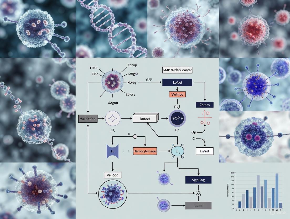
Automated Cell Counting Workflow in GMP Manufacturing
The following diagram illustrates the complete workflow for implementing and validating an automated cell counting method in a GMP environment:
Essential Research Reagent Solutions
Successful implementation of cell counting in GMP environments requires specific reagents and systems designed for regulatory compliance:
Table 3: Essential Research Reagents and Systems for GMP Cell Counting
| Reagent/System | Function | GMP Relevance |
|---|---|---|
| NucleoCounter NC-3000 | Advanced image cytometer for cell cycle analysis | Standardized results between different users, no calibration required [11] |
| BD FACSLyric Flow Cytometer | Flow cytometry with BD FACSuite Application | Supports 21 CFR Part 11 compliance with password protection, electronic signatures [12] |
| Countess II FL Automated Cell Counter | Fluorescence-based cell counting | Enables rapid assessment of cell concentration and viability [8] |
| Trypan Blue Stain | Viability staining for manual counting | Distinguishes live/dead cells via dye exclusion [9] |
| LIVE/DEAD Fixable Dead Cell Stains | Fluorescence-based viability markers | Avoids potential fluorescence quenching artifacts vs. trypan blue [8] |
| BD RUO (GMP) Reagents | Research use only reagents manufactured under GMP | Lot-to-lot consistency for standardized manufacturing QC assays [12] |
| DAPI-based Cell Cycle Assays | DNA content quantification | No RNase treatment required, enables cell cycle phase analysis [11] |
Detailed Experimental Protocols
Protocol 1: Validation of Automated Cell Counting Methods for GMP Compliance
This protocol outlines the comprehensive validation of an automated cell counting system according to ICH Q2(R1) guidelines [7] [1].
Materials
- Automated cell counter (e.g., NucleoCounter NC-100 or Countess II FL)
- Reference method materials (Bürker chamber, microscope, trypan blue)
- Cell samples (hiPSCs, MNCs, or MSCs at various concentrations)
- Dilution series materials (PBS, tubes, pipettes)
Procedure
Accuracy Assessment:
- Prepare cell suspensions at 3-5 different concentrations covering the expected working range
- Count each sample using both the automated system and the reference Bürker chamber method
- Perform statistical analysis comparing results from both methods
- Calculate percentage agreement, with acceptable accuracy being >95% agreement
Precision Testing:
- Prepare a homogeneous cell suspension at mid-range concentration
- For intra-operator precision: Have one operator perform 10 replicate counts of the same sample
- For inter-operator precision: Have 3 different operators each perform 5 replicate counts of the same sample
- Calculate mean, standard deviation, and coefficient of variation for each data set
- Acceptable precision: CV <10% for total cells, <5% for viable cells [1]
Linearity and Range Evaluation:
- Prepare a 1:1 dilution series covering the entire expected concentration range (e.g., 1:2 to 1:128 dilutions)
- Count each dilution in triplicate using the automated system
- Plot measured concentration against expected concentration and perform linear regression analysis
- Acceptable linearity: R² value >0.95 [10]
Specificity Testing:
- Prepare samples with known ratios of viable and non-viable cells (e.g., via heat treatment or ethanol fixation)
- Compare viability measurements against expected values
- Validate ability to distinguish different cell types in mixed populations if applicable
Protocol 2: Routine Cell Counting in GMP Manufacturing Using Automated Systems
This protocol details the standardized procedure for routine cell counting once the automated method has been validated.
Materials
- Validated automated cell counter (e.g., Countess II FL)
- Appropriate cell counting chamber slides
- Trypan blue stain or fluorescent viability dyes
- PBS for dilutions
Procedure
Sample Preparation:
- For adherent cells: Trypsinize, neutralize with serum-containing media, and prepare single-cell suspension
- For suspension cells: Ensure homogeneous suspension by gentle mixing
- Remove 10 μL of cell suspension and mix with 10 μL of trypan blue (for brightfield systems) or appropriate viability dye [8]
Loading and Counting:
- Pipette 10 μL of stained sample into a counting chamber slide
- Insert slide into the automated cell counter
- Allow instrument to autofocus and acquire images
- Press "Count" to initiate automated analysis
Data Analysis and Recording:
- Review automated gating for size, brightness, and circularity parameters
- Adjust gates if necessary based on histogram displays
- Record concentrations and percentages of total, live, and dead cells
- Use built-in dilution calculator if needed to determine appropriate cell dilution for downstream processes
Quality Control Measures:
- Perform regular instrument calibration according to manufacturer specifications
- Include control samples with known concentrations in each counting session
- Maintain complete documentation including sample identification, operator, date/time, and all counting parameters
The critical role of cell counting in process control and product release for ATMPs necessitates implementation of validated, automated counting methods in GMP environments. Automated cell counting systems provide the precision, accuracy, and reproducibility required for dose determination and product characterization, significantly reducing the variability inherent in manual hemocytometer methods. Through rigorous validation following ICH Q2(R1) guidelines and implementation of standardized protocols, manufacturers can ensure the quality, safety, and efficacy of cell therapy products while maintaining regulatory compliance.
In the development and quality control (QC) of advanced therapy medicinal products (ATMPs), such as human induced pluripotent stem cells (hiPSCs), adherence to a robust regulatory framework is not just a formality but a fundamental requirement for ensuring product safety, efficacy, and consistent quality [6]. The validation of critical analytical procedures, like cell counting, sits at the heart of this framework. It provides the assurance that the methods used to determine cell dose—a key potency indicator—are reliable and reproducible. This document delineates the integration of three cornerstone regulatory guidelines—ICH Q2(R1), EudraLex, and the European Pharmacopoeia (Ph. Eur.)—specifically in the context of validating cell counting methods for Current Good Manufacturing Practice (cGMP) compliant production [1] [6].
The journey of a cell-based product from the laboratory to the clinic is governed by these stringent regulations. For instance, hiPSCs destined for therapeutic applications must be manufactured as ATMPs, making their production process subject to cGMP standards [6]. Within this process, cell counting is more than a simple quantification step; it is an in-process control for monitoring cell expansion and a batch release test for determining the final therapeutic dose. The European Pharmacopoeia provides the primary legal and scientific standard for the cell count method, typically describing the manual hemocytometer as a reference [6]. However, the ICH Q2(R1) guideline provides the international benchmark for validating any analytical procedure, whether manual or automated, to demonstrate its suitability for the intended purpose. Furthermore, EudraLex Volume 4, particularly its Annexes 15 and Part IV, provides the enforceable GMP requirements for ATMP manufacturing within the European Union, mandating that all analytical methods be thoroughly validated [6]. This application note synthesizes these guidelines into a coherent strategy for validating cell counting methods, using a comparative study between an automated NucleoCounter system and a manual Bürker hemocytometer as a case study.
The Regulatory Pillars of Analytical Method Validation
ICH Q2(R1): Validation of Analytical Procedures
The ICH Q2(R1) guideline, entitled "Validation of Analytical Procedures: Text and Methodology," is the globally accepted standard for validating analytical methods. It defines the key validation parameters that must be evaluated to ensure an analytical procedure is suitable for its intended use [6]. For a potency test like cell counting, the following parameters are of critical importance:
- Accuracy: This expresses the closeness of agreement between the value found and a reference value. In cell counting, the accuracy of a new method (e.g., an automated system) is often demonstrated by comparing its results to those obtained from the pharmacopoeial reference method (e.g., Bürker hemocytometer) [1].
- Precision: This parameter evaluates the closeness of agreement between a series of measurements from multiple samplings of the same homogeneous sample. Precision has three tiers:
- Repeatability (Intra-assay Precision): Assesses precision under the same operating conditions over a short interval of time, typically performed by a single analyst.
- Intermediate Precision (Inter-Operator Reproducibility): Evaluates the influence of variations within the laboratory, such as different analysts, different days, or different equipment.
- Linearity and Range: Linearity is the ability of the method to obtain test results that are directly proportional to the analyte concentration (in this case, cell concentration) within a given range. The range is the interval between the upper and lower concentrations for which linearity, accuracy, and precision have been demonstrated [1].
EudraLex Volume 4: GMP Requirements
EudraLex, the "Rules Governing Medicinal Products in the European Union," is the comprehensive regulatory framework for medicinal products in the EU. Volume 4 of EudraLex contains the principles and guidelines of Good Manufacturing Practice (GMP) [13]. For ATMPs, which include many cell therapies, the requirements outlined in Annex 15 ("Qualification and Validation") and the dedicated Part IV ("GMP requirements for Advanced Therapy Medicinal Products") are directly applicable [6]. These documents mandate that the manufacturing process, including all in-process and batch release QC tests, must be validated. They emphasize that equipment, instruments, and software used in a cGMP facility must undergo Installation Qualification (IQ) and Operational Qualification (OQ) to ensure they function according to specifications. Furthermore, any computerized system, including that of an automated cell counter, must comply with Annex 11 on "Computerised Systems," ensuring data integrity and security [6].
European Pharmacopoeia: Legal Quality Standards
The European Pharmacopoeia (Ph. Eur.) is a single, legally binding collection of quality standards for medicines and their ingredients in all signatory states (currently 39 European countries and the EU). Its standards are a legal requirement for marketing authorizations. The Ph. Eur. provides monographs and general chapters that describe specific analytical methods. For cell counting, the general chapter 2.7.29 details the reference method using a hemocytometer [6]. It is important to note that as of June 2025, the Ph. Eur. transitioned to an online-only format with its 12th Edition, published in three cumulative issues per year (.1, .2, .3). Users must subscribe via a 365-day licence to access the current, legally binding standards [14] [15]. When validating an alternative counting method (like an automated system), its results must be demonstrated to be at least as reliable as those generated by the Ph. Eur. reference method.
Interplay of Guidelines in Practice
In a practical cGMP setting, these guidelines are not applied in isolation but are deeply intertwined. A validation strategy for a cell counting method must be designed to satisfy ICH Q2(R1) parameters, operate within the quality system defined by EudraLex Volume 4, and use the Ph. Eur. method as the benchmark for comparison. The overarching goal is to generate validated, reliable data that supports the quality of the ATMP throughout its lifecycle.
Experimental Validation: A Case Study on Automated Cell Counting
Aim and Design
A pivotal 2022 study provides a clear template for validating an automated cell counting method for cGMP manufacturing of hiPSCs [6] [2]. The study's aim was to validate the NucleoCounter NC-100, a fluorescence imaging-based automated system, against the reference Bürker hemocytometer method described in the Ph. Eur. 10th Edition [6]. The validation was designed to comply with ICH Q2(R1), EudraLex Volume 4 (Annex 15 and Part IV), and relevant ISO standards [6].
The experimental design was rigorous to account for biological and operational variability:
- Cell Type: Three independent research-grade hiPSC batches were used to incorporate biological variability.
- Operators: Two independent analysts performed the counts to assess inter-operator reproducibility.
- Replication: For each batch, three independent runs of analysis were performed, with samples prepared independently for each run [6].
Prerequisites: GMP Compliance Foundations
Before initiating the analytical validation, several cGMP prerequisites were ensured, as required by EudraLex:
- Instrument Qualification: The NucleoCounter NC-100 system underwent Installation (IQ) and Operational Qualification (OQ) to confirm it was installed correctly and functioned according to manufacturer specifications [6].
- Computerized System Compliance: The instrument's software was compliant with EudraLex Annex 11 on "Computerised Systems" [6].
- Reagent and Personnel Control: All reagents were of appropriate quality and verified before use. Personnel were adequately qualified, trained, and had relevant practical experience [6].
The following workflow diagram illustrates the key stages of the validation process, from sample preparation to data analysis for both counting methods.
Detailed Experimental Protocols
Sample Preparation Protocol
- Cell Culture: hiPSCs were expanded on Matrigel-coated surfaces in TeSR-E8 medium at 37°C, 20% O₂, 5% CO₂ [6].
- Cell Harvesting: Cells were dissociated into a single-cell suspension using accutase incubation for 5 minutes at 37°C [6].
- Cell Washing: The cell suspension was pelleted by centrifugation at 300 ×g for 10 minutes and the resulting pellet was resuspended in Dulbecco's Phosphate Buffered Saline (d-PBS) without Ca²⁺ and Mg²⁺ [6].
- Sample Dilution: The cell suspension was diluted to fall within the optimal analytical range for both the manual (50,000–550,000 cells/mL) and automated (5,000–2,000,000 cells/mL) counting methods [6].
Manual Cell Counting Protocol (Bürker Hemocytometer)
- Loading: A volume of 10 µL of cell suspension was loaded into each chamber of the hemocytometer [6].
- Counting: For each sampling, counts were performed in duplicate by two independent analysts using a microscope. Viable cells were identified and counted based on morphology [6].
- Calculation: Cell concentration was calculated based on the counted cells and the known volume of the hemocytometer chamber.
Automated Cell Counting Protocol (NucleoCounter NC-100)
- Principle: The method is based on the fluorescence detection of propidium iodide (PI). PI is a DNA dye that only enters cells with compromised membranes (non-viable cells) [6].
- Viable Cell Count: A sample is analyzed without pretreatment. The software automatically identifies nuclei and calculates the starting cell concentration [6].
- Total Cell Count: For a total cell count, 100 µL of cell suspension was pretreated with a mixture of 100 µL of lysis buffer and 100 µL of stabilizing buffer to permeabilize all cells before measurement [6].
Key Validation Parameters and Results
The validation of the automated cell counting method focused on critically assessing the parameters defined by ICH Q2(R1). The quantitative results from the case study are summarized in the table below, which provides a clear, side-by-side comparison of the performance of the manual and automated methods.
Table 1: Summary of Validation Parameters and Results for Manual vs. Automated Cell Counting
| Validation Parameter | Experimental Procedure | Manual Bürker Method | Automated NucleoCounter NC-100 Method |
|---|---|---|---|
| Accuracy | Comparison of results to the Ph. Eur. reference method (Bürker) | (Reference Method) | Showed close agreement with the reference method [6] |
| Precision (Repeatability) | Multiple counts of the same sample by a single analyst in one session | Higher operator-dependent variability [6] | Higher precision (lower CV%) than manual method [6] |
| Precision (Intermediate Precision) | Multiple counts by different analysts on different days | Significant inter-operator variation observed [6] | Higher inter-operator reproducibility (lower CV%) [6] |
| Specificity | Analysis of sample matrix (d-PBS) to check for interference | Not formally evaluated as a significant risk [6] | No interference from the matrix was detected [6] |
| Linearity & Range | Analysis of a series of sample dilutions | Optimal range: 50,000–550,000 cells/mL [6] | Demonstrated linearity across a wider range: 5,000–2,000,000 cells/mL [6] |
The data conclusively demonstrated that the automated NucleoCounter NC-100 method met all validation criteria. It showed higher precision (both intra- and inter-operator) than the manual method and excelled in linearity across a significantly wider working range [6]. This makes it particularly suitable for processes where cell concentrations can vary greatly.
The Scientist's Toolkit: Essential Reagents and Materials
The successful execution of a validated cell counting method relies on the use of specific, high-quality materials. The following table details the key reagents and solutions used in the featured case study and their critical functions in the protocol.
Table 2: Key Research Reagent Solutions for Cell Counting Validation
| Item / Reagent | Function / Purpose in the Protocol | Example / Note |
|---|---|---|
| Bürker Hemocytometer | The reference counting chamber as per European Pharmacopoeia. Provides a grid for manual cell enumeration under a microscope [6]. | An "Improved Neubauer" chamber is a common variant [16]. |
| NucleoCounter NC-100 | Automated, fluorescence-based cell counter. Proposed method for validation; reduces analyst-dependent variability [6]. | System includes the instrument and disposable Via2-Cassettes [6]. |
| d-PBS (without Ca²⁺ and Mg²⁺) | A balanced salt solution used to wash and resuspend cells without triggering differentiation or clumping. Serves as the sample matrix [6]. | Used to ensure cells are in a non-activating, stable buffer for counting. |
| Accutase | A enzymatic cell detachment solution used to dissociate adherent hiPSC cultures into a robust single-cell suspension [6]. | Critical for obtaining an accurate and representative count of hiPSCs. |
| Propidium Iodide (PI) | A fluorescent DNA dye used by the NucleoCounter system. It is excluded by viable cells but enters dead cells, staining their nuclei [6]. | Allows for automated differentiation between viable and non-viable cells. |
| Lysis & Stabilizing Buffer | Specific reagents for the NucleoCounter. Used to lyse all cells in a sample for the total cell count protocol [6]. | Proprietary reagents provided with the instrument. |
The integration of ICH Q2(R1), EudraLex, and the European Pharmacopoeia provides a complete and non-negotiable roadmap for the validation of analytical methods in a cGMP environment for ATMPs. The case study on validating the NucleoCounter NC-100 for hiPSC counting demonstrates a practical application of this framework. The study confirmed that the automated method was not only successful in meeting all regulatory validation parameters but also offered significant advantages over the traditional manual method, including superior precision, reduced operator dependency, and a broader linear range [6].
For researchers and drug development professionals, this validation paradigm is essential. It moves cell counting from a basic laboratory technique to a fully controlled, documented, and validated analytical procedure. This rigor is mandatory for generating the high-quality data required for regulatory submissions, ensuring that the cell-based therapies delivered to patients are safe, effective, and of consistent quality. Adopting such a meticulous approach paves the way for the successful clinical translation of innovative regenerative medicine products.
In the development and quality control of cell therapy products, cell counting is a fundamental measurement technique crucial for assessing the number, viability, and purity of these living therapeutics [17]. Unlike conventional drugs, cell therapy products—such as CAR-T cells, mesenchymal stem cells (MSCs), and human induced pluripotent stem cells (hiPSCs)—leverage the properties of living cells for their therapeutic effects, making accurate cell counting indispensable for evaluating potency and effectiveness [17]. Despite its widespread use, manual hemocytometer counting presents significant limitations that can compromise data integrity, particularly in current Good Manufacturing Practice (cGMP) environments where precision and standardization are paramount. This application note examines the critical challenges of operator dependency and time consumption associated with manual hemocytometer counting, providing quantitative data and experimental protocols to support method validation and transition to automated systems.
Key Limitations of Manual Hemocytometer Counting
Operator Dependency and Variability
Manual cell counting using a hemocytometer is highly susceptible to human interpretation, leading to significant variability even among trained personnel. This subjectivity manifests in multiple aspects of the counting process:
- Cell Identification Criteria: Individual operators adhere to different criteria for defining a cell versus cell debris or other particles, including varying thresholds for stain intensity to classify cells as viable or dead [18]. This variation in human perception can be extremely detrimental to experimental setup and analysis.
- Viability Assessment Challenges: When using trypan blue for viability assessment, the intensity of the stain can vary within a sample, making it difficult to determine whether a cell stains positive [18]. The toxic nature of trypan blue also means viable cells are eventually stained if not analyzed within 5-30 minutes, potentially leading to viability underestimation [18].
- Statistical Limitations: The standard practice of manually counting approximately 100 cells introduces substantial statistical uncertainty. According to Poisson distribution, counting only 100 cells yields a minimum standard variation of 10% even without human-introduced variations [18].
Table 1: Sources of Operator-Induced Error in Manual Hemocytometer Counting
| Error Source | Impact on Results | Quantitative Effect |
|---|---|---|
| Cell Recognition Subjectivity | Inconsistent cell vs. debris discrimination | Inter-operator CV ≥ 15% common [18] |
| Viability Interpretation | Variable live/dead cell classification | Trypan blue toxicity alters viability over time [18] |
| Statistical Sampling Error | Low cell count increases variance | ~10% SD when counting 100 cells [18] |
| Grid Selection Bias | Non-uniform cell distribution sampling | Underestimates or overestimates concentration [19] |
Validation studies conducted in cGMP facilities demonstrate that when multiple analysts count the same sample using hemocytometers, the inter-operator variation typically exceeds 15% coefficient of variation (CV) [18]. This level of variability is particularly problematic for cell therapy products where precise dosing is critical for patient safety and therapeutic efficacy.
Time Consumption and Procedural Inefficiencies
The manual hemocytometer counting process is inherently time-consuming and labor-intensive, creating bottlenecks in cell manufacturing workflows:
- Labor-Intensive Process: Manual counting requires careful sample preparation, chamber loading, microscopic examination, and manual recording of counts for large numbers of cells [6] [20]. This process becomes particularly burdensome in medium- to high-throughput environments where multiple samples require processing.
- Post-Counting Cleanup: Unlike automated systems with disposable consumables, hemocytometers must be thoroughly cleaned between samples to prevent cross-contamination, adding significant time to the overall process [20].
- Workflow Disruption: The time-sensitive nature of cell processing means that delays in counting can compromise cell health. Studies show that diluents such as phosphate-buffered saline can lower cell viability by 25% after just five minutes of incubation [21].
Table 2: Time and Efficiency Comparison of Cell Counting Methods
| Process Step | Manual Hemocytometer | Automated System |
|---|---|---|
| Sample Preparation | 2-5 minutes (staining, dilution) | ~1 minute (direct loading) |
| Instrument Setup | 1-2 minutes (cover slip placement) | Minimal (instrument ready) |
| Measurement Time | 5-10 minutes per sample | 30-60 seconds per sample |
| Data Recording | Manual transcription | Automated digital recording |
| Cleanup | 2-3 minutes (cleaning, drying) | Minimal (disposal or quick clean) |
| Total Time/Sample | 10-20 minutes | ~2-3 minutes |
The cumulative effect of these inefficiencies becomes substantial in manufacturing settings where multiple batches require regular monitoring, ultimately impacting production timelines and resource allocation.
Experimental Protocols for Method Validation
Protocol 1: Inter-Operator Variability Assessment
Purpose: To quantify the inter-operator variability of manual hemocytometer counting within a laboratory setting.
Materials:
- Hemocytometer (Bürker or Neubauer)
- Microscope with 20X objective
- Trypan blue solution or AO/DAPI stains
- Cell suspension (recommended concentration: 1-2×10^6 cells/mL)
- Timer
- Data recording sheets
Procedure:
- Prepare a homogeneous cell suspension from a standard cell line (e.g., hiPSCs expanded in defined culture conditions [6]).
- Aliquot five 200 μL samples of the same cell suspension into separate tubes. Thoroughly mix the sample before aliquoting.
- Engage five trained operators to count one aliquot each without communication or data sharing.
- Each operator performs counting using their normal standards and typically counted squares (do not count more squares or cells than normal).
- Operators record viable cell count based on morphology or staining, and calculate concentration using standard hemocytometer formulas.
- Calculate the arithmetic mean, standard deviation, and coefficient of variation (CV) for the five results.
Validation Parameters:
- Precision: Calculate inter-operator CV; CV ≥ 15% indicates significant variability typical of manual counting [18].
- Accuracy: Compare mean operator values to automated reference method if available.
Protocol 2: Time-Motion Efficiency Analysis
Purpose: To quantitatively compare the time investment required for manual versus automated cell counting methods.
Materials:
- Hemocytometer setup
- Automated cell counter (e.g., NucleoCounter NC-100 or similar)
- Cell suspensions (n=5 samples of identical origin)
- Timer
- Data collection spreadsheet
Procedure:
- Prepare five identical cell samples from a homogeneous suspension.
- For manual counting:
- Record time for sample staining/mixing, hemocytometer loading, microscopic counting, data recording, and hemocytometer cleaning.
- Repeat for all five samples with one operator.
- For automated counting:
- Record time for sample loading, measurement initiation, result acquisition, and system preparation for next sample.
- Repeat for all five samples.
- Calculate total time investment and average time per sample for both methods.
- Compare cell viability results between methods, noting that extended time in suspension can impact viability measurements [21].
Validation Parameters:
- Efficiency: Time savings with automated systems typically demonstrate 70-80% reduction in hands-on time [6] [20].
- Viability Impact: Assess correlation between time-in-suspension and viability measurements.
Essential Research Reagents and Materials
Table 3: Key Research Reagent Solutions for Cell Counting Validation
| Reagent/Material | Function | Application Notes |
|---|---|---|
| Bürker Hemocytometer | Standardized chamber for manual cell counting | Reference method described in European Pharmacopoeia 10th ed. [6] |
| Trypan Blue Solution | Exclusion dye for viability assessment | Toxic to cells; requires rapid analysis (5-30 min) [18] |
| AO/DAPI Stains | Fluorescent nucleic acid binding dyes | AO stains total cells; DAPI identifies dead cells via membrane integrity [18] |
| Propidium Iodide (PI) | Membrane-impermeable DNA dye | Used in automated systems like NucleoCounter for dead cell identification [6] |
| Accutase Enzyme | Gentle cell dissociation | Maintains cell viability for accurate counting [6] |
| Phosphate-Buffered Saline (PBS) | Cell suspension and washing medium | May reduce cell viability during extended incubation [21] |
| Dimethyl Sulfoxide (DMSO) | Cryoprotectant in cell preservation | Can interfere with fluorescence staining at higher concentrations [17] |
Workflow and Decision Pathways
Figure 1: Decision pathway for evaluating cell counting methods in GMP environments. The workflow highlights how identifying limitations of manual counting leads to automated method validation and implementation.
Figure 2: Comparative workflow analysis of manual versus automated cell counting processes. The automated pathway demonstrates significant efficiency advantages with fewer steps and reduced hands-on time.
The limitations of manual hemocytometer counting—particularly operator dependency and time consumption—present significant challenges in GMP-compliant manufacturing of cell therapies. Quantitative validation studies consistently demonstrate inter-operator variability exceeding 15% CV and processing times 5-10 times longer than automated systems. These limitations directly impact product quality, manufacturing efficiency, and regulatory compliance. Implementation of validated automated cell counting methods, such as fluorescence-based image cytometry systems, addresses these challenges by standardizing cell enumeration, improving precision, and integrating more effectively with cGMP requirements for advanced therapy medicinal products. The experimental protocols provided herein enable systematic evaluation of counting methods to support data-driven decisions in method selection and validation.
The transition from manual hemocytometer-based cell counting to automated methods represents a critical evolution in Good Manufacturing Practice (GMP) for cell and gene therapy products. Manual counting, while historically the reference method, is inherently prone to operator-dependent variability and is time-consuming, making standardization across manufacturing protocols challenging [6]. Automated cell counting systems, such as the NucleoCounter series, offer a paradigm shift by enhancing precision, ensuring data integrity, and supporting full traceability—attributes that are indispensable for manufacturing Advanced Therapy Medicinal Products (ATMPs) under current GMP standards [6] [22]. This application note details the validation and implementation of an automated cell counting method within a GMP framework, providing a direct comparative analysis against the manual hemocytometer and outlining a standardized protocol for ensuring compliance.
Comparative Analysis: Automated vs. Manual Cell Counting
The validation of an analytical method must demonstrate its suitability for the intended purpose, focusing on key performance metrics as outlined in ICH Q2(R1) and ISO 20391 guidelines [6] [23]. A study validating the NucleoCounter NC-100 system for counting human induced pluripotent stem cells (hiPSCs) provides a compelling quantitative comparison against the manual Bürker hemocytometer, which is the reference method described in the European Pharmacopoeia [6].
Table 1: Performance Comparison of Cell Counting Methods for hiPSCs
| Performance Metric | Manual Hemocytometer | Automated NucleoCounter NC-100 |
|---|---|---|
| Principle | Visual identification and counting via microscope [24] | Fluorescence-based imaging with propidium iodide staining [6] |
| Accuracy (Comparison to Reference) | Reference Method | High accuracy demonstrated versus manual method [6] |
| Intra-Assay Precision (CV) | Higher variability | Significantly higher precision (Lower CV) [6] |
| Inter-Operator Precision | Dependent on analyst expertise and experience [6] [24] | Minimal variability between different analysts [6] |
| Sample Analysis Time | Time-consuming (e.g., 5-10 minutes per sample) [6] | Rapid (results in minutes) [6] |
| Standardization | Difficult to standardize; requires strict SOPs and multiple operators for validation [6] [24] | High level of standardization through automated protocols [6] |
| Data Traceability | Manual recording in logbook; prone to transcription errors | Direct digital capture and storage; full audit trail [25] |
| Key Advantage | Low-cost equipment; described in pharmacopeia | Precision, speed, and compliance for GMP manufacturing [6] |
The data underscores the operational advantages of automation. The precision of the NucleoCounter system, evidenced by lower coefficients of variation (CV), translates to more reliable and consistent data for critical decisions, from in-process controls to final product dose determination [6]. Furthermore, the significant reduction in inter-operator variability ensures that cell counts are reproducible regardless of the staff performing the analysis, a fundamental requirement for robust manufacturing processes.
GMP Compliance and Regulatory Framework
Implementing an automated cell counter in a GMP environment requires more than just instrument procurement; it necessitates a rigorous qualification and validation process aligned with regulatory guidance.
Foundational Standards
The ISO 20391-1 standard provides the essential framework for cell counting quality control, defining core concepts such as accuracy (closeness to the true value), precision (consistency of repeated measurements), and measurement uncertainty [23]. Adherence to this standard ensures data reliability and international credibility [23].
The Qualification Process: IQ, OQ, PQ
A structured qualification process is mandatory for GMP compliance [6] [23]:
- Installation Qualification (IQ): Verifies the instrument is correctly installed as per manufacturer specifications in the intended environment [6] [23].
- Operational Qualification (OQ): Confirms the instrument operates according to its functional specifications, often using built-in tests or standard reference materials [6] [23].
- Performance Qualification (PQ): Demonstrates the instrument performs as expected for its specific analytical application using actual cell samples, verifying accuracy, precision, and linearity over the intended range [6] [23].
Regulatory Guidance for Cell and Gene Therapies
Manufacturers must also consult relevant regulatory guidances. The U.S. FDA provides numerous documents specific to cellular and gene therapy products, covering aspects from Chemistry, Manufacturing, and Control (CMC) and potency assurance to manufacturing changes and comparability [26]. Following these guidelines, coupled with a comprehensive Quality Management Program, is crucial for maintaining safety and reproducible product quality from material procurement to product administration [27].
The following workflow diagram illustrates the complete pathway from sample preparation to GMP-compliant data output using an automated system.
Detailed Experimental Protocol: Validation of an Automated Cell Counting Method
This protocol is adapted from a published study validating the NucleoCounter NC-100 for hiPSCs in a cGMP environment [6]. It can be tailored for other automated systems and cell types.
Aim and Scope
To validate the accuracy, precision, and linearity of an automated cell counting system against the manual hemocytometer reference method for a specific cell type (e.g., hiPSCs, T cells, PBMCs).
Materials and Equipment
The Scientist's Toolkit: Essential Materials for cGMP Cell Counting Validation
| Item | Function / Description | GMP Consideration |
|---|---|---|
| NucleoCounter NC-100/250 | Automated, fluorescence-based cell counter for concentration and viability [6] | Requires IQ/OQ/PQ; software compliant with 21 CFR Part 11 [6] |
| Via1-Cassette | Disposable cassette with integrated fluorescent dyes (e.g., Acridine Orange, DAPI) for staining nuclei [28] | Single-use, sterile, reduces cross-contamination |
| Bürker Hemocytometer | Manual counting chamber as the reference method [6] | Must be included in method validation; requires strict SOP [24] |
| Trypan Blue Solution | Vital dye for distinguishing live (unstained) from dead (blue) cells in manual counts [24] | Quality-controlled reagent; note cytotoxic nature limits staining time [24] |
| Single-Cell Suspension | Sample prepared using enzyme (e.g., Accutase) to dissociate cell clusters [6] | Critical for accuracy in both manual and automated counts [22] |
| cGMP-Qualified Reagents | Cell culture media, enzymes, buffers (e.g., DPBS) | Sourced from qualified vendors, appropriate for human-use products [27] |
Step-by-Step Procedure
Sample Preparation (for both methods):
- Culture and expand the target cells (e.g., hiPSCs on Matrigel) following standard cGMP-compliant protocols [6].
- Wash cells and dissociate into a single-cell suspension using a validated enzyme (e.g., Accutase incubation at 37°C for 5 minutes) [6].
- Quench the enzyme activity with an appropriate buffer, pellet cells by centrifugation (e.g., 300 ×g for 10 min), and resuspend the pellet in DPBS.
- Prepare a dilution series of the cell suspension (e.g., 1:1, 1:2, 1:3, 1:4, 1:5) to assess the linearity of the counting methods over a range of concentrations [22].
Automated Counting (NucleoCounter):
- Ensure the NucleoCounter instrument has a valid IQ/OQ/PQ status.
- Pipette 100 µL of the cell suspension into a Via1-Cassette. The cassette contains lytic and staining reagents.
- Insert the cassette into the instrument. The software will automatically acquire images of fluorescently stained nuclei and calculate the total and viable cell concentration (cells/mL) and viability (%).
Manual Counting (Hemocytometer):
- Mix 50 µL of the cell suspension with 50 µL of 0.4% Trypan Blue solution (1:2 dilution) [24]. Note: Count within 5 minutes due to trypan blue cytotoxicity [24].
- Carefully load approximately 10 µL of the mixture into both chambers of a clean Bürker hemocytometer using a pipette.
- Using a microscope with a 10x objective, count the viable (unstained) and non-viable (blue) cells in the four large corner squares of each chamber (total of 8 squares) [24].
- Follow standardized counting rules: cells touching the top and left boundary lines are counted; cells touching the bottom and right lines are not counted [24].
Data Analysis and Validation:
- Precision (Intra-assay): Calculate the Coefficient of Variation (CV = Standard Deviation / Mean) for replicate measurements (e.g., n=3) of the same sample for both methods. The automated method should demonstrate a lower CV [6].
- Accuracy & Linearity: Use linear regression analysis to compare the cell concentrations obtained by the automated method (y-axis) against those from the manual method (x-axis) across the dilution series. A slope close to 1.0 and a high coefficient of determination (R²) indicate good agreement and linearity [6].
- Specificity: Analyze a blank sample (DPBS) with the automated system to confirm it does not detect particulate background interference [6].
The move from manual hemocytometry to automated cell counting is a strategic imperative for any GMP-focused operation. As validated by rigorous studies, automated systems like the NucleoCounter deliver the superior precision, standardized output, and embedded traceability required for the manufacturing of ATMPs. By following the structured validation protocol and regulatory framework outlined in this application note, researchers and manufacturers can effectively justify and implement automated cell counting, thereby strengthening the foundation of quality, safety, and efficacy for their cell and gene therapy products.
Implementing Automated Counting: From Sample Prep to Data Management
The NucleoCounter platform represents a significant advancement in automated cell counting and viability analysis, utilizing fluorescence-based image cytometry to provide highly precise and objective measurements. This technology is particularly vital in Good Manufacturing Practice (GMP) environments, such as cell therapy production and biopharmaceutical manufacturing, where accurate cell dosing is a critical potency test and manual methods like hemocytometers are prone to human error and subjectivity [29] [1]. By employing a standardized, cassette-based system with immobilized fluorescent dyes, the NucleoCounter eliminates pipetting errors and human counting variation, ensuring consistency across instruments, users, and sites [29].
The core principle of the system relies on the distinct staining characteristics of two fluorescent dyes: acridine orange (AO) and 4′,6-diamidino-2-phenylindole (DAPI). AO is a membrane-permeable dye that stains all nucleated cells by binding to nucleic acids, while DAPI can only penetrate cells with compromised membranes, selectively staining non-viable cells [29]. This differential staining allows the instrument's detection system to automatically distinguish between live and dead cell populations while excluding debris, providing a comprehensive assessment of total cell count, viability percentage, and concentration in less than one minute [29].
Principles of Fluorescence-Based Viability Assessment
The NucleoCounter system employs a sophisticated biochemical approach to cell viability assessment through its proprietary Via2-Cassette, which contains immobilized fluorescent dyes in a pre-calibrated, single-use chamber. The fundamental mechanism operates on the integrity of the cell membrane, a key indicator of cellular health, using two DNA-binding dyes with different membrane permeability properties [29].
Acridine Orange (AO) Staining Principle: As a membrane-permeable vital dye, AO readily enters all cells regardless of viability status and intercalates with nucleic acids (both DNA and RNA). When excited by the appropriate wavelength, it emits fluorescence that allows the detection of the total cell population in the sample. This provides the denominator for viability calculations.
DAPI Staining Principle: DAPI is a membrane-impermeant dye that can only enter cells with damaged or compromised plasma membranes – a characteristic feature of dead or dying cells. Once inside non-viable cells, DAPI binds strongly to AT-rich regions in DNA and produces a distinct fluorescent signal upon excitation. This selectively identifies the non-viable cell population.
The instrument's optical system and analysis software automatically detect and quantify the signals from both dyes, applying sophisticated algorithms to distinguish intact viable cells (AO-positive only) from non-viable cells (both AO and DAPI positive), while excluding acellular debris that doesn't stain with either dye [29]. This comprehensive approach provides objective viability assessment without the subjectivity inherent in manual trypan blue exclusion methods.
Table 1: Fluorescent Dyes Used in NucleoCounter Viability Assessment
| Dye Name | Membrane Permeability | Nucleic Acid Target | Cell Population Stained | Excitation/Emission Characteristics |
|---|---|---|---|---|
| Acridine Orange (AO) | Permeable to all cells | DNA and RNA | Total cell population | Blue excitation/Green emission |
| DAPI | Impermeant (enters only damaged cells) | DNA | Non-viable cells only | UV excitation/Blue emission |
Experimental Validation & Performance Data
Extensive validation studies demonstrate that NucleoCounter technology provides superior precision and reproducibility compared to manual counting methods, with significantly lower coefficients of variation (CV). According to manufacturer specifications and independent studies, the NC-202 model typically provides a CV below 5%, far exceeding the precision of manual hemocytometer counting which often exhibits CVs of 10-30% depending on operator experience [29] [1].
A 2025 study validating cell counting methods for corneal endothelial cell therapy directly compared manual hemocytometry with automated cell counters, including the NucleoCounter system, and found "comparable accuracy and reproducibility" between the methods, supporting the transition to automated systems for critical therapeutic applications [30]. The precision of NucleoCounter instruments is maintained through multiple technological innovations: fixed focus and exposure settings to eliminate adjustment variability, individual calibration of each cassette to ensure volume accuracy, and instrument calibration against reference standards to guarantee consistency across devices [29].
In a recent methodological validation study using the Countess 3 FL automated cell counter (a similar fluorescence-based system) for tear-derived cell analysis, researchers reported an intra-assay CV of just 4.7%, demonstrating the exceptional repeatability achievable with automated fluorescence counting systems [31]. This level of precision is particularly crucial in GMP environments where cell counting constitutes a potency test, and validated methods must demonstrate high reproducibility for batch release purposes [1].
Table 2: Performance Metrics of NucleoCounter Technology vs. Manual Counting
| Performance Parameter | NucleoCounter NC-202 | Manual Hemocytometer | Significance in GMP Context |
|---|---|---|---|
| Coefficient of Variation (Precision) | Typically <5% [29] | Often 10-30% [1] | Essential for reliable potency testing |
| Sample Processing Time | ~1 minute per sample [29] | 5-10 minutes per sample | Improved efficiency in quality control |
| Operator Dependency | Minimal inter-operator variation [29] | High inter-operator variability [1] | Critical for method transfer and multi-site studies |
| Sample Volume Requirements | Standardized via pre-calibrated cassettes | Variable depending on loading technique | Eliminates pipetting errors in sample preparation |
| 21 CFR Part 11 / GMP Compliance | Built-in compliance features [29] [32] | Requires extensive documentation and validation | Streamlines regulatory compliance |
Detailed Experimental Protocols
Universal Cell Counting and Viability Protocol
The standard procedure for determining cell count and viability using the NucleoCounter system consists of three simplified steps that can be completed in approximately one minute:
Sample Collection: Gently homogenize the cell suspension to ensure even distribution. Using the attached piston, draw approximately 50 μL of cell suspension into the Via2-Cassette. The cassette automatically mixes the sample with the pre-dried fluorescent dyes (Acridine Orange and DAPI) in a standardized ratio [29].
Sample Analysis: Insert the loaded Via2-Cassette into the instrument's cassette port. Select the appropriate counting protocol based on cell type (universal protocol is suitable for most mammalian cells). Initiate the measurement cycle, which typically completes within 30-60 seconds [29].
Data Collection and Interpretation: View the results on the instrument display or connected computer running NC-View software. The system automatically displays total cell concentration (cells/mL), viable cell concentration (cells/mL), and viability percentage (%). The software also allows visualization of fluorescent images showing viable (green) and non-viable (blue) cells for quality control verification [29] [33].
Specialized Protocol for Complex Samples: PBMCs and Aggregated Cells
For challenging sample types such as peripheral blood mononuclear cells (PBMCs) or aggregated cells, modified protocols optimize counting accuracy:
PBMC Protocol: Select the dedicated PBMC application protocol which incorporates specific parameters for the size distribution and staining characteristics of lymphocyte populations. The system's lysing solution can be employed to dissociate subtle aggregates without damaging cell integrity [33].
Aggregated Cell Protocol: For stem cells or other cultures prone to aggregation, use the specialized dissociation protocol that combines chemical dissociation with optimized analysis algorithms that can accurately identify individual cells within clusters. This approach has been specifically validated for mesenchymal stem cells and induced pluripotent stem cells [29] [32].
Protocol for Fluorescence-Based Immunophenotyping
Adapting the NucleoCounter for additional fluorescence parameters enables basic immunophenotyping capabilities alongside viability assessment, as demonstrated in a 2025 study analyzing HLA-DR and CD3 expression in tear-derived cells:
Sample Preparation: Centrifuge cells and resuspend pellet in 1% BSA in PBS at approximately 1×10⁶ cells/mL [31].
Antibody Staining: Incubate cell suspension with fluorescently-conjugated antibodies (e.g., PE anti-HLA-DR at 1:50 dilution and FITC anti-CD3 at 1:100 dilution) for 1 hour at 37°C in the dark [31].
Analysis: Load stained cells into Via2-Cassette and select multi-fluorescence protocol. The system will quantify total cells (brightfield), viable/dead cells (AO/DAPI), and antigen-positive cells (additional fluorescence channels) simultaneously [31].
Research Reagent Solutions and Essential Materials
The NucleoCounter system employs a standardized set of reagents and consumables designed to optimize performance and maintain consistency across applications:
Table 3: Essential Research Reagents and Materials for NucleoCounter Experiments
| Reagent/Material | Function | Application Specifics |
|---|---|---|
| Via2-Cassette | Single-use sample chamber | Pre-loaded with immobilized AO and DAPI dyes; individually volume-calibrated for precision [29] |
| Acridine Orange (AO) | Fluorescent vital dye | Membrane-permeable nucleic acid stain; labels total cell population [29] |
| DAPI | Fluorescent dead cell stain | Membrane-impermeant dye; selectively labels non-viable cells with compromised membranes [29] |
| PBS or Appropriate Buffer | Sample diluent | Maintains cell viability and prevents aggregation during analysis |
| Lysis Buffer (Optional) | Dissociation of aggregates | Specifically formulated for challenging samples like PBMCs or stem cell aggregates [33] |
| Trypan Blue (Validation) | Reference method comparison | For method validation against traditional viability assessment [1] |
| Specific Antibody Conjugates (Advanced Applications) | Immunophenotyping | Fluorescently-labeled antibodies for simultaneous surface marker analysis [31] |
Compliance with Regulatory Standards
The NucleoCounter platform is designed to meet rigorous regulatory requirements for therapeutic product manufacturing. The NC-200, NC-202, NC-250, and NC-3000 instruments are Annex-11 and 21 CFR Part 11-ready, featuring electronic signature capabilities, audit trails, and data security measures essential for GMP compliance [29] [32]. This regulatory alignment is further strengthened by the system's inherent validation-friendly characteristics, including minimal operator-induced variability and comprehensive documentation features.
For cell-based therapies, where cell counting constitutes a critical potency test, the validation of counting methods must follow ICH Q2 guidelines and European Pharmacopoeia standards, assessing accuracy, precision, repeatability, linearity, and range [1]. The NucleoCounter system facilitates this validation through its consistent performance and low variability. Furthermore, the technology aligns with the principles of ISO 20391-2, which provides a framework for validating cell counting methods through statistical evaluation of repeatability, reproducibility, and proportionality using metrics such as Coefficient of Variation (CV), Coefficient of Determination (R²), and Proportionality Index (PI) [34].
This compliance framework ensures that cell counting data generated using NucleoCounter technology maintains the integrity and reliability required for regulatory submissions in advanced therapy medicinal products (ATMPs), supporting the translation of cell-based research from bench to bedside [30] [1].
Sample Preparation Protocols for hiPSCs and Other Sensitive Cell Types
For researchers working with human induced pluripotent stem cells (hiPSCs) and other sensitive cell types, robust sample preparation is the foundational step that determines the success of downstream applications, particularly in Current Good Manufacturing Practice (cGMP) environments. Sample preparation for cell counting is not merely a technical prerequisite but a critical analytical procedure that directly impacts data reliability, process validation, and ultimately, the safety and efficacy of advanced therapy medicinal products (ATMPs) [7] [6]. The unique biological properties of hiPSCs—including their sensitivity to mechanical and chemical stresses, tendency to form aggregates, and critical viability requirements for therapeutic applications—demand specialized protocols that go beyond standard cell culture practices [6].
Within cGMP frameworks, the accuracy of cell counting directly influences critical quality attributes, including potency assessments and dosing accuracy for cell-based therapies [6]. Studies have demonstrated that inconsistent sample preparation introduces significant variability in cell counting results, with errors often reaching 20-30% when using traditional hemocytometer methods [16]. This technical brief provides detailed, validated protocols for preparing hiPSCs and other sensitive cell types for cell counting, with specific emphasis on method validation suitable for GMP-compliant manufacturing and research.
Fundamental Principles for Handling Sensitive Cells
Successful sample preparation for sensitive cell types adheres to three core principles: maintaining viability, ensuring sample representativeness, and maintaining phenotype. Physical and chemical stresses during preparation can induce apoptosis, differentiation, or phenotypic changes that compromise both counting accuracy and cell functionality [35] [36].
Recent research has revealed that physical properties of the microenvironment, including substrate stiffness and topography, significantly influence stem cell behavior and differentiation [36]. While these factors are primarily considered during culture, they highlight the importance of gentle handling during preparation to minimize unintended signaling. Furthermore, the choice of detachment reagents and their exposure time must be optimized to preserve surface proteins and membrane integrity, which are critical for both cell counting and subsequent therapeutic function [35] [37].
Detailed Sample Preparation Protocols
Protocol 1: Standard Preparation of hiPSCs for Cell Counting
This protocol is validated for hiPSCs expanded under defined conditions and is suitable for in-process monitoring during cGMP manufacturing [6].
Materials and Reagents
Table 1: Essential Reagents for hiPSC Sample Preparation
| Reagent/Material | Specification | Function | Notes |
|---|---|---|---|
| Accutase | GMP-grade, if available | Enzymatic detachment | Gentler alternative to trypsin; preserves cell surface markers [6] |
| DPBS (without Ca2+/Mg2+) | GMP-grade | Washing and dilution | Maintains osmotic balance without disrupting detachment |
| Trypan Blue | 0.4% solution | Viability staining | Distinguishes viable/non-viable cells; slightly cytotoxic [38] |
| hiPSC Qualification | Validated banking system | Starting material | Ensure pluripotency markers and normal karyotype |
Step-by-Step Procedure
Pre-harvest Assessment: Confirm hiPSCs are 70-80% confluent with typical morphology before harvesting. High confluence increases differentiation risk and aggregation [6].
Culture Vessel Preparation:
- Aspirate and discard spent culture medium.
- Wash gently with pre-warmed DPBS (without Ca2+/Mg2+) to remove residual serum and debris.
Cell Detachment:
- Add sufficient GMP-grade Accutase to cover the culture surface (e.g., 1-2 mL for a T75 flask).
- Incubate at 37°C for 5 minutes or until cells detach. Monitor detachment visually; avoid extended incubation [6].
- Gently tap the vessel side to facilitate detachment if needed.
Neutralization and Collection:
- Transfer cell suspension to a sterile tube containing an equal volume of cold DPBS with 10% knockout serum replacement (KSR).
- Rinse the culture surface with DPBS to recover residual cells and pool.
Centrifugation:
- Pellet cells at 300 × g for 10 minutes at 4°C [6].
- Carefully decant supernatant without disturbing the pellet.
Resuspension:
- Gently resuspend the cell pellet in an appropriate volume of DPBS using a wide-bore pipette tip to minimize shear stress.
- For accurate counting, ensure a single-cell suspension by pipetting up and down 3-5 times gently.
Sample Preparation for Counting:
- For manual counting: Mix cell suspension with 0.4% trypan blue at a 1:1 ratio (or appropriate dilution). Incubate less than 5 minutes before counting to avoid dye toxicity effects [38].
- For automated counting (NucleoCounter NC-100): Follow manufacturer's instructions for sample loading and reagent use [6].
Protocol 2: Enzyme-Free Detachment for Ultra-Sensitive Applications
For cells requiring maximum surface protein preservation, such as those for CAR-T therapies or when using animal-derived reagents is undesirable, this novel electrochemical method provides an alternative.
Materials and Special Equipment
Table 2: Specialized Equipment for Enzyme-Free Detachment
| Equipment/Material | Specification | Function |
|---|---|---|
| Conductive Polymer Nanocomposite Surface | Biocompatible | Culture surface allowing electrical modulation |
| Alternating Current Source | Low-frequency | Generates electrochemical redox cycling |
| Specialized Buffer Solutions | Biocompatible electrolyte | Maintains physiological conditions during detachment |
Step-by-Step Procedure
Culture Surface Preparation: Seed and culture cells on specialized conductive polymer nanocomposite surfaces until target confluence is reached.
Buffer Exchange: Replace culture medium with a biocompatible electrolyte solution compatible with electrochemical processing.
Electrochemical Detachment:
- Apply low-frequency alternating voltage to the culture surface.
- Optimal parameters: 5-10 minute exposure at frequencies that achieve 95% detachment efficiency while maintaining >90% viability [35].
- The process disrupts cell-surface adhesion through electrochemical redox cycling at the biointerface without enzymatic action.
Cell Collection: Gently collect detached cells by pipetting and transfer to collection tubes.
Post-Processing: Centrifuge and resuspend in appropriate counting medium as in Protocol 1.
This method eliminates enzyme-induced damage to delicate cell membranes and surface proteins, particularly beneficial for primary cells and sensitive immune cells [35].
Protocol 3: Preparation of Fresh Cells for Chromatin Analysis
While focused on CUT&RUN applications, this protocol exemplifies gentle handling for molecular analyses where preserving native chromatin state is paramount [37].
Key Modifications for Sensitivity
- Gentle Detachment: Use Accutase at 37°C for minimal time required for detachment.
- Reduced Centrifugation Force: Pellet cells at 400 × g for 5 minutes to minimize mechanical stress [37].
- Immediate Processing: Process cells immediately after detachment; avoid prolonged storage in suspension.
- Specialized Buffers: Use washing buffers containing 10% knockout serum replacement (KSR) to maintain cell viability and function [37].
Essential Research Reagent Solutions
Table 3: Key Reagent Solutions for hiPSC Sample Preparation
| Reagent Category | Specific Examples | Function in Sample Prep | Considerations for Sensitive Cells |
|---|---|---|---|
| Detachment Reagents | Accutase, TrypLE, Enzyme-free electrochemical | Release cells from substrate | Accutase is gentler than trypsin; enzyme-free preserves surface markers [35] [6] |
| Viability Stains | Trypan blue, Propidium iodide, Acridine orange | Distinguish live/dead cells | Trypan blue is cytotoxic with prolonged exposure (>5 min) [38] |
| Washing Buffers | DPBS (Ca2+/Mg2+-free), XL-Wash Buffer | Remove enzymes, maintain osmolarity | Ca2+/Mg2+-free prevents re-aggregation; specialized buffers for specific applications [37] |
| Counting Assay Kits | Via2-Cassette, NucleoCounter Assay Kits | Standardized viability assessment | Fluorescence-based methods often more accurate than dye exclusion [10] |
Method Validation in GMP Environments
For cGMP manufacturing, sample preparation methods require rigorous validation to ensure accuracy, precision, and reproducibility. The International Conference on Harmonisation (ICH) Q2(R1) guidelines provide the framework for analytical procedure validation [6].
Key Validation Parameters
Table 4: Method Validation Parameters for hiPSC Sample Preparation
| Validation Parameter | Acceptance Criteria | Experimental Approach |
|---|---|---|
| Accuracy | >90% recovery compared to reference | Compare with validated standard method |
| Precision (Repeatability) | CV <10% for intra-operator | Multiple counts of same sample by same operator |
| Intermediate Precision | CV <15% across operators/days | Multiple counts by different operators on different days |
| Linearity | R² >0.95 across working range | Serial dilutions across expected concentration range |
| Specificity | No interference from matrix | Analyze buffer alone for contaminating particles |
GMP Compliance Considerations
- Documentation: Maintain complete records of all validation activities, including deviations and corrective actions.
- Operator Training: Validate that different operators can consistently execute the sample preparation protocol with minimal variability [6] [38].
- Reagent Qualification: Use reagents with appropriate certification levels (research-grade vs. GMP-grade) based on application stage.
- Equipment Validation: Ensure all equipment (centrifuges, pipettes) undergo installation qualification (IQ), operational qualification (OQ), and performance qualification (PQ) [6].
Comparative Analysis of Detachment Methods
Table 5: Quantitative Comparison of Cell Detachment Methods
| Method | Detachment Efficiency | Cell Viability | Processing Time | cGMP Compatibility | Key Advantages |
|---|---|---|---|---|---|
| Enzymatic (Accutase) | 85-95% | 85-90% | 15-20 min | High with qualification | Gentle, effective, well-established |
| Traditional Trypsin | 90-98% | 70-85% | 10-15 min | Moderate | Fast, effective but harsher |
| Enzyme-Free Electrochemical | 95% | >90% | 5-10 min | High (animal component-free) | No enzyme damage, automatable, scalable [35] |
| Mechanical Scraping | 70-90% | 60-80% | 10-15 min | Low | Simple but significantly reduces viability |
Workflow Visualization
The following diagram illustrates the complete sample preparation workflow for hiPSCs, highlighting critical decision points and quality control checkpoints:
Figure 1: Comprehensive Workflow for hiPSC Sample Preparation
Troubleshooting Common Sample Preparation Issues
Table 6: Troubleshooting Guide for hiPSC Sample Preparation
| Problem | Potential Causes | Solutions |
|---|---|---|
| Low Cell Viability | Over-exposure to enzymes, excessive centrifugation force, prolonged trypan blue exposure | Optimize detachment time, reduce centrifugation force, limit trypan blue exposure to <5 minutes [38] |
| Cell Clumping/Aggregation | Incomplete dissociation, calcium/magnesium in buffer, high cell density | Use wide-bore pipette tips, ensure Ca2+/Mg2+-free buffers, optimize initial confluence before harvesting |
| Poor Counting Reproducibility | Inconsistent sampling, improper mixing, variable operator technique | Standardize resuspension protocol, train multiple operators, implement double-sampling procedures [38] |
| High Variability Between Counts | Non-representative sampling, insufficient cell numbers counted, instrument calibration | Always resuspend by pipetting before sampling, count minimum 150 cells per replicate, regular instrument maintenance [10] [16] |
Robust sample preparation protocols for hiPSCs and other sensitive cell types require careful consideration of detachment methods, handling conditions, and validation approaches. The protocols presented here—from standard enzymatic processing to innovative enzyme-free techniques—provide a foundation for reliable cell counting in both research and cGMP environments. As the field advances toward increased automation and standardization, particularly for therapeutic applications, implementing validated, reproducible sample preparation methods becomes increasingly critical for generating accurate, reliable cell counting data that supports both scientific discovery and clinical application.
Step-by-Step Operational Procedure for GMP Compliance
For researchers, scientists, and drug development professionals, ensuring Good Manufacturing Practice (GMP) compliance in cell counting is not optional—it is a fundamental regulatory requirement that directly impacts product quality, patient safety, and data integrity. This document provides detailed application notes and protocols for the validation of cell counting methods, specifically framing them within the critical context of a broader thesis on cell counting method validation comparing NucleoCounter systems and traditional hemocytometer methodologies. The core principles of GMP, including quality management, rigorous documentation, personnel training, and equipment qualification, form the foundation of all procedures outlined herein [39]. Adherence to these validated procedures ensures that cell counting data generated during pharmaceutical development and manufacturing is reliable, accurate, and defensible during regulatory audits.
Fundamental GMP Principles for the Laboratory
Implementing GMP in a research and development setting requires a holistic system of quality assurance, not merely the execution of isolated techniques.
- Quality Management: A robust Quality Management System (QMS) is the backbone of GMP compliance. This system encompasses all aspects of production and testing, including detailed Standard Operating Procedures (SOPs), a formal change control process, and a Corrective and Preventive Action (CAPA) system to address deviations and prevent their recurrence [39].
- Personnel and Training: All laboratory staff must receive thorough and continuous GMP training. This includes initial training for new employees, regular refresher courses, and role-specific instruction. Effectiveness of training must be demonstrated through competency assessments, and all training activities must be meticulously documented with employee and trainer signatures [40].
- Data Integrity: Regulatory bodies place significant emphasis on data integrity. This requires that all data generated, including electronic records from automated cell counters, are attributable, legible, contemporaneous, original, and accurate. This is supported by strict access controls, validated audit trails that track all data changes, and regular data validation checks [39].
- Validation and Qualification: A core GMP principle is proving that processes and equipment consistently produce valid results. This involves equipment qualification for all instruments and process validation for critical methods like cell counting to ensure they are fit for their intended purpose [39].
GMP Compliance Protocol for Cell Counting Method Validation
This protocol provides a step-by-step operational procedure for validating a cell counting method, such as for a NucleoCounter system or hemocytometer, under GMP guidelines.
Pre-Validation Requirements
- Objective: To ensure all prerequisites for the validation study are met.
- Procedure:
- SOP Development: Draft and approve a detailed SOP for the routine cell counting operation. This SOP will form the basis for the validation.
- Equipment Qualification: Confirm that the cell counting instrument (e.g., NucleoCounter, automated cell counter, or microscope) has a current and valid status for Installation Qualification (IQ), Operational Qualification (OQ), and Performance Qualification (PQ) [41].
- Reagent Qualification: Use only qualified reagents. For example, ensure dyes like Acridine Orange/Propidium Iodide or Trypan Blue are within their expiration dates and have been received from approved suppliers [42].
- Analyst Training and Certification: Ensure all personnel involved in the validation study have been trained and certified as competent on the specific cell counting method and the principles of GMP [10].
Experimental Validation of Method Parameters
The meticulous researcher validates key performance parameters of the cell counting method as outlined below [10].
Accuracy and Precision
- Objective: To validate that the method is both accurate (close to the true value) and precise (repeatable).
- Protocol:
- Prepare a cell suspension of known concentration using a reference standard, if available.
- Perform a minimum of n=10 replicate counts of this same suspension.
- Calculate the mean concentration and coefficient of variation (CV).
- Acceptance Criterion: The mean should be within ±10% of the expected concentration, and the CV should be less than 5-10%, depending on the criticality of the application.
Linearity and Working Range
- Objective: To determine the range of cell concentrations over which the method provides linear and accurate results.
- Protocol:
- Prepare a high-concentration cell suspension and perform a 1:1 serial dilution across the expected operating range (e.g., from 1 x 10^7 cells/mL to 1 x 10^5 cells/mL).
- Count each dilution in duplicate.
- Plot the measured concentration against the expected (theoretical) concentration.
- Perform linear regression analysis.
- Acceptance Criterion: The coefficient of determination (R²) should be ≥ 0.95, and the line should pass through or near the origin [10].
Viability Linearity
- Objective: To validate that the method correctly identifies the percentage of living and dead cells across different viability levels.
- Protocol:
- Mix living and dead cells (e.g., heat-killed or ethanol-fixed) at known ratios (e.g., 90%, 70%, 50%, 30% viability) [10].
- Count each mixture in duplicate to determine the measured viability.
- Plot the measured viability against the expected viability.
- Acceptance Criterion: The measured viability should be within ±5% of the expected value across the range.
Intermediate Precision (Instrument and Analyst Variation)
- Objective: To assess the impact of instrument-to-instrument and analyst-to-analyst variability.
- Protocol:
- Instrument Variability: Using the same cell suspension and a single analyst, perform counts on three different instruments of the same model.
- Analyst Variability: Using the same cell suspension and instrument, have three different qualified analysts each perform counts.
- Analyze the data using an appropriate statistical model (e.g., ANOVA).
- Acceptance Criterion: The variability introduced by different instruments or analysts should not be statistically significant (p > 0.05), or should fall within a pre-defined, acceptable range [10].
Sample Analysis and Data Recording
- Objective: To execute the cell counting procedure in a GMP-compliant manner.
- Protocol:
- Aseptic Sampling: Using proper aseptic technique, obtain a representative sample of the cell suspension. For products in a bag, mix by inverting side-to-side for at least 5 rotations before sampling [42].
- Sample Preparation: Mix the sample thoroughly by pipetting up and down at least 5 times. Dilute the sample as required by the counting method [42].
- Staining: Combine the sample with the appropriate dye (e.g., an equal volume of Trypan Blue or a proprietary dual fluorescence dye) and incubate as per the SOP [42].
- Loading and Counting: Load the chamber (hemocytometer or proprietary cassette) and perform the count according to the manufacturer's instructions.
- Data Recording: Record all raw data directly in a bound notebook or electronic laboratory notebook. Entries must be made contemporaneously, signed, and dated. Any deviations from the SOP must be documented and investigated [40].
Calculations
- For Hemocytometer with Trypan Blue [42]:
- Cell Concentration (cells/mL) = (Total viable cells counted / Number of quadrants counted) x Dilution Factor x 10^4
- Cell Viability (%) = (Number of viable cells / Total number of cells (viable + dead)) x 100
Table 1: Key Performance Criteria for Cell Counting Method Validation
| Validation Parameter | Experimental Approach | Typical Acceptance Criterion |
|---|---|---|
| Accuracy | Replicate counts (n=10) of known standard | Mean within ±10% of expected value |
| Precision | Replicate counts (n=10) of a homogeneous sample | Coefficient of Variation (CV) < 5-10% |
| Linearity | 1:1 Serial dilution series | Coefficient of determination R² ≥ 0.95 |
| Viability Linearity | Mixing live/dead cells at known ratios | Measured viability within ±5% of expected |
| Intermediate Precision | Multiple analysts/instruments | No statistically significant difference (p > 0.05) |
The Scientist's Toolkit: Essential Research Reagent Solutions
The following reagents and materials are critical for executing GMP-compliant cell counting.
Table 2: Essential Materials for GMP Cell Counting and their Functions
| Item | Function in GMP Context |
|---|---|
| Automated Cell Counter / NucleoCounter | Qualified instrument for automated, reproducible cell count and viability analysis, reducing human error [10]. |
| Hemacytometer | Validated manual counting chamber; serves as a reference method or for low-throughput applications. |
| Dual Nucleation Dye (e.g., AO/PI) | GMP-grade dye for fluorescent differentiation of viable (acridine orange) and non-viable (propidium iodide) cells [42]. |
| Trypan Blue (0.4%) | A well-characterized, simple dye exclusion test for viability; must be used within stability limits [42]. |
| Dulbecco's Phosphate Buffered Saline (DPBS) | A qualified buffer for diluting cell suspensions to within the linear range of the counting method [42]. |
| Qualification Kits | Standardized materials provided by instrument vendors for ongoing performance qualification (PQ) to ensure continuous GMP compliance [41]. |
Compliance Documentation and Audit Preparedness
Preparing for an audit is an ongoing process integral to GMP operations.
- The Audit Trail: Regulatory (e.g., FDA) inspections assess compliance with GMP regulations. Audits can last from one day for simple assessments to ten days for complex sites [40]. Be prepared for a review of all validation documentation, training records, and raw data.
- The Audit War Room: Establish a dedicated room for audits, stocked with essential documents: organizational charts, the Quality System Manual, approved SOPs, staff training records, equipment logs, and batch records. A designated War Room Coordinator should manage document requests [40].
- Internal Audits and Mock Inspections: Schedule internal audits at least annually to proactively identify gaps. Conduct mock inspections where supervisors act as inspectors, testing staff knowledge and procedures under realistic conditions [40].
Workflow and Signaling Pathways
The following diagram illustrates the logical workflow for establishing and maintaining a GMP-compliant cell counting process, from core principles to audit readiness.
For researchers and drug development professionals working under Good Manufacturing Practices (GMP), 21 CFR Part 11 establishes the Food and Drug Administration (FDA) requirements for electronic records and electronic signatures [43]. This regulation ensures that electronic records are trustworthy, reliable, and equivalent to paper records, which is critical for maintaining data integrity in cell therapy manufacturing and biopharmaceutical development [44]. In the context of cell counting method validation, compliance affects every stage from initial equipment validation to final product release testing.
The regulation applies to electronic records that are maintained in place of paper format, submitted to the FDA electronically, or used to perform regulated activities [43]. For cell counting laboratories, this encompasses data generated by automated cell counters, associated software, and any electronic system used to create, modify, maintain, or transmit critical data such as cell concentration and viability results [44].
Core Regulatory Requirements
System Validation
The FDA mandates that all electronic systems used to create, modify, maintain, or transmit electronic records must be validated to ensure accuracy, reliability, and consistent performance [44]. This is particularly crucial for automated cell counters used in GMP environments.
- Purpose: Demonstrate that the computerized system operates according to its intended purpose and meets all predefined specifications [43]
- Risk-Based Approach: Validation efforts should be proportionate to the system's potential impact on product quality, safety, and record integrity [43]
- Instrument Qualification: As demonstrated in cGMP validation studies for cell counting systems, this includes Installation Qualification (IQ), Operational Qualification (OQ), and ongoing performance qualification in accordance with manufacturer specifications [6]
Audit Trail Requirements
A fundamental requirement of 21 CFR Part 11 is the implementation of secure, computer-generated, time-stamped audit trails that allow for reconstruction of events relating to the creation, modification, or deletion of electronic records [43].
Regulatory Requirements for Audit Trails [43]:
- Record the date and time of operator entries and actions
- Be secure and protected from unauthorized access
- Be stored to allow for easy retrieval and review
- Capture changes to critical data, including cell culture conditions and results
Electronic Signatures and User Management
21 CFR Part 11 establishes criteria for electronic signatures to be considered equivalent to handwritten signatures [43]. Effective user management is essential for compliance.
Key User Management Requirements [43]:
- Unique user IDs and passwords for all system users
- Role-based access controls (RBAC) aligned with job responsibilities
- Regular review and updating of user access rights
- Immediate revocation of access for terminated or transferred employees
Implementation in Cell Counting Method Validation
Compliant Cell Counting Systems
Automated cell counting systems designed for GMP environments incorporate built-in features to facilitate 21 CFR Part 11 compliance. The table below summarizes key compliance features available in commercial systems:
Table 1: 21 CFR Part 11 Compliance Features in Automated Cell Counting Systems
| System/Software | Audit Trail Capabilities | User Management Features | Electronic Signature Support | Validation Support |
|---|---|---|---|---|
| Mateo FL Digital Microscope [43] | Built-in, detailed audit trails; time-stamped entries | Role-based user access; unique user IDs | Not specified | System validation guidance |
| Logos Biosystems CountWire Software [44] | Comprehensive audit trail of all assay actions | Secure user authentication | Compliant electronic signatures | Full validation services |
| NucleoCounter Systems [41] [25] | Not specified | Not specified | Not specified | On-site validation services; specialized qualification kits |
Data Integrity in Method Validation
When validating cell counting methods according to GMP principles, electronic data management must align with regulatory expectations. The validation of an automated cell counting method for human induced pluripotent stem cells (hiPSCs) demonstrates this approach, following ICH Q2(R1) guidelines for validation of analytical procedures [6].
Critical Validation Parameters with Data Integrity Considerations:
- Accuracy: Comparison against reference method (hemocytometer) with electronic record keeping
- Precision: Intra- and inter-operator reproducibility with user-attributed results
- Specificity: Electronic documentation of method selectivity for target cells
- Linearity and Range: Automated recording of results across measurement range
Experimental Protocols for Compliant Cell Counting
Cell Counting Method Validation Protocol
This protocol outlines the validation of an automated cell counting system for GMP-compliant use, based on published validation approaches for hiPSCs [6].
Materials and Reagents:
- Automated cell counter (e.g., NucleoCounter NC-100, LUNA-FX7)
- Reference standard: Bürker hemocytometer
- Cell suspension (appropriate cell line)
- Staining reagents (e.g., propidium iodide, acridine orange)
- Dilution buffer (d-PBS without Ca²⁺ and Mg²⁺)
Table 2: Research Reagent Solutions for Cell Counting Validation
| Reagent/Material | Function | Example Specifications |
|---|---|---|
| Bürker Hemocytometer [6] | Reference method for manual cell counting | European Pharmacopoeia 10th ed. compliant |
| Propidium Iodide (PI) [6] | Fluorescent viability stain; DNA intercalating dye | Distinguishes non-viable cells in automated systems |
| Acridine Orange [45] | Fluorescent nucleic acid stain | Viability assessment in fluorescence-based counting |
| Trypan Blue [45] | Traditional viability stain | Manual counting and some automated systems |
| Validation Slides [44] | System performance verification | Pre-spotted patterns or fluorescent beads with known counts |
| d-PBS without Ca²⁺ and Mg²⁺ [6] | Sample dilution matrix | Maintains cell integrity during counting procedures |
Experimental Procedure:
Sample Preparation
- Harvest cells using appropriate dissociation reagent (e.g., accutase)
- Pellet cells at 300 ×g for 10 minutes and resuspend in d-PBS
- Prepare dilution series covering the analytical measurement range
Instrument Preparation
- Verify system compliance with 21 CFR Part 11 requirements
- Ensure audit trail functionality is enabled
- Confirm user authentication is active
- Perform system qualification using validation slides [44]
Method Comparison
- Analyze samples in parallel using automated system and hemocytometer
- For automated system: Follow manufacturer's protocol for sample loading and analysis
- For hemocytometer: Load 10 μL of cell suspension into each chamber; count in duplicate by two independent analysts [6]
- Record all results directly in electronic format
Precision Assessment
- Analyze multiple replicates (n ≥ 3) of the same sample within a single run (intra-assay precision)
- Analyze the same sample across different days (inter-assay precision)
- Ensure all iterations are tracked through the system's audit trail
Linearity and Range Evaluation
- Prepare and analyze serial cell dilutions across the claimed measurement range
- Document results electronically with user attribution
- Calculate linearity (R²) using the system's automated statistics
Data Review and Approval
- Designated supervisor reviews complete electronic dataset
- Apply electronic signature to approve final results
- Export and archive records in compliant format (PDF, XML)
Figure 1: Cell counting method validation workflow for 21 CFR Part 11 compliance.
Routine Cell Counting Under 21 CFR Part 11
For daily cell counting operations in a GMP environment, the following protocol ensures ongoing compliance:
Materials:
- Qualified automated cell counting system
- Appropriate disposable counting chambers or slides
- Viability staining reagents
- Electronic logbook or LIMS system
Procedure:
- User Authentication: Log into the system using unique user credentials
- Sample Identification: Enter or scan sample identifiers
- Sample Analysis: Load sample and perform counting according to SOP
- Electronic Recording: All results automatically captured with timestamp and user attribution
- Data Verification: Review results for compliance with acceptance criteria
- Electronic Approval: Apply electronic signature to final results
- Secure Archiving: Export and store records in designated repository
Best Practices for Sustainable Compliance
Organizational Implementation Strategies
Comprehensive Training Programs [43]:
- Provide ongoing training for all personnel on 21 CFR Part 11 requirements
- Focus on audit trail and user management protocols
- Include specific training on equipment operation and software use
Regular Audits and Monitoring [43]:
- Conduct periodic internal audits to identify compliance gaps
- Review user access logs for unauthorized activities
- Perform regular audit trail reviews for critical data changes
- Engage third-party auditors for unbiased assessment
Documentation Management [43]:
- Maintain comprehensive documentation of all compliance activities
- Document system validations, user access reviews, and audit trail audits
- Establish and maintain Standard Operating Procedures (SOPs) for electronic record management
Technical Implementation Considerations
System Architecture:
- Implement role-based access controls (RBAC) to limit system functionality based on user roles [43]
- Utilize multi-factor authentication for additional security where appropriate [43]
- Ensure systems can produce copies of records in common electronic formats (PDF, XML) while preserving content and meaning [43]
Data Lifecycle Management:
- Establish record retention policies based on risk assessment and regulatory requirements [43]
- Implement secure archival processes for long-term record preservation
- Define procedures for record destruction at end of retention period
Figure 2: Electronic record lifecycle management under 21 CFR Part 11.
Implementation of robust data management and traceability systems in accordance with 21 CFR Part 11 is essential for cell counting applications in GMP environments. By integrating compliant technologies, establishing comprehensive validation protocols, and maintaining rigorous operational practices, organizations can ensure data integrity throughout the cell counting process. The convergence of proper technology implementation, thorough staff training, and ongoing monitoring creates a sustainable framework for regulatory compliance while supporting the critical quality assessments necessary for cell therapy products and biopharmaceutical manufacturing.
Instrument Qualification (IQ/OQ/PQ) and Ongoing Performance Verification
In the current Good Manufacturing Practice (cGMP) environment for Advanced Therapy Medicinal Products (ATMPs), proper Instrument Qualification (IQ/OQ/PQ) and ongoing performance verification are not merely regulatory checkboxes but fundamental requirements for ensuring product quality. Cell counting, as a critical analytical procedure directly impacting potency and dosing of cell-based therapies, demands particular attention to method validation and instrument qualification [6]. The FDA CGMP regulations emphasize that systems must be properly designed, monitored, and controlled, with equipment adequately maintained and calibrated [46]. This application note details comprehensive protocols for qualifying and verifying automated cell counting systems—specifically the NucleoCounter platform—against the traditional hemocytometer, providing a framework for researchers and drug development professionals to ensure data integrity and regulatory compliance throughout the cell therapy product lifecycle.
Regulatory Framework and Critical Definitions
The Foundation of Data Integrity in cGMP
The CGMP requirements establish systems that assure proper design, monitoring, and control of manufacturing processes and facilities [46]. For cell counting instruments used in ATMP manufacturing, this translates to formal qualification protocols that demonstrate suitability for intended use. The European Medicines Agency and FDA regulations require that equipment be subjected to Installation Qualification (IQ), confirming proper installation according to manufacturer specifications, and Operational Qualification (OQ), verifying operational performance within specified limits [6] [47]. The International Council for Harmonisation (ICH) Q2(R1) guideline provides the foundational framework for validation of analytical procedures, emphasizing that objective validation must demonstrate suitability for intended purpose [6] [48].
Distinguishing Accuracy from Precision
For cell counting method validation, understanding metrological principles is crucial:
- Accuracy refers to the closeness of agreement between measured value and true value [10]. For automated cell counters, this is typically validated through linearity studies using serial dilutions with R² close to 1.0 indicating high accuracy [10].
- Precision describes the closeness of agreement between a series of measurements from multiple sampling under prescribed conditions, encompassing repeatability (same conditions) and reproducibility (different conditions) [6] [48].
- Specificity in cell counting context refers to the ability to accurately distinguish and quantify target cells in complex biological matrices [6].
- Robustness represents the capacity to maintain performance despite minor variations in operational parameters [48].
Comprehensive Qualification Protocol for Automated Cell Counters
Installation Qualification (IQ) Requirements
The IQ process verifies that the instrument is received as specified, installed properly, and meets all manufacturer specifications in the intended environment [47].
Table: Installation Qualification (IQ) Verification Elements
| Verification Category | Specific Requirements | Documentation Evidence |
|---|---|---|
| Physical Inspection | No visible damage, all components present | Packing list, visual inspection report |
| Installation Environment | Proper power requirements, clean stable surface, appropriate temperature/humidity | Environmental monitoring records |
| Software Installation | Correct version installed, data storage functionality | Installation logs, software version reports |
| Peripheral Connectivity | Network access, printer functionality if applicable | Connection verification records |
Operational Qualification (OQ) Protocol
OQ demonstrates that the instrument operates according to specifications under defined operational ranges [47]. For the NucleoCounter systems, manufacturers provide specific OQ kits containing reference materials for this purpose [47].
Table: Operational Qualification (OQ) Test Parameters
| Performance Parameter | Acceptance Criteria | Test Methodology |
|---|---|---|
| Fluorescence Sensitivity | CV < 5% for viable cell counts | Analysis of standardized fluorescence beads |
| Brightfield Resolution | Clear distinction of 5μm particles | Imaging of reference slides |
| Volume Accuracy | ≤ 2% deviation from reference | Gravimetric measurement of dispensed volume |
| Temperature Control | ±0.5°C of set point | External temperature probe verification |
| Incubation Timing | ±5% of programmed time | Independent timer measurement |
Performance Qualification (PQ) and Method Validation
PQ confirms the instrument performs appropriately for its specific application using actual test samples [6] [47]. For cell counting in cGMP environments, PQ should follow ICH Q2(R1) validation parameters as demonstrated in the validation of the NucleoCounter NC-100 for human induced pluripotent stem cells (hiPSCs) [6].
Table: Performance Qualification Parameters for hiPSC Counting
| Validation Parameter | Experimental Design | Acceptance Criteria | Reported Results (NucleoCounter vs. Hemocytometer) |
|---|---|---|---|
| Accuracy | Comparison to hemocytometer reference | Mean difference < 5% | Significantly higher accuracy with automated system [6] |
| Intra-operator Precision | 10 replicates by single analyst | CV < 5% | CV < 2% for automated vs. CV 5-15% for manual [6] |
| Inter-operator Precision | Multiple analysts, different days | CV < 10% | Minimal variability between operators with automated system [6] |
| Linearity & Range | Serial dilutions (1:1) from 5×10⁴–5×10⁷ cells/mL | R² > 0.95 | Linear correlation R² = 0.99 across working range [6] [10] |
| Specificity | Viability assessment via PI exclusion | Correlation with reference method | Accurate discrimination of viable/non-viable cells [6] |
The experimental data from hiPSC manufacturing demonstrates that automated cell counting systems can be successfully validated for cGMP applications, showing higher precision compared to manual hemocytometer methods while significantly reducing operator-dependent variability [6].
Experimental Protocols for Ongoing Performance Verification
Monthly Linearity and Working Range Verification
Purpose: Verify instrument maintains linear response across specified concentration range.
Materials:
- Reference cell line (documented passage number)
- Appropriate culture medium
- Serial dilution tubes
- Via2-Cassette or appropriate disposable slides
Procedure:
- Harvest reference cells at mid-log growth phase
- Prepare serial 1:1 dilutions in triplicate across expected working range (e.g., 5×10⁴–5×10⁷ cells/mL)
- Analyze each dilution in triplicate using standardized protocol
- Plot measured concentration against expected concentration
- Calculate regression statistics (slope, intercept, R²)
Acceptance Criteria: R² ≥ 0.95, slope = 1.0 ± 0.1, intercept = 0 ± 5% of maximum concentration [10]
Quarterly Operator Variability Assessment
Purpose: Quantify inter-operator and intra-operator precision.
Materials:
- Standardized cell suspension
- Three qualified analysts
- Data recording forms
Procedure:
- Prepare single large volume of standardized cell suspension
- Each analyst performs triplicate counts using identical protocol
- Repeat with different cell preparations on three separate days
- Calculate within-run (repeatability), between-run (intermediate precision), and between-operator (reproducibility) coefficients of variation
Acceptance Criteria: CV < 5% for intra-operator, CV < 10% for inter-operator [6]
Viability Measurement Accuracy Assessment
Purpose: Verify accuracy of viability determinations.
Materials:
- Healthy reference cell culture
- Ethanol or DMSO for creating non-viable cell controls [10]
- Propidium iodide or appropriate viability dye
Procedure:
- Prepare mixtures with known viability ratios (100%, 75%, 50%, 25%, 0% viable cells)
- Analyze each mixture in triplicate using both brightfield and fluorescence modes
- Compare measured viability to expected values
- Calculate accuracy as percent difference from expected
Acceptance Criteria: Mean difference < 5% across viability range [10]
Essential Research Reagent Solutions
Table: Critical Materials for Cell Counting Method Validation
| Reagent/Material | Specification Requirements | Application Context |
|---|---|---|
| Reference Cell Line | Documented origin, stable characteristics, consistent growth properties | System suitability testing, longitudinal performance tracking |
| Viability Stains (PI/DAPI) | >95% purity, validated staining concentration, documented storage conditions | Discrimination of viable/non-viable cells, apoptosis assessment |
| Quality Control Beads | Consistent size (5-15μm), stable fluorescence, low lot-to-lot variability | Fluorescence sensitivity verification, daily QC checks |
| Via2-Cassette/Disposable Slides | Sterile, volume-calibrated, minimal background fluorescence | Standardized sample presentation, minimal pipetting error |
| Lysis/Stabilization Buffers | GMP-grade if applicable, consistent composition, endotoxin-free | Total cell count protocols, sample preparation standardization |
Workflow Visualization
Instrument Qualification Pathway
Instrument Qualification and Verification Workflow showing the sequential relationship between IQ, OQ, and PQ phases, with continuous ongoing verification during routine operation.
Performance Verification Protocol
Ongoing Performance Verification Protocol illustrating the multi-parameter assessment required for maintaining instrument qualification status.
Successful implementation of Instrument Qualification (IQ/OQ/PQ) and ongoing performance verification for cell counting in GMP environments requires meticulous planning, execution, and documentation. The validation data from hiPSC manufacturing demonstrates that automated cell counting systems like the NucleoCounter platform can provide superior precision and reduced operator dependence compared to manual hemocytometer methods [6]. By adopting the comprehensive protocols outlined in this application note, researchers and drug development professionals can establish robust, defensible qualification programs that meet regulatory expectations while ensuring the integrity of critical cell counting data throughout the therapeutic product lifecycle. The framework supports compliance with FDA CGMP requirements [46], ICH Q2(R1) validation principles [48], and specific ATMP manufacturing guidelines [6], ultimately contributing to the development of safe and effective cell-based therapies.
Optimizing Precision and Troubleshooting Common Cell Counting Challenges
Establishing Your Linear Range and Optimal Cell Concentration
In the field of biopharmaceuticals and advanced therapy medicinal product (ATMP) development, determining the linear range and optimal cell concentration for your counting method is a fundamental requirement for generating reliable, reproducible, and GMP-compliant data. Cell counting is not merely a preliminary step; it is an critical analytical procedure that impacts every subsequent decision, from seeding cultures for expansion to determining final therapeutic doses [7] [2]. A properly defined linear range ensures that your cell concentration measurements are accurate, proportional, and dependable across the entire spectrum of concentrations encountered in your process.
This application note provides detailed protocols and data analysis techniques to establish the linear range and optimal working concentration for your cell counting method, with a specific focus on compliance with good manufacturing practice (GMP) principles. We will explore experimental designs for validating both manual hemocytometer and automated NucleoCounter systems, which are central to modern cGMP manufacturing of sensitive cell types like human induced pluripotent stem cells (hiPSCs) [7] [2].
Experimental Design for Linearity and Range Determination
The validation of a cell counting method's linear range involves preparing a series of cell dilutions across the expected operating concentration and demonstrating that the measured results are directly proportional to the expected theoretical concentrations [10] [49]. The following protocol outlines a standardized approach for this determination.
Materials and Equipment
- Cell culture: A stable cell line relevant to your process (e.g., CHO-K1, U937, or hiPSCs) [49].
- Diluent: Phosphate-buffered saline (PBS) or serum-free culture medium.
- Viability Stain: Trypan Blue (0.4%) or proprietary fluorescent stains (e.g., Via2-Cassette for NucleoCounter systems) [50] [51].
- Counting Instruments:
- General Lab Equipment: Micropipettes, microcentrifuge tubes, and a vortex mixer.
Step-by-Step Protocol
Preparation of Cell Stock Suspension: Begin with a high-density, homogenous single-cell suspension of your target cell line. Accurately determine the approximate concentration of this stock suspension using a pre-qualified method [49].
Serial Dilution Series: Perform a 1:1 serial dilution to create a minimum of five concentration levels across the expected operating range. For instance, if your stock is approximately 8 × 10^6 cells/mL, prepare standards of 8, 4, 2, 1, and 0.5 × 10^6 cells/mL [10] [49].
- Critical Note: Use precise pipetting technique and mix each dilution thoroughly to ensure homogeneity. Inaccuracies in dilution will directly impact linearity results.
Sample Analysis:
- Analyze each dilution level in triplicate with the test method (e.g., NucleoCounter) [49].
- For comparative validation, analyze the same dilutions with the reference hemocytometer method, following a standardized counting protocol [50] [9].
- Randomize the order of sample analysis to avoid systematic bias.
Data Recording: Record the measured concentration and viability for each replicate at every dilution level.
Data Analysis and Interpretation
Statistical Evaluation
Once data is collected, statistical analysis is used to define the linear range.
- Linearity Plot: Plot the mean measured concentration for each dilution level against the theoretical (expected) concentration.
- Linearity Equation and R²: Calculate the linear regression equation (y = mx + c) and the coefficient of determination (R²). An R² value ≥ 0.98 is typically indicative of strong linearity [10]. The slope (m) should be close to 1, and the intercept (c) close to 0.
- Precision Assessment: Calculate the coefficient of variation (CV) for the replicate measurements at each concentration level. A CV of < 10% is generally acceptable for automated methods, while manual hemocytometer counts may show higher variability [52] [49].
Defining Optimal Cell Concentration
The "optimal" concentration is not a single point but a range within the linear spectrum where the method demonstrates the highest precision (lowest CV). Automated systems typically maintain high precision across a wider range, while manual counting precision deteriorates at very low and very high concentrations [52] [53].
The workflow for the experiment and the decision pathway for method selection are summarized below.
Performance Comparison of Counting Methods
The table below summarizes typical performance characteristics of different cell counting methods, illustrating the enhanced performance of automated systems in a GMP environment.
Table 1: Comparative Performance of Cell Counting Methods for Linearity and Precision
| Counting Method | Typical Linear Range (cells/mL) | Typical Precision (CV) | Key Advantages for GMP | Key Limitations |
|---|---|---|---|---|
| Manual Hemocytometer [52] [49] | 2.5 × 10⁵ – 8.0 × 10⁶ | 5% – 30% (User-dependent) | Low cost; considered a reference method; versatile for different cell types [9] [54] | Time-consuming; high inter-operator variation; subjective [7] [52] |
| Fluorescence-Based Automated (NucleoCounter) [7] [52] | 1 × 10⁴ – 1 × 10⁸ (instrument dependent) | < 5% | High precision and accuracy; reduced operator dependency; built-in data integrity [7] [2] | Higher initial instrument cost; limited to compatible stains and consumables |
| Impedance-Based Automated (Coulter Principle) [51] [53] | 1 × 10⁴ – 1 × 10⁷ | < 3% | Highly precise for cell size and concentration; no staining required [53] | Cannot distinguish viable/dead cells without modification; coincidence error [51] |
| Vision-Based Automated (e.g., Vi-CELL XR) [49] | 5 × 10⁴ – 1 × 10⁷ | 3% – 8% | Good throughput; combines imaging with trypan blue exclusion | Constrained by staining options; may struggle with aggregated cells [49] |
The Scientist's Toolkit: Essential Reagents and Materials
Successful validation requires high-quality, consistent reagents. The following table lists key materials and their critical functions in the validation process.
Table 2: Essential Research Reagent Solutions for Cell Counting Validation
| Reagent / Material | Function in Validation | GMP-Relevance & Notes |
|---|---|---|
| Trypan Blue (0.4%) [50] [9] | Viability stain for dye exclusion methods. Penetrates compromised membranes of dead cells. | Potential toxicity can overestimate viability; staining must be consistent in timing [52]. |
| Fluorescent Viability Stains (e.g., Acridine Orange/Propidium Iodide or proprietary dyes) [7] [51] | Provides superior viability assessment by differentially staining nucleic acids of live/dead cells. | More reliable and less toxic than Trypan Blue; often used in automated fluorescence counters [51]. |
| PBS or Serum-Free Medium [50] [49] | Serves as a diluent for creating serial dilutions. | Must be sterile and particle-free to avoid background interference in counts. |
| Reference Beads (ViaCheck) [49] | Precisely characterized particles used as external controls to validate instrument accuracy and precision. | Crucial for periodic performance qualification (PQ) of automated systems in a GMP context. |
| Standardized Hemocytometer (Improved Neubauer) [9] [54] | Provides a reference counting chamber with defined volume for manual counting. | Must be meticulously cleaned and loaded to minimize volume errors. |
Implementing a GMP-Compliant Validation Protocol
For cGMP manufacturing, such as the production of hiPSCs as ATMPs, the validation of the analytical method must be rigorous and documented [7] [2]. The International Council for Harmonisation (ICH) Q2(R1) guideline provides a framework for this validation.
Your validation study should encompass the following parameters, with linearity and range as a core component:
- Accuracy: Demonstrate that the measured value is close to the true value, often assessed by comparison to a reference standard or method [7] [49].
- Precision: Evaluate both intra-operator repeatability and inter-operator reproducibility to quantify measurement variability [7] [52]. Automated methods significantly reduce this variability [7] [2].
- Specificity: Ensure the method can reliably distinguish between viable cells, dead cells, and debris, which is a key advantage of fluorescence-based methods [7] [49].
Establishing a well-defined linear range and optimal cell concentration is a critical component of cell counting method validation, directly impacting the reliability of data generated in research and cGMP production. By following the systematic protocol outlined in this application note—employing serial dilutions, rigorous statistical analysis, and comparative performance assessment—researchers and drug development professionals can ensure their methods are fit-for-purpose. The move towards automated, fluorescence-based counting technologies, such as the NucleoCounter, offers a clear path to achieving the high levels of precision, accuracy, and data integrity required for the development and manufacture of next-generation advanced therapies.
In the context of Good Manufacturing Practice (GMP) for advanced therapy medicinal products (ATMPs), validating analytical methods is a regulatory requirement to ensure product quality, safety, and efficacy [55] [6]. Cell counting, as a critical potency test, must be rigorously validated to guarantee accurate dose determination [55]. This application note details the use of serial dilution studies to validate key parameters of accuracy and precision, specifically comparing the automated NucleoCounter NC-100 system to the manual Bürker hemocytometer, a reference method described in the European Pharmacopoeia [6].
Accuracy and precision are fundamental, distinct characteristics of a reliable analytical method [10]:
- Accuracy expresses the closeness of agreement between a value accepted as a conventional true value or an accepted reference value and the value found [55].
- Precision expresses the closeness of agreement between a series of measurements obtained from multiple sampling of the same homogeneous sample under prescribed conditions [55]. This includes repeatability (intra-assay precision) and reproducibility (inter-operator precision).
Serial dilution studies provide a robust experimental framework to simultaneously assess the linearity, accuracy, and precision of a cell counting method. A method that produces linear results across a range of dilutions demonstrates high accuracy, while low variability between replicate measurements at each dilution level demonstrates high precision [56] [10].
Key Concepts and Validation Parameters
For cell counting method validation in a GMP environment, compliance with ICH Q2(R1) guidelines and relevant pharmacopoeial chapters is mandatory [55] [6]. The table below summarizes the critical validation characteristics and their relevance to serial dilution studies.
Table 1: Key Validation Parameters for Cell Counting Methods
| Validation Parameter | Definition | Assessment via Serial Dilution |
|---|---|---|
| Accuracy [55] | Closeness of agreement between the value found and a reference value. | Comparison of measured cell concentrations against expected values calculated from the dilution factor. |
| Precision [55] | Closeness of agreement between a series of measurements. | Calculated from the coefficient of variation (% CV) of replicate counts at each dilution level [6] [57]. |
| Linearity [55] | Ability to obtain results directly proportional to analyte concentration. | Dilution series should yield an R² value close to 1.0 when plotted against expected concentrations [10]. |
| Range [55] [6] | Interval between upper and lower analyte concentrations with suitable precision, accuracy, and linearity. | Established by identifying the dilution range (e.g., 1:8 to 1:128) over which all parameters are met [55]. |
Experimental Protocol: Serial Dilution for Linearity, Accuracy, and Precision
This protocol is designed to validate the linearity, accuracy, and precision of a cell counting method, such as the NucleoCounter NC-100 or a hemocytometer, using human induced pluripotent stem cells (hiPSCs) or similar cell therapy products [6].
Materials and Equipment
Table 2: The Scientist's Toolkit: Essential Materials and Reagents
| Item | Function / Description |
|---|---|
| NucleoCounter NC-100 | Automated, fluorescence-based cell counter using propidium iodide for viability assessment [6]. |
| Bürker Hemocytometer | Manual counting chamber, reference method per European Pharmacopoeia [6]. |
| hiPSC Single Cell Suspension | Cell therapy product; prepared using accutase digestion [6]. |
| D-PBS (without Ca²⁺ and Mg²⁺) | Diluent and suspension buffer for cell counting [6]. |
| Trypan Blue or Propidium Iodide (PI) | Vital dyes to exclude non-viable cells from counts [55] [6]. |
| Lysis & Stabilizing Buffer | Used with NucleoCounter for total cell count protocol [6]. |
Sample Preparation
- Culture and Harvest: Expand hiPSCs on Matrigel-coated surfaces in TeSR-E8 medium. At the desired confluence, dissociate cells into a single-cell suspension using accutase [6].
- Primary Suspension: Pellet cells at 300 ×g for 10 minutes and resuspend in d-PBS to a high, known concentration (e.g., 2 × 10⁶ cells/mL) to create the "neat" sample [6].
- Serial Dilution Scheme: Perform a linear 1:2 serial dilution in d-PBS across a minimum of 5 dilution points. For example, dilute the neat suspension 1:2, 1:4, 1:8, 1:16, and 1:32 [10] [57]. Mix each dilution thoroughly to ensure homogeneity.
Cell Counting Procedure
- Automated Counting (NucleoCounter NC-100):
- For viable cell count, load 100 µL of cell suspension directly into a Via2-Cassette or equivalent [6] [10].
- For total cell count, mix 100 µL of cell suspension with 100 µL lysis buffer and 100 µL stabilizing buffer before measurement [6].
- The proprietary software automatically calculates the cell concentration.
- Manual Counting (Bürker Hemocytometer):
- Load 10 µL of the cell suspension into each chamber of the hemocytometer [55] [6].
- Count viable cells based on morphology or trypan blue exclusion in the designated squares under a microscope [55] [6].
- Perform all counts in duplicate or triplicate by at least two independent operators to assess inter-operator precision [55].
Data Analysis
- Linearity: For each dilution, plot the mean measured concentration (from either method) against the expected concentration (calculated from the dilution factor of the neat sample). Calculate the coefficient of determination (R²); a value ≥ 0.95 is typically acceptable [10].
- Accuracy: At each dilution level, calculate the % recovery [56]: % Recovery = (Mean Measured Concentration / Expected Concentration) × 100% Acceptable recovery is generally within 80-120% [56].
- Precision: For each dilution, calculate the % Coefficient of Variation (CV) for the replicate measurements [6] [57]: % CV = (Standard Deviation / Mean) × 100% For cell counting, a CV of less than 10% for total cells and under 5% for viable cells is often the acceptance criterion [55].
Workflow and Data Analysis Visualization
The following diagram illustrates the logical workflow for the serial dilution validation study, from sample preparation through data analysis and acceptance criteria evaluation.
Serial Dilution Validation Workflow
Expected Results and Data Interpretation
The following table summarizes typical quantitative data obtained from a validation study comparing an automated system (NucleoCounter NC-100) to a manual hemocytometer [6].
Table 3: Exemplary Data from a Serial Dilution Validation Study
| Dilution Factor | Expected Conc. (10⁴ cells/mL) | NucleoCounter Mean ± SD (10⁴ cells/mL) | NucleoCounter % Recovery | NucleoCounter % CV | Hemocytometer Mean ± SD (10⁴ cells/mL) | Hemocytometer % Recovery | Hemocytometer % CV |
|---|---|---|---|---|---|---|---|
| 1:2 (Neat) | 200.0 | 198.5 ± 5.5 | 99.3 | 2.8 | 195.0 ± 12.0 | 97.5 | 6.2 |
| 1:4 | 100.0 | 98.2 ± 2.8 | 98.2 | 2.9 | 96.5 ± 7.5 | 96.5 | 7.8 |
| 1:8 | 50.0 | 49.5 ± 1.2 | 99.0 | 2.4 | 48.0 ± 3.5 | 96.0 | 7.3 |
| 1:16 | 25.0 | 24.3 ± 0.6 | 97.2 | 2.5 | 23.8 ± 1.9 | 95.2 | 8.0 |
| 1:32 | 12.5 | 12.0 ± 0.4 | 96.0 | 3.3 | 11.5 ± 1.2 | 92.0 | 10.4 |
Interpretation:
- Linearity: Both methods should demonstrate high linearity (R² > 0.95) across the dilution range. The automated system typically shows a slope closer to 1 [6].
- Accuracy: The % Recovery for the automated method remains close to 100% across a wider range, while the manual method may show a decline at higher dilutions [6] [57].
- Precision: The automated system demonstrates superior precision, evidenced by consistently lower % CV values, due to the elimination of operator-dependent variability inherent in manual counting [6]. The manual method's precision may fall below acceptance criteria (% CV ≥ 10%) at higher dilutions where cell counts are lower [55].
Serial dilution studies are a powerful, integrated approach for validating the linearity, accuracy, and precision of cell counting methods. As demonstrated, automated systems like the NucleoCounter NC-100 offer higher precision and maintain accuracy over a wider working range compared to manual hemocytometry [6]. For GMP-compliant manufacturing of ATMPs, employing a validated, precise, and accurate cell counting method is not just a regulatory requirement but a critical factor in ensuring the consistent quality, potency, and safety of the final cell therapy product [55] [6].
Assessing and Minimizing Inter-Operator and Intra-Operator Variation
In the context of Good Manufacturing Practice (GMP) for cell therapies, cell counting is a critical potency test that directly influences product dosing and quality [1]. The validation of analytical methods, including cell counting, must comply with stringent regulatory guidelines such as the International Conference on Harmonisation (ICH) Q2(R1) to ensure accuracy, precision, and reliability [1] [2]. Inter-operator (variation between different operators) and intra-operator (variation by the same operator across multiple trials) variability represent significant sources of measurement uncertainty that can compromise data integrity and product quality [10] [58]. This application note provides detailed protocols and analytical frameworks for quantifying, assessing, and minimizing these variations, with specific application to cell counting methods including hemocytometers and automated systems like the NucleoCounter.
Quantitative Assessment of Operator Variation
Data from method validation studies provide critical benchmarks for acceptable levels of operator variation.
Table 1: Typical Coefficients of Variation (CV%) from Cell Counting Method Validations
| Cell Counting Method | Cell Type | Inter-Operator CV% | Intra-Operator CV% | Reference/Acceptance Criterion |
|---|---|---|---|---|
| Fast Read 102 (Disposable Hemocytometer) | Mononuclear Cells (MNCs) & Mesenchymal Stem Cells (MSCs) | <10% (Total Cells) | Not Explicitly Reported | Validation complied with ICH Q2; CV <10% established as acceptance criterion [1] |
| NucleoCounter NC-100 (Automated) | Human Induced Pluripotent Stem Cells (hiPSCs) | Higher precision than manual method | Higher precision than manual method | Validation complied with IudraLex cGMP and ICH Q2 [2] |
| General Immunoassay Performance | N/A | <15% (Generally Acceptable) | <10% (Generally Acceptable) | Common benchmarks for assay precision [59] |
Experimental Protocol for Assessing Operator Variation
This protocol outlines a systematic approach to evaluate both inter- and intra-operator variability for a cell counting method, based on ICH Q2(R1) guidelines and industry best practices [1] [10] [2].
Experimental Design and Workflow
The following diagram illustrates the overall experimental workflow for assessing operator variation.
Materials and Reagents
Table 2: Essential Research Reagents and Materials
| Item | Function/Description | Example |
|---|---|---|
| Cell Sample | A homogeneous, well-characterized cell suspension to minimize sample-intrinsic variability [1]. | Mononuclear Cells (MNCs), Mesenchymal Stem Cells (MSCs), or hiPSCs [1] [2]. |
| Viability Stain | Distinguishes live from dead cells for viability assessment [9]. | Trypan Blue (0.4% solution) for dye exclusion [1] [9]. |
| Counting Device | The platform being validated. | Bürker chamber, disposable Fast Read 102 slide, or NucleoCounter NC-100 automated system [1] [2]. |
| Diluent | Isotonic solution to prepare appropriate dilution series for counting. | Phosphate Buffered Saline (PBS) [1]. |
| Lysing Solution | For counting nucleated cells in whole blood or bone marrow by lysing red blood cells [1]. | Tuerk solution [1]. |
Step-by-Step Procedure
- Sample Preparation: Generate a single, homogeneous master cell suspension from a relevant cell type (e.g., hiPSCs, MNCs). Determine the optimal dilution factor to ensure cell counts fall within the linear range of the method (e.g., between 1:8 and 1:128 for the Fast Read 102) [1]. Prepare the dilution series for analysis.
- Operator Training: All operators involved in the study must undergo standardized training on the exact counting protocol. This includes:
- Detailed instructions on sample handling and dilution.
- Standardized pipetting techniques to minimize technical error [59].
- For manual hemocytometers: establish and enforce consistent rules for counting cells on grid lines (e.g., "count cells on the right and bottom lines, ignore those on the left and top") [9].
- Data Acquisition:
- Intra-Operator Precision (Repeatability): Have each operator perform the cell count on the same homogeneous sample at least 3-5 times in independent, repeated measurements. A short time interval should separate these replicates to minimize temporal drift [1].
- Inter-Operator Precision (Reproducibility): Have all operators (at least 2-3) perform the cell count on the same homogeneous sample independently, following the exact same protocol [1].
- Data Analysis:
- For each set of replicates, calculate the mean, standard deviation (SD), and coefficient of variation (CV%) (CV% = (SD / Mean) × 100) [1] [59].
- Intra-Operator CV% is calculated from the replicates of each individual operator.
- Inter-Operator CV% is calculated using the mean values obtained by each different operator. The reproducibility standard deviation (sR) can be computed as defined by standards like ASTM E691 [60].
- Use statistical tests like the Intraclass Correlation Coefficient (ICC) or Analysis of Variance (ANOVA) to further quantify the proportion of variance attributable to operators [61].
Strategies for Minimizing Operator Variation
A multi-faceted approach is required to effectively control and reduce operator-induced variability.
- Implement Comprehensive, Standardized Training: Develop a detailed, hands-on training program for all operators. Evidence from medical imaging shows that face-to-face training sessions lead to significantly lower interoperator variability compared to self-training using only written materials [61]. This training must include practical pipetting competency assessment, as poor technique is a major source of error [59].
- Automate Where Possible: Transition from manual to automated methods. A 2022 validation study demonstrated that the fluorescence-based NucleoCounter NC-100 system provided higher precision for counting hiPSCs compared to the manual Bürker hemocytometer, thereby reducing operator dependency [2]. Automated image analysis can also remove subjectivity associated with manual hemocytometer counting.
- Develop and Adhere to Robust SOPs: Create exhaustive Standard Operating Procedures (SOPs) that leave no room for interpretation. These should cover every step, from sample preparation and dilution to the exact rules for counting cells on a hemocytometer grid [9]. Consistent application of the SOP is critical.
- Utilize Disposable Counting Chambers: In GMP environments, disposable devices like the Fast Read 102 chamber eliminate variability introduced by the cleaning and drying processes of traditional glass hemocytometers, ensuring a consistent counting surface for every use [1].
- Establish Ongoing Monitoring: Operator performance should be monitored periodically. This can be achieved by having operators count a standardized control sample at regular intervals, allowing for the tracking of their intra- and inter-operator CV% over time to detect and correct any drift in technique.
Rigorous assessment and control of inter- and intra-operator variation are fundamental to the validation of any cell counting method in a GMP environment. By employing a structured experimental protocol to quantify this variability and implementing a strategic combination of standardized training, automation, robust SOPs, and appropriate materials, laboratories can significantly improve the precision and reliability of their cell counting results. This ensures the quality, safety, and efficacy of advanced therapy medicinal products, ultimately supporting their successful development and regulatory approval.
Addressing Inter-Instrument Variability in Multi-Instrument Facilities
In current Good Manufacturing Practice (cGMP) environments for cell therapy and bioprocessing, maintaining consistency across multiple cell counting instruments is paramount. Inter-instrument variability—the variation in results between different instruments of the same model—poses a significant risk to data integrity and process control, especially in facilities using several instruments across production and quality control processes [10]. For advanced therapies like human induced pluripotent stem cells (hiPSCs), which are manufactured as advanced therapy medicinal products (ATMPs), a consistent, rapid, and reliable cell counting method is essential for in-process control of cell growth kinetics and final product dose determination [6]. This application note provides a detailed protocol for quantifying and addressing inter-instrument variability, ensuring data consistency across multi-instrument facilities operating under cGMP standards.
Quantifying Inter-Instrument Variability: Experimental Protocol
Aim and Design
The objective of this experiment is to determine the variability in cell concentration and viability measurements between multiple instruments of the same model (e.g., multiple NucleoCounter units) by analyzing identical cell samples. Validation should comply with ICH Q2(R1) guidelines and cGMP requirements for ATMP manufacturing [6].
Materials and Reagents
- Cell Lines: Use a stable, well-characterized cell line relevant to your process (e.g., CHO-K1, U937, or hiPSCs) [49] [6].
- Culture Media: Appropriate for the cell line.
- PBS: Dulbecco's Phosphate Buffered Saline (d-PBS) without Ca²⁺ and Mg²⁺ [6].
- Dissociation Agent: Such as accutase or trypsin-EDTA.
- Viability Stain: Trypan blue or instrument-specific fluorescent dyes (e.g., Propidium Iodide for NucleoCounter systems) [49] [6].
- Control Beads (Optional): ViaCheck Control beads or similar with known concentration and viability for initial instrument assessment [49] [62].
Sample Preparation
- Cell Culture: Expand cells using standard protocols to achieve a robust and viable population.
- Harvesting: Detach adherent cells using a standardized dissociation method (e.g., 5-minute incubation with accutase at 37°C) [6].
- Suspension: Quench the reaction with culture media, pellet cells (e.g., 300 ×g for 10 minutes), and resuspend in d-PBS or an appropriate buffer [6].
- Sample Aliquoting: Create a single, large, homogenous master cell suspension. Gently mix and aliquot this suspension into identical, pre-labeled tubes for each instrument to be tested. This ensures all instruments analyze the same sample, isolating instrument variability from sample preparation variability [10].
Instrumental Analysis
- Instrument Preparation: Ensure all instruments have undergone installation qualification (IQ) and operational qualification (OQ) and are maintained under a calibrated state as per cGMP requirements [6].
- Analysis: Analyze each sample aliquot with its designated instrument. Perform all analyses within a short, defined timeframe (e.g., within 2 hours) to minimize viability changes [63].
- Replication: For statistical rigor, perform the experiment with n ≥ 3 independent biological replicates (different cell batches). For each biological replicate, conduct a minimum of 3 technical replicates per instrument [6].
- Data Recording: Record the total cell concentration (cells/mL) and percentage viability for each measurement.
Data Analysis and Acceptance Criteria
Statistical Calculations
For each biological replicate, calculate the following for both cell concentration and viability:
- Mean (
x̅) and Standard Deviation (σ): Calculate for the technical replicates of each instrument. - Coefficient of Variation (CV%): A key metric for precision, calculated as
CV% = (σ / x̅) × 100[52]. - Overall Mean and Pooled Standard Deviation: Calculate the grand mean and a pooled standard deviation across all instruments.
- Inter-Instrument CV: Determine the CV across the mean values obtained from each instrument.
Acceptance Criteria
Establish pre-defined acceptance criteria based on regulatory guidance and process requirements. For automated cell counting, typical precision targets are:
- Intra-Instrument Precision (CV): < 5% for concentration, < 10% for viability.
- Inter-Instrument Precision (CV): < 10% for concentration, < 15% for viability.
Data should demonstrate that variability between instruments falls within an acceptable, pre-defined range, showing no statistically significant difference (e.g., p > 0.05 in a one-way ANOVA) between the results from different instruments [62].
Performance Data Comparison
The table below summarizes typical performance characteristics of common cell counting methods, highlighting the precision advantages of automated systems.
Table 1: Comparison of Cell Counting Method Performance
| Method | Typical Precision (CV for Concentration) | Key Sources of Variability | Throughput |
|---|---|---|---|
| Manual Hemocytometer | 5% - 30% [52] | Subjective counting, pipetting errors, sample handling, chamber overloading [49] [52] | Low |
| Image-Based Automated Counter | < 5% | Cell-type settings, focus, staining consistency [49] | Medium |
| Fluorescence-Based Automated Counter (e.g., NC-100) | < 5% [6] | Reagent mixing, pipetting for sample load | High |
| Flow Cytometer | < 5% (for counts >30 cells/μL) [64] | Instrument calibration, sample stream stability | High |
Essential Reagents and Materials
A standardized toolkit is critical for reproducible validation studies and routine monitoring.
Table 2: Research Reagent Solutions for Validation Studies
| Item | Function | Example & Notes |
|---|---|---|
| Characterized Cell Bank | Biological reference material | Use a stable, well-defined cell line (e.g., CHO-K1, hiPSCs) to simulate production samples [49] [6]. |
| Viability Stains | Differentiate viable/non-viable cells | Trypan Blue: For manual and some automated systems [49]. Propidium Iodide (PI): Fluorescent dye for systems like NucleoCounter [6]. |
| Control Beads | Instrument accuracy verification | ViaCheck beads with certified concentration/viability; eliminate biological variability for initial checks [49] [62]. |
| Buffer Solutions | Sample dilution and resuspension | d-PBS without Ca²⁺/Mg²⁺ to prevent cell clumping [6]. |
| Single-Cell Dissociation Reagent | Generate uniform cell suspensions | Accutase or trypsin-EDTA; critical for accurate counting of adherent cells [6]. |
Experimental Workflow Visualization
The following diagram outlines the logical flow and key decision points in the validation protocol for addressing inter-instrument variability.
Proactively managing inter-instrument variability is not merely a technical exercise but a cGMP necessity for ensuring the quality, safety, and efficacy of ATMPs. The systematic application of the protocols described herein—using homogeneous samples, adequate replication, and rigorous statistical analysis—enables facilities to qualify their instrument fleet, ensure data comparability across different stages of production, and maintain robust control over critical manufacturing processes.
Managing Sample-Specific Interferences and Contaminant Effects
Accurate cell counting is a foundational requirement in biomanufacturing and pharmaceutical development, directly impacting dose determination, process control, and final product quality. However, sample-specific interferences and contaminants frequently compromise result accuracy, leading to potential risks in product safety and efficacy. Within Current Good Manufacturing Practice (cGMP) environments, particularly for Advanced Therapy Medicinal Products (ATMPs) like human induced pluripotent stem cells (hiPSCs), managing these analytical errors is not just good practice—it is a regulatory necessity [3] [7]. This application note details standardized protocols for identifying, quantifying, and mitigating common interferents, thereby ensuring data integrity throughout the cell counting workflow.
Common Interferences in Cell Counting and Their Signatures
Understanding the source and impact of common interferents is the first step in effective management. The following table summarizes key interference types, their effects on counting parameters, and recommended corrective actions.
Table 1: Common Cell Counting Interferences and Management Strategies
| Interference Type | Common Causes | Impact on Cell Counting Parameters | Recommended Corrective Action |
|---|---|---|---|
| Cold Agglutinins | Autoantibodies causing RBC clumping at room temperature [65]. | Spurious ↓ in RBC count; ↑ in MCV; MCHC > 365 g/L [65]. | Utilize optical methods (e.g., RBC-O on Sysmex XN-10); pre-warm sample [65]. |
| Lipemia / Turbidity | High lipid content in plasma [65]. | False ↑ in hemoglobin (HGB) measurement via photometry; ↑ MCHC [65]. | Use optical hemoglobin (HGB-O) methods; sample dilution or replacement of lipid-rich plasma [65]. |
| Leukocytosis | Extremely high WBC counts (e.g., ≥90 x 10⁹/L) [65]. | ↑ RBC and HCT; MCHC < 320 g/L; impact visible on RBC histogram [65]. | Employ optical RBC count (RBC-O) to negate impedance errors [65]. |
| Cell Clumping & Aggregation | Inadequate dissociation; apoptosis; certain cell culture conditions [51]. | Underestimation of total cell count; overestimation of cell viability [51]. | Optimize dissociation protocols; use of image cytometers that can identify clusters [51]. |
| Microbial Contamination | Bacterial or fungal infection in cell culture [66]. | Altered culture metabolism; unreliable viability counts; culture failure [66]. | Use of rapid detection methods like flow cytometry with DNA dyes (e.g., thiazole orange) [66]. |
| Debris from Cryopreservation or Cell Death | Dead cell fragments; cryoprotectant precipitates [67]. | Overestimation of total cell count; inaccurate cell sizing [67]. | Use of fluorescence-based viability stains (e.g., PI, Acridine Orange) to distinguish intact cells [3] [51]. |
| High Immunoglobulin Levels | Paraproteinemias; therapeutic antibodies [65]. | ↑ HGB and MCHC via photometric interference [65]. | Utilize alternative hemoglobin channels (HGB-O) not affected by plasma turbidity [65]. |
Quantitative Management of Interferences: A Case Study on RBC Parameters
The development of decision-making flowcharts based on alternative measurement parameters has proven highly effective. A study utilizing the Sysmex XN-10 haematology analyzer established deviation thresholds between standard impedance/photometry methods and alternative optical parameters to automatically detect and correct for interferences affecting Red Blood Cell (RBC) parameters [65]. The following table presents key deviation thresholds identified in this study.
Table 2: Deviation Thresholds for Interference Detection on Sysmex XN-10 [65]
| Parameter Comparison | Measured Deviation (Δ%) | Interference Indicated |
|---|---|---|
| ΔRBC% (Impedance vs. Optical) | > 4.8% | Cold agglutinins, Cryoglobulin |
| ΔHGB% (Photometry vs. Optical) | > 2.5% | Lipemia, Immunoglobulin, Sickle cell disease |
| ΔMCHC% (Calculated vs. Corrected) | > 4.5% | Cold agglutinins, Leukocytosis, Sickle cell disease |
| ΔMCH% (Calculated) | > 3.5% | Leukocytosis, Sickle cell disease |
The data in Table 2 formed the basis for flowcharts that successfully corrected 97% (63/65) of historical interference cases and identified 18 additional interferents in 901 prospective samples [65]. This data-driven approach ensures objective and reproducible management of analytical errors.
Experimental Protocol: Implementing an Interference Management Workflow
This protocol outlines the steps for detecting and correcting sample interferences using a combination of primary counts and alternative parameters, as validated in [65].
1. Equipment and Reagents:
- Sysmex XN-10 haematology analyzer (or equivalent with multiple detection channels).
- K₂-EDTA whole blood samples.
- Quality control materials.
2. Procedure: 1. Sample Analysis: Run the sample through the analyzer to obtain a complete dataset from all channels (impedance, photometry, fluorescence/optical). 2. Flag Review: Check for instrument flags indicating potential interferences like "RBC Agglutination?" or "Turbidity/HGB Interference?". 3. MCHC Check: - If MCHC > 365 g/L: This suggests possible cold agglutinins, lipemia, or immunoglobulin interference [65]. Proceed to compare ΔHGB% (|HGB - HGB-O|/HGB). A deviation >2.5% confirms plasma-related interference, and HGB-O should be reported. - If MCHC < 320 g/L with high WBC: This suggests leukocytosis interference [65]. Proceed to compare ΔRBC% (|RBC - RBC-O|/RBC). A deviation >4.8% confirms interference, and RBC-O should be reported. 4. Result Reporting: Report the corrected parameters (e.g., RBC-O, HGB-O, and derived MCHCc) in the final clinical or analytical report.
3. Logical Workflow Diagram: The following diagram visualizes the decision-making process for managing interferences based on MCHC values and parameter deviations.
GMP- Compliant Cell Counting Method Validation
Transitioning from a manual method like the hemocytometer to an automated, validated system is critical for cGMP production. The validation of an automated cell counting method must comply with International Council for Harmonisation (ICH) Q2(R1) guidelines and cGMP regulations [3] [7] [1].
Table 3: Key Validation Parameters for Cell Counting Methods in GMP [3] [1]
| Validation Parameter | ICH Q2(R1) Definition | Experimental Approach for Cell Counting | Acceptance Criterion (Example) |
|---|---|---|---|
| Accuracy | Closeness of agreement between accepted reference value and value found. | Compare results from the new method (e.g., NucleoCounter NC-100) against the reference method (e.g., Bürker hemocytometer or flow cytometry) across multiple samples [7] [1]. | Coefficient of Variation (CV) < 10% for total cells; <5% for viable cells [1]. |
| Precision (Repeatability) | Closeness of agreement under the same operating conditions over a short interval. | Perform multiple counts of the same homogeneous sample by the same operator in one session (intra-assay) [3] [1]. | CV < 10% for intra-operator precision [3]. |
| Precision (Intermediate Precision) | Within-laboratories variations: different days, analysts, equipment. | Perform counts on the same sample by different operators or on different days [3]. | CV < 10% for inter-operator precision [3]. |
| Linearity | Ability to obtain results directly proportional to analyte concentration. | Serially dilute a high-concentration cell sample and count each dilution. Plot observed count vs. expected count [1]. | Coefficient of determination (R²) > 0.95 [3]. |
| Range | Interval between upper and lower concentration with suitable precision, accuracy, and linearity. | Established from the linearity study, defining the minimum and maximum concentrations for reliable counting [3]. | e.g., A dilution range from 1:8 to 1:128 yielding a slope near 1 [1]. |
| Specificity | Ability to assess the analyte unequivocally in the presence of other components. | Assess the method's ability to distinguish viable cells from dead cells and debris using fluorescent dyes (e.g., PI) [3]. | Clear discrimination of stained (non-viable) and unstained (viable) cell populations. |
Experimental Protocol: Validation of an Automated Cell Counter
This protocol summarizes the key steps for validating an automated cell counter (e.g., NucleoCounter NC-100) for hiPSC counting according to cGMP principles, as detailed in [3] and [7].
1. Equipment and Reagents:
- Automated cell counter (e.g., NucleoCounter NC-100).
- Reference method (e.g., Bürker hemocytometer or flow cytometry compliant with European Pharmacopoeia 2.7.29).
- hiPSC culture.
- Appropriate fluorescent stains (e.g., Propidium Iodide (PI) for the automated system, Trypan Blue for hemocytometer).
- Disposable counting cassettes.
2. Procedure: 1. Sample Preparation: Harvest and prepare a homogeneous hiPSC suspension. Determine the approximate concentration to define the validation range. 2. Linearity and Range: - Create a series of dilutions (e.g., 1:2, 1:4, 1:8... 1:128) from the stock cell suspension. - Count each dilution in triplicate using the automated counter. - Plot the observed cell concentration against the expected concentration and calculate the R² value. The range is defined by dilutions where R² > 0.95 and the slope is close to 1. 3. Accuracy: - Select at least 5 samples covering the defined range. - Count each sample using both the automated counter and the reference method. - Calculate the % difference for each sample and the overall CV. The method is accurate if the CV is <10%. 4. Precision (Repeatability & Intermediate Precision): - Repeatability: One analyst counts a single sample 10 times in one session. Calculate the CV for total and viable cell counts. - Intermediate Precision: A second analyst repeats the repeatability study on a different day. Compare the results between analysts. 5. Specificity: - Intentionally stress a portion of the cell sample (e.g., by heat) to create a mix of viable and non-viable cells. - Analyze the sample. The instrument's software should clearly differentiate and accurately count the two populations based on fluorescence.
3. Validation Workflow Diagram: The following diagram outlines the key stages in the validation lifecycle of a cell counting method for GMP.
The Scientist's Toolkit: Key Reagents and Materials
Table 4: Essential Research Reagent Solutions for Cell Counting and Interference Management
| Item | Function / Application |
|---|---|
| Fluorescent Viability Stains (e.g., Propidium Iodide, Acridine Orange) | Bind to nucleic acids; used in automated systems (e.g., NucleoCounter) to accurately differentiate viable and non-viable cells, improving reliability over Trypan Blue [3] [51]. |
| Trypan Blue | Azo dye used in manual hemocytometer counts for dye exclusion viability testing. Being phased out in the EU due to toxicity and potential overestimation of viability [16] [51]. |
| DNA Stains for Microbial Detection (e.g., Thiazole Orange) | Bind to nucleic acids of bacteria; used in flow cytometric methods for rapid, sensitive detection of microbial contamination in cell culture media [66]. |
| Disposable Counting Cassettes/Slides (e.g., for NC-100, Fast Read 102) | Pre-loaded with fluorescent dyes; provide a single-use, sterile environment for cell counting, eliminating cleaning steps and reducing cross-contamination risk in GMP [3] [1]. |
| Bürker Hemocytometer | A manual counting chamber with a specific ruling pattern, often described as a reference method in pharmacopoeias like the European Pharmacopoeia [1]. |
| Standardized Beads/Controls | Used for calibration and quality control of automated cell counters (e.g., flow cytometers, impedance counters) to ensure day-to-day and inter-instrument precision [51] [67]. |
Head-to-Head Validation: NucleoCounter vs. Hemocytometer Performance Data
Within current Good Manufacturing Practice (cGMP) environments, particularly for the production of advanced therapy medicinal products (ATMPs) like human induced pluripotent stem cells (hiPSCs), the validation of analytical methods is not merely a regulatory formality but a critical cornerstone of product quality and patient safety [6]. Cell counting, as a fundamental potency test that directly informs dosing decisions, requires a rigorously validated method to ensure reliability [1]. This application note provides a detailed framework for the validation of key performance parameters—accuracy, specificity, and linearity—for cell counting methods, with a specific focus on comparing automated systems like the NucleoCounter against the traditional manual hemocytometer [6]. The strategy and protocols herein are aligned with the International Council for Harmonisation (ICH) Q2(R1) guideline, EudraLex GMP regulations, and relevant parts of the European Pharmacopoeia, providing a clear path for researchers and drug development professionals to generate defensible validation data [6] [48].
Theoretical Foundations of Key Validation Parameters
The ICH Q2(R1) guideline defines several performance characteristics that collectively demonstrate a method is suitable for its intended use [68] [48]. For cell counting, accuracy, specificity, and linearity are of paramount importance.
- Accuracy is defined as the closeness of agreement between a test result and an accepted reference value [68] [48]. In the context of cell counting, it expresses the ability of the method to recover the true concentration of viable cells in a sample. It is typically reported as the percentage recovery of a known value or the difference between the mean result from the test method and the reference method [68].
- Specificity is the ability to assess the analyte unequivocally in the presence of components that may be expected to be present [48]. For viable cell counting, this means the method must accurately distinguish and quantify intact, viable cells from dead cells, debris, and other non-cellular particles in the suspension [6].
- Linearity of an analytical procedure is its ability to elicit test results that are directly, or through a known mathematical transformation, proportional to the concentration of analyte in the sample within a given range [68]. The range is the interval between the upper and lower concentrations for which suitability has been demonstrated [68]. A linear relationship ensures that cell concentrations can be reliably compared across different dilutions.
Experimental Protocols
The following protocols are adapted from validated methods used in cGMP-compliant settings for hiPSCs, but the principles are applicable to other mammalian cell types [6].
Sample Preparation
- Cell Line: Human induced pluripotent stem cells (hiPSCs).
- Culture Conditions: Maintain hiPSCs on hESC-qualified Matrigel-coated surfaces in complete TeSR-E8 medium at 37°C, 20% O2, 5% CO2 [6].
- Harvesting: Incubate cells with accutase for 5 minutes at 37°C to obtain a single-cell suspension. Neutralize the enzyme with an appropriate buffer.
- Washing: Pellet the cells at 300 × g for 10 minutes and carefully resuspend the pellet in Dulbecco's Phosphate Buffered Saline (d-PBS) without Ca2+ and Mg2+ [6].
- Dilution: Perform serial dilutions of the cell suspension to generate samples that cover the intended validation range for both the automated and manual methods.
Protocol for Assessing Accuracy
The objective is to demonstrate that the results from the NucleoCounter system do not differ significantly from those obtained by the reference hemocytometer method.
- Prepare a homogeneous cell suspension as described in Section 3.1.
- Analyze the sample using the automated NucleoCounter system according to the manufacturer's instructions. For viable cell count, the instrument typically uses a propidium iodide (PI) stain to identify non-viable cells [6].
- Simultaneously, perform a manual count using a Bürker hemocytometer.
- Load 10 µL of the same cell suspension into each chamber of the hemocytometer.
- Under a microscope with a 20X objective, count viable cells in each of the four large corner squares (1 mm² each) based on morphology (e.g., bright, smooth membrane) or via Trypan Blue exclusion [6] [1].
- Calculate the manual cell concentration using the standard formula.
- Repeat this process for a minimum of nine independent determinations over at least three different concentration levels covering the specified range (e.g., low, medium, high) [68]. These replicates should be performed by two analysts on different days to incorporate intermediate precision into the accuracy assessment.
Protocol for Assessing Specificity
The objective is to confirm that the automated method correctly identifies and counts only the target viable cells without interference from the sample matrix or debris.
- Matrix Blank: Analyze d-PBS (the sample matrix) using the NucleoCounter to verify that the instrument does not detect particulate matter as cells [6].
- Specificity against Debris: Prepare a sample containing a known concentration of cells. Intentionally introduce a level of cellular debris, for example, by subjecting a portion of the cell suspension to freeze-thaw cycles.
- Compare Methods: Analyze the "debris-spiked" sample using both the NucleoCounter and the hemocytometer. The specificity of the fluorescent method in the NucleoCounter (e.g., via DNA-binding dyes like PI) should allow it to better discriminate intact nuclei from non-nuclear debris compared to bright-field microscopy [6].
- Viability Assessment: To test the specificity of viability measurement, create samples with known ratios of living and dead cells. The latter can be generated by heat-treating or ethanol-fixing a portion of cells [10]. The ability of the instrument to correctly identify the cells as living or dead should be tested and compared to a validated method [10].
Protocol for Assessing Linearity and Range
The objective is to demonstrate that the automated cell counter provides results directly proportional to the cell concentration within a defined operating range.
- Prepare a high-concentration cell stock as per Section 3.1.
- Perform a 1:1 serial dilution to create a series of samples covering a wide range of concentrations, from below the expected lower limit to above the expected upper limit of the working range [10].
- Analyze each dilution in the series using the NucleoCounter. Perform each measurement in triplicate.
- Plot the measured cell concentration (or the instrument's output) against the expected concentration (calculated based on the dilution factor from the stock). The stock concentration should be determined by the reference hemocytometer method or from a pre-validated reading of the stock itself.
- Statistically analyze the data using linear regression. Calculate the coefficient of determination (R²), the y-intercept, and the slope of the regression line [10] [68]. An R² value close to 1.0 (e.g., >0.98) indicates excellent linearity [10].
Data Presentation and Analysis
The following table summarizes typical results from a validation study comparing an automated fluorescence-based system (NucleoCounter NC-100) to the manual hemocytometer for hiPSC counting [6].
Table 1: Exemplary Validation Data for an Automated Cell Counter vs. Hemocytometer
| Validation Parameter | Experimental Outcome | Acceptance Criterion Met? | Key Implication |
|---|---|---|---|
| Accuracy (Recovery) | >95% recovery compared to hemocytometer reference [6] | Yes | High confidence in measurement correctness |
| Precision: Intra-operator (Repeatability) | Coefficient of Variation (CV) <5% for viable cells [6] [1] | Yes | Excellent consistency for a single user in one session |
| Precision: Inter-operator (Intermediate Precision) | CV <10% for total cells [6] | Yes | Robust against minor variations in technique between analysts |
| Specificity | Effectively discriminates viable cells from debris via fluorescence; minimal background from matrix [6] | Yes | Reliable counts in complex samples |
| Linearity | R² value >0.98 across a wide dilution series [10] | Yes | Results are proportional to concentration within the range |
| Range | Demonstrated reliability from 5x10³ to 2x10⁶ cells/mL [6] | Yes | Covers typical concentrations for hiPSC expansion and dosing |
Workflow and Parameter Relationships
The diagram below illustrates the logical workflow and relationships between the core components of the validation study design.
The Scientist's Toolkit: Essential Research Reagent Solutions
The following table details key materials and reagents required to execute the validation protocols described in this document.
Table 2: Key Reagents and Materials for Cell Counting Validation
| Item | Function / Purpose | Example & Notes |
|---|---|---|
| Bürker Hemocytometer | Reference method for manual cell counting; described in the European Pharmacopoeia [6] [1]. | A standardized chamber with a specific grid pattern for microscopic cell enumeration. |
| NucleoCounter System | Automated cell counter and analyzer. The method under validation [6]. | Fluorescence-based system (e.g., NC-100) that uses propidium iodide to identify non-viable cells for viability counting [6]. |
| Propidium Iodide (PI) | Fluorescent DNA dye for viability assessment. | Used in systems like the NucleoCounter to stain nuclei of dead/damaged cells; enables specific automated counting [6]. |
| Trypan Blue | Vital dye for manual viability assessment. | Stains dead cells blue, allowing for differentiation under a bright-field microscope during manual hemocytometer counting [1]. |
| d-PBS (without Ca2+/Mg2+) | Diluent and washing buffer. | Used to resuspend and dilute cell samples without causing aggregation or activation [6]. |
| Accutase | Enzyme for cell dissociation. | Generates a single-cell suspension from adherent hiPSC cultures, which is critical for accurate and reproducible counting [6]. |
| hESC-qualified Matrigel | Substrate for cell culture. | Provides a defined surface for the attachment and growth of hiPSCs prior to harvesting for counting [6]. |
Comparative Analysis of Intra-Operator and Inter-Operator Reproducibility
In the context of cell counting method validation under Good Manufacturing Practice (GMP) guidelines, the reproducibility of analytical results is a fundamental requirement for product quality and patient safety. This is especially critical for Advanced Therapy Medicinal Products (ATMPs), where the cell count directly indicates the product dose, a key potency test [1]. This application note provides a detailed comparative analysis of intra-operator (repeatability) and inter-operator (intermediate precision) reproducibility between a conventional manual hemocytometer and an automated cell counter, the NucleoCounter NC-202, within a GMP-compliant framework [2] [7]. The data and protocols herein are designed to support scientists and drug development professionals in the validation of analytical methods for clinical cell manufacturing.
The comparative analysis was structured to evaluate two cell counting methods using primary cells and cell lines, including human induced pluripotent stem cells (hiPSCs), mononuclear cells (MNCs), and mesenchymal stem cells (MSCs) [1] [2]. The experiments were performed by multiple operators to assess both intra-operator repeatability (precision under the same operating conditions over a short time) and inter-operator reproducibility (agreement between different operators) in accordance with ICH Q2(R1) guidelines [1] [2].
Table 1: Summary of Reproducibility Metrics for Manual vs. Automated Cell Counting
| Method | Metric | Cell Type | Performance Outcome | Key Statistical Result |
|---|---|---|---|---|
| Bürker Hemocytometer (Manual) | Intra-operator Repeatability | General Ophthalmic | High Repeatability | ICC: 0.985 and 0.935 (p < 0.001) [69] |
| Inter-operator Reproducibility | General Ophthalmic | High Reproducibility | ICC: 0.979 (p < 0.01) [69] | |
| NucleoCounter (Automated) | Intra-operator Repeatability | hiPSCs | Higher precision than manual method [2] [7] | |
| Inter-operator Reproducibility | hiPSCs | Higher precision than manual method [2] [7] | ||
| Precision (Viable Cells) | MNCs & MSCs | High Precision | Coefficient of Variation (CV) < 5% [1] | |
| Precision (Total Cells) | MNCs & MSCs | High Precision | Coefficient of Variation (CV) < 10% [1] |
Table 2: Key Reagent Solutions for Cell Counting Validation
| Reagent / Solution Name | Function in Experiment |
|---|---|
| Trypan Blue | A vital dye used in manual counting with a hemocytometer to identify non-viable cells [1]. |
| Acridine Orange (AO) & DAPI | Fluorescent stains within Via2-Cassette for automated counting; AO stains all cells, DAPI stains dead cells [70]. |
| Solution 17 (Blood Lysis Buffer) | Lyses red blood cells (non-nucleated) to enable precise counting of nucleated cells in whole blood samples [70]. |
| Tuerk Solution | A lysing solution used as a vital dye for counting white blood cells in a hemocytometer [1]. |
| Via2-Cassette | A disposable, self-staining cassette that eliminates pipetting errors and exposure to reagents [70] [71]. |
Detailed Experimental Protocols
Protocol 1: Manual Cell Counting Using a Bürker Hemocytometer
The Bürker hemocytometer is a non-disposable glass chamber ruled with grids, described in the European Pharmacopoeia as a reference method [1] [2].
Procedure:
- Cell Suspension Preparation: Ensure the cell suspension is thoroughly mixed. For viability assessment, mix a sample of the cell suspension with an equal volume of Trypan Blue solution [1].
- Chamber Loading: Using a micropipette, carefully draw 10 µl of the prepared cell suspension. Gently expel the suspension into the chamber of the clean Bürker hemocytometer, allowing capillary action to draw the liquid under the cover glass. Avoid overfilling or creating air bubbles [1].
- Microscopy and Counting: Place the hemocytometer on the stage of a bright-field microscope. Using a magnification of 100x (10x objective), focus on the grid lines. Count the cells within each of the four large corner squares (each 1 mm²), as identified by the triple lines. Viable cells (which exclude Trypan Blue) and non-viable cells (which are blue) should be counted separately [1].
- Calculation:
- Calculate the average number of cells per large square from the four counts.
- Apply the following formula to determine the cell concentration:
Cell concentration (cells/ml) = Average count per square x Dilution Factor x 10⁴ - The dilution factor is 2 if Trypan Blue was used. The factor of 10⁴ accounts for the chamber volume (0.1 mm³ per square equals 10⁻⁴ ml) [1].
Protocol 2: Automated Cell Counting Using the NucleoCounter NC-202
The NucleoCounter system is an automated, fluorescence-based cell counter designed for high precision and GMP compliance, utilizing pre-filled, disposable cassettes to minimize human error [70] [71].
Procedure:
- Sample Preparation: Take a representative sample of the homogeneous cell suspension. For the NC-202, no pre-staining is required as the dyes are pre-loaded in the Via2-Cassette [70] [71].
- Cassette Loading:
- Press the piston of the Via2-Cassette fully down. This prepares the internal chamber for sample intake.
- Submerge the tip of the cassette into the cell suspension and slowly release the piston to draw a precise 60 µl sample into the cassette. The cassette will automatically mix the sample with the internal fluorescent stains, Acridine Orange (AO) and DAPI [70].
- Analysis:
- Insert the loaded cassette into the slot of the NucleoCounter NC-202 instrument.
- Initiate the analysis via the touchscreen interface. The instrument automatically acquires images in two fluorescent channels (green for AO-stained total cells, blue for DAPI-stained dead cells) and uses advanced algorithms to count the cells and calculate viability. The analysis is typically completed within 30-40 seconds [70] [71].
- Data Review and Export: Results for total cell concentration, viability, and other parameters are displayed on the screen and can be exported in multiple formats (.csv, .pdf). The integrated NC-View software, which is 21 CFR Part 11/GMP-ready, maintains a secure audit trail [70] [71].
Workflow and Data Analysis Diagrams
The following diagram illustrates the logical flow of the comparative validation study, from experimental setup to data analysis and conclusion, as derived from the cited methodologies.
The statistical analysis of reproducibility relies on key metrics calculated from the experimental data, as visualized below.
The data from this comparative analysis demonstrate that while the manual hemocytometer can achieve high reproducibility in skilled hands [69], the automated NucleoCounter system provides superior precision with a lower coefficient of variation [2] [7]. The disposable cassette system of the automated counter standardizes sample preparation, a major source of variability in manual methods, thereby enhancing inter-operator reproducibility and ensuring consistency across different users and sites [70]. Furthermore, the integration of GMP-ready software with full audit trail capabilities directly addresses the need for improved transparency and data integrity in preclinical and manufacturing research, helping to bridge the "valley of death" in drug development [72] [73].
In conclusion, for GMP manufacturing of critical cell-based products like hiPSCs, the validation and implementation of automated cell counting methods are strongly recommended. These methods offer a faster, more precise, and more reproducible analytical process, which is essential for ensuring the quality, potency, and safety of advanced therapy medicinal products [2] [7].
Evaluating Method Precision and Range for hiPSC Manufacturing
The transition of human induced pluripotent stem cells (hiPSCs) from research tools to clinical-grade Advanced Therapy Medicinal Products (ATMPs) necessitates manufacturing processes that are robust, reproducible, and compliant with current Good Manufacturing Practices (cGMP) [6]. A critical yet challenging aspect of this process is obtaining fast and reliable cell counts for in-process monitoring and final dose determination [6]. Conventional manual counting using a hemocytometer, while described in the European Pharmacopeia, is operator-dependent, time-consuming, and difficult to standardize [49] [6]. This application note details the validation of an automated cell counting method for hiPSC manufacturing, focusing on the evaluation of precision and working range, and providing a direct comparison with the manual hemocytometer method to support its adoption in cGMP environments.
Experimental Design and Validation Strategy
Aim and Scope
The primary aim of this validation was to evaluate the precision and range of an automated cell counting method (NucleoCounter NC-100 system) against the reference manual method (Bürker hemocytometer) for hiPSCs expanded under conditions translatable to cGMP manufacturing [6]. Validation was planned and executed in accordance with ICH Q2(R1) guidelines, considering also relevant parts of the European Pharmacopoeia (10th ed.) and EudraLex Volume 4, Annex 15 [6].
Cell Culture and Sample Preparation
Research-grade hiPSC batches (n=3) were expanded on Matrigel-coated surfaces in TeSR-E8 medium at 37°C, 20% O2, and 5% CO2 to account for biological variability [6]. For cell counting, cells were dissociated into single-cell suspensions using accutase, pelleted by centrifugation, and resuspended in Dulbecco's Phosphate Buffered Saline (d-PBS) without Ca2+ and Mg2+ [6]. Sample concentrations were adjusted to fall within the analytical range of each counting method prior to analysis.
Cell Counting Methods
- Reference Method (Manual): A Bürker hemocytometer was used. A 10 µL aliquot of cell suspension was loaded into each chamber of the hemocytometer. For each of two samplings, counts were performed in duplicate by two independent analysts under a microscope with a 20X objective. Viable cells were identified and enumerated based on morphology. Cell counts were considered valid within the range of 50,000–550,000 cells/mL [6].
- Proposed Automated Method: The NucleoCounter NC-100 system, which is based on fluorescence imaging and propidium iodide (PI) incorporation, was used. For a total cell count, 100 µL of cell suspension was mixed with 100 µL of lysis buffer and 100 µL of stabilizing buffer before measurement. The viable cell count was derived from the determination of non-viable cells analyzed without pre-treatment. The proprietary software automatically calculated the cell concentration. The instrument's validated range is 5,000–2,000,000 cells/mL [6].
Key Experiments for Precision and Range
The validation comprised the following core experiments, each performed with three independent hiPSC batches and repeated in three analytical runs by two analysts [6]:
- Intra-operator Precision: Evaluation of repeatability by a single analyst performing multiple counts on the same sample set within a single run.
- Inter-operator Precision: Evaluation of intermediate precision by two different analysts performing counts on the same sample sets in different runs.
- Linearity and Range: Analysis of serially diluted cell samples across the specified working range of both methods to confirm response linearity and the validated concentration interval.
Results and Data Analysis
Comparative Performance of Cell Counting Methods
Table 1: Summary of validation results for manual and automated cell counting methods applied to hiPSCs.
| Validation Parameter | Manual Method (Hemocytometer) | Automated Method (NucleoCounter NC-100) |
|---|---|---|
| Working Range | 50,000 - 550,000 cells/mL [6] | 5,000 - 2,000,000 cells/mL [6] |
| Intra-operator Precision (CV) | Higher variability [6] | < 5% [6] |
| Inter-operator Precision (CV) | Significant variability [6] | < 10% [6] |
| Specificity | Relies on morphological assessment [6] | Based on PI fluorescence and system algorithms [6] |
| Sample Volume | 10 µL [6] | 100 µL (for total count) [6] |
| Analysis Time | Time-consuming (minutes per sample) [49] [6] | < 1 minute per sample [6] |
| Key Advantage | Reference method in Pharmacopeia [6] | High precision, speed, and reduced operator dependence [6] |
| Key Limitation | Operator-dependent, low throughput [49] [6] | Requires specific reagents and equipment [6] |
The automated NucleoCounter NC-100 system demonstrated significantly higher precision compared to the manual hemocytometer, with intra-operator and inter-operator coefficients of variation (CV) below 5% and 10%, respectively [6]. Furthermore, it offered a substantially wider working range (5,000–2,000,000 cells/mL) compared to the hemocytometer (50,000–550,000 cells/mL), reducing the need for sample dilutions and associated error [6].
Essential Reagents and Materials
Table 2: Research reagent solutions and key materials for automated hiPSC counting validation.
| Item | Function / Application | Example / Note |
|---|---|---|
| NucleoCounter NC-100 | Automated cell counter & analyzer | Fluorescence-based system using propidium iodide [6]. |
| Via2-Cassette | Disposable sample chamber | Integrated with NucleoCounter system to eliminate handling error [10]. |
| Lysis & Stabilizing Buffer | Sample preparation for total cell count | Proprietary reagents used with the NucleoCounter system [6]. |
| Propidium Iodide (PI) | Fluorescent viability dye | Stains DNA of non-viable (membrane-compromised) cells [6]. |
| Bürker Hemocytometer | Reference manual counting chamber | Standardized grid for microscopic cell enumeration [6]. |
| d-PBS (without Ca2+/Mg2+) | Cell suspension and dilution buffer | Matrix for sample preparation and analysis [6]. |
| Accutase | Enzymatic cell dissociation | Generates single-cell suspensions from hiPSC cultures [6]. |
| Matrigel | hPSC-quality substrate | Coats culture surfaces for hiPSC expansion [6]. |
Visualized Experimental Workflow
hiPSC Counting Validation Workflow
The diagram below outlines the core experimental workflow for validating the automated cell counting method.
Validation Strategy for Precision and Range
This diagram illustrates the logical framework for assessing the key validation parameters of precision and range.
Discussion
The data generated from this validation protocol demonstrates that the automated NucleoCounter NC-100 system offers a superior alternative to the manual hemocytometer for hiPSC counting in a cGMP-aligned manufacturing context. The significantly higher precision, combined with a wider working range and faster analysis time, directly addresses the major limitations of the manual method [6]. This enhanced performance supports more reliable in-process controls during hiPSC expansion and ensures greater accuracy in the final cell dose determination for ATMP release.
The principles of method validation extend beyond the specific equipment used here. As underscored in general cell counting methodology, a thorough validation should include testing the linearity of the cell count through a dilution series (ensuring an R² value close to 1.0) and confirming the linearity of viability measurements by mixing living and dead cells at known ratios [10]. Furthermore, in facilities where multiple instruments are used, confirming low instrument-to-instrument variability is crucial for data comparability across different stages of the production process [10].
Adopting a validated automated cell counting method is a critical step towards standardizing hiPSC manufacturing. It reduces analyst-induced variability, increases throughput, and provides the robustness and reliability required for the production of clinical-grade cell therapies, ultimately paving the way for their broader application in regenerative medicine.
This case study details the successful validation of an automated cell counting method for the current Good Manufacturing Practice (cGMP) manufacturing of human induced pluripotent stem cells (hiPSCs). The validation demonstrated that the fluorescence-based NucleoCounter NC-100 system offers superior precision and efficiency compared to the manual hemocytometer, meeting stringent regulatory requirements for Advanced Therapy Medicinal Products (ATMPs). This work provides a validated, fit-for-purpose analytical procedure critical for in-process control and final dose determination in clinical-grade hiPSC production [6].
The transition of hiPSCs from research tools to clinical-grade Advanced Therapy Medicinal Products (ATMPs) necessitates radical paradigm shifts in manufacturing and quality control. A consistent, rapid, and reliable cell counting method is indispensable for monitoring cell growth during expansion and for determining the final cell dose for administration. Conventional manual counting with a hemocytometer, while described in the European Pharmacopeia, is plagued by high operator-dependency, time consumption, and difficulty in standardization, with errors often reaching 20-30% [6] [16]. Automated cell counters promise reduced variability and faster analysis, but their use in cGMP environments requires rigorous validation to ensure data integrity and product quality [6] [74].
This case study outlines the experimental design and results of a validation study for the NucleoCounter NC-100 system for hiPSC counting, following international guidelines for analytical procedures in a cGMP-compliant facility.
Materials and Methods
Aim and Design of the Validation Protocol
The validation aimed to confirm that the automated NucleoCounter NC-100 system is a suitable and superior method for counting hiPSCs in a cGMP context. The validation strategy was designed in compliance with:
- The ICH Q2(R1) guideline for the validation of analytical procedures.
- EudraLex, Volume 4, Annex 15 and Part IV, concerning cGMP for ATMPs.
- Relevant parts of the ISO 20391 standard for cell counting [6] [74].
The study was conducted on three independent research-grade hiPSC batches to account for biological variability. For each batch, three independent runs were performed by two analysts.
Cell Culture and Sample Preparation
- hiPSC Lines: Research-grade hiPSCs were used, generated from cord blood-derived mesenchymal stromal cells [6].
- Culture Conditions: hiPSCs were expanded on Matrigel-coated surfaces in TeSR-E8 medium and maintained at 37°C with 5% CO₂ [6].
- Sample Preparation: Cells were dissociated into single-cell suspensions using accutase, pelleted by centrifugation, and resuspended in Dulbecco's Phosphate Buffered Saline (d-PBS) without calcium and magnesium. Samples were diluted to fall within the analytical range of both the manual and automated counting methods [6].
Reference and Proposed Cell Counting Methods
- Reference Method (Manual): A Bürker hemocytometer was used according to the principles described in the European Pharmacopoeia. A 10 μL cell suspension was loaded into the chamber, and viable cells were counted based on morphology under a microscope with a 20X objective. Counts were performed in duplicate by two analysts [6] [16].
- Proposed Automated Method: The NucleoCounter NC-100 system (Chemometec) was used. This instrument is based on propidium iodide (PI) incorporation and the analysis of nuclei. For total cell count, 100 μL of cell suspension was mixed with 200 μL of lysis and stabilizing buffers. Viable cell count was calculated after the determination of non-viable cells analyzed without pre-treatment. The proprietary software automatically calculated the cell concentration [6] [75].
Validation Parameters and Experimental Protocols
The validation focused on the following parameters, derived from ICH Q2(R1) and ISO 20391 standards [6] [74]:
- Specificity: The interference of the sample matrix (d-PBS) was evaluated by analyzing it with the automated system to ensure no contaminating particles were misidentified as cells [6].
- Accuracy: The agreement between the test results from the automated method and the accepted reference value (manual method) was assessed. As there is no certified reference material for live cells, accuracy was evaluated indirectly through comparative analysis [6] [74].
- Precision: This was evaluated at two levels:
- Repeatability (Intra-operator Precision): The precision of each analyst under the same operating conditions over a short interval was measured.
- Intermediate Precision (Inter-operator Precision): The precision between the two different analysts was assessed [6].
- Linearity and Range: The ability of the method to obtain results directly proportional to the cell concentration within a specified range was demonstrated by analyzing serially diluted samples [6].
Results and Data Analysis
The performance of the NucleoCounter NC-100 was systematically evaluated against the reference manual method across key validation parameters. The quantitative results are summarized in the table below.
Table 1: Summary of Validation Results for the Automated NucleoCounter NC-100 vs. Manual Hemocytometer
| Validation Parameter | NucleoCounter NC-100 (Automated) | Bürker Hemocytometer (Manual) | Assessment |
|---|---|---|---|
| Specificity | No interference from matrix (d-PBS) detected [6]. | Not formally evaluated for misidentification [6]. | Pass |
| Precision (Repeatability) | CV < 5% [6] | Higher variability typical of manual methods [6]. | Superior |
| Precision (Intermediate Precision) | CV < 10% (between two analysts) [6] | Significant inter-operator variation observed [6]. | Superior |
| Linearity | R² > 0.99 across the tested range [6]. | Linearity is sample and analyst-dependent [16]. | Excellent |
| Range | 5.0 × 10³ - 2.0 × 10⁶ cells/mL [6] | ~5.0 × 10⁴ - 5.5 × 10⁵ cells/mL (optimal) [6] [16] | Wider |
| Analysis Time | < 2 minutes per sample (including preparation) [49] [6] | ~5-10 minutes per sample (counting and calculation) [49] [16] | Faster |
Key Findings and Interpretation
- Enhanced Precision and Reduced Variability: The automated system demonstrated significantly lower coefficients of variation (CV) for both repeatability and intermediate precision compared to the manual method. This directly addresses a critical weakness of hemocytometer counting, where inter- and intra-operator variation can adversely impact data reliability and process control [10] [6].
- Superior Performance with Problematic Samples: A key advantage of the NucleoCounter system for hiPSC workflows is its inherent compatibility with aggregated cells. The method lyses cells and counts nuclei using a fluorescent DNA stain (e.g., DAPI or propidium iodide), ensuring that individual cells within clumps are quantified accurately. This is a known challenge for bright-field-based systems, including both manual hemocytometers and some other automated counters [75].
- Efficiency and Standardization: The automated method drastically reduced the hands-on time and total analysis time per sample. Furthermore, by minimizing human intervention in the counting and calculation processes, the NucleoCounter system enhances standardization, data integrity, and overall operational efficiency in a cGMP setting [6].
The Scientist's Toolkit: Essential Research Reagents and Materials
The following table lists key materials and reagents essential for replicating this cell counting validation study in a cGMP-aligned environment.
Table 2: Key Research Reagent Solutions for hiPSC Cell Counting Validation
| Item | Function & Rationale |
|---|---|
| hiPSC Master Cell Bank | A well-characterized, stable source of cells for validation. Using multiple batches accounts for biological variability [6] [76]. |
| Defined, Xeno-Free Culture Medium (e.g., TeSR-E8) | Supports robust and reproducible hiPSC expansion under defined conditions, critical for process consistency [6] [76]. |
| Enzymatic Dissociation Reagent (e.g., Accutase) | Generates a high-quality single-cell suspension from hiPSC cultures, which is essential for accurate and representative cell counting [6]. |
| NucleoCounter NC-100 System & Reagent Kits | The validated automated system. The integrated lysis/staining buffers (e.g., containing PI) are crucial for the specific, fluorescence-based nucleus counting principle [6] [75]. |
| Bürker Hemocytometer & Microscope | The reference manual counting system. Required for the comparative accuracy assessment as per ICH Q2(R1) [6] [16]. |
| Dulbecco's Phosphate Buffered Saline (d-PBS) | An isotonic buffer used for washing and resuspending cells without interfering with cell viability or the counting process [6]. |
This case study successfully demonstrates that the NucleoCounter NC-100 automated cell counting system is a precise, accurate, and fit-for-purpose method for the cGMP manufacturing of hiPSCs. The validation data confirmed its superiority over the traditional hemocytometer in terms of precision, speed, and robustness, particularly in handling the challenges associated with stem cell cultures, such as aggregation.
The implementation of this validated method provides a higher degree of confidence in cell counting data, which is fundamental for in-process controls during hiPSC expansion and for the accurate dosing of the final ATMP product. This work serves as a model for other cGMP facilities pursuing the clinical translation of hiPSC-based therapies and underscores the importance of modernizing and validating critical quality control assays.
In the current Good Manufacturing Practice (cGMP) manufacturing of advanced therapy medicinal products (ATMPs), such as human induced pluripotent stem cells (hiPSCs), a fast and reliable cell counting method is a critical in-process control for cell growth kinetics and final product release [6]. The conventional manual method using a hemocytometer is the reference method described in the European Pharmacopeia. However, it is heavily dependent on the analyst's expertise, is time-consuming, and is hard to standardize [6]. In contrast, automated cell counting methods reduce analyst-dependent variability and analysis time, presenting a methodology that can be easily validated for ATMP manufacturing [6]. This application note provides a detailed, data-driven comparison of the time efficiency and precision of automated cell counting using the NucleoCounter NC-100 system versus manual counting with a Bürker hemocytometer, within a GMP-compliant validation framework.
The validation data, compiled from multiple studies, highlights significant differences in performance between the automated and manual cell counting methods. The tables below summarize the key quantitative findings.
Table 1: Comparative Analysis of Counting Method Performance Metrics
| Performance Metric | Automated Method (NucleoCounter) | Manual Method (Hemocytometer) |
|---|---|---|
| Precision (CV) | Shows a lower, more consistent CV [52]. | Typically 5-15%; CV can be >20-30% due to inherent errors [52]. |
| Inter-Operator Variation | Nearly eliminated; high inter-operator reproducibility [6]. | Can be as high as 20% [52]. |
| Data Accuracy | Data accuracy approaches 99% [77]. | Susceptible to calculation and subjectivity errors [52]. |
| Typical Process Time | Minutes per sample (rapid analysis) [6]. | Time-consuming [6]. |
Table 2: Documented Time Savings from Workflow Automation in Related Fields
| Task and Context | Time Cost (Manual) | Time Cost (Automated) | Reference |
|---|---|---|---|
| Scheduling & Assigning Work (Accounting Firms) | >5 hours/week (53.8% of firms) | ≤5 hours/week (75.8% of firms) [78] | |
| Daily Repetitive Tasks (Sales Teams) | N/A | 2-3 hours saved daily per rep [77] | |
| Administrative Workflows (General Industry) | N/A | 40% reduction in task time [79] |
Experimental Protocols for Method Validation
The following section outlines the core experimental protocols used to generate the comparative data, focusing on validation parameters required for cGMP compliance.
Sample Preparation Protocol
A standardized sample preparation protocol is crucial for a fair comparison.
- Cell Line: Human induced pluripotent stem cells (hiPSCs) [6].
- Culture Conditions: Expand hiPSCs on hESC-qualified Matrigel-coated surfaces in complete TeSR-E8 medium at 37°C, 20% O2, 5% CO2 [6].
- Harvesting: Incubate cells with accutase for 5 minutes at 37°C to obtain a single-cell suspension [6].
- Washing: Pellet cells at 300 × g for 10 minutes and resuspend in d-PBS without Ca2+ and Mg2+ [6].
- Dilution: Dilute samples to fall within the analytical range of both the automated (e.g., 5,000–2,000,000 cells/mL for NucleoCounter) and manual (50,000–550,000 cells/mL) methods [6].
Manual Cell Counting Protocol (Bürker Hemocytometer)
- Loading: Pipette 10 µL of cell suspension into each chamber of a Bürker hemocytometer [6].
- Counting: Under a microscope with a 20X objective, count viable cells based on morphology in both chambers. Each count should be performed in duplicate by two independent analysts [6].
- Calculation: Calculate cell concentration and viability manually, applying the appropriate dilution factor and hemocytometer calculation formula.
Automated Cell Counting Protocol (NucleoCounter NC-100)
- Viable Cell Count (Non-viable cell analysis): Load a small sample (≈30 µL) into a single-use Via2-Cassette without pretreatment. The instrument uses propidium iodide (PI) to stain non-viable cells [6] [10].
- Total Cell Count: For total cell count, mix 100 µL of cell suspension with 100 µL of lysis buffer and 100 µL of stabilizing buffer before loading into the cassette [6].
- Analysis: Insert the cassette into the NucleoCounter NC-100. The proprietary software automatically calculates the cell concentration and viability.
Core Validation Experiments
Validation should comply with ICH Q2(R1) guidelines and focus on the following parameters [6] [10]:
- Accuracy: Demonstrate the agreement between the automated method's results and the manual reference method. This is typically shown through a high correlation coefficient (R²) in a linearity study [10].
- Precision:
- Linearity and Range: Perform a 1:1 dilution series of a cell sample. The results should be linear (R² close to 1.0) across the instrument's specified operating range, proving results are proportional to cell concentration [10].
- Specificity: Analyze the sample matrix (e.g., d-PBS) to ensure it does not interfere with or produce false-positive counts in the automated system [6].
Workflow Visualization
The fundamental difference between the two methods lies in their workflow structure and level of human intervention. The automated workflow is consolidated and software-driven, while the manual process is sequential and heavily reliant on analyst skill.
The Scientist's Toolkit: Essential Research Reagents and Materials
The following table details key materials and reagents essential for executing the cell counting validation protocols described in this note.
Table 3: Key Research Reagent Solutions for Cell Counting Validation
| Item | Function / Description |
|---|---|
| Bürker Hemocytometer | A precise counting chamber with a grid, used for manual cell enumeration under a microscope [6]. |
| NucleoCounter NC-100 | An automated, fluorescence imaging-based system for cell counting and viability analysis. It uses pre-filled, single-use cassettes to minimize error [6]. |
| Via2-Cassette | A single-use disposable cassette for the NucleoCounter system. It incorporates the fluorescent dye propidium iodide (PI) for viability assessment and ensures consistent sample volume [10]. |
| Propidium Iodide (PI) | A fluorescent, membrane-impermeant dye that stains DNA in dead cells with compromised membranes. Used in the NucleoCounter system [6]. |
| Trypan Blue | A vital dye traditionally used for manual viability counting. It stains non-viable cells blue. Note: It can be toxic to cells and lead to staining inconsistencies [52]. |
| d-PBS (without Ca2+/Mg2+) | Dulbecco's Phosphate Buffered Saline. Used as a buffer for washing and resuspending cells during sample preparation [6]. |
| Accutase | A cell detachment solution used to harvest hiPSCs into a single-cell suspension for accurate counting [6]. |
| Lysis & Stabilizing Buffers | Proprietary reagents used with the NucleoCounter system for the total cell count protocol [6]. |
Conclusion
Transitioning from manual hemocytometer to automated NucleoCounter systems represents a critical advancement for cGMP-compliant cell therapy manufacturing. Validation studies consistently demonstrate that automated counting offers superior precision, reduced operator-dependent variability, and significant time savings, addressing key challenges in ATMP production. By following a structured validation framework aligned with ICH Q2(R1) and other regulatory guidelines, manufacturers can effectively implement this technology to enhance the robustness and reliability of their processes. This shift not only supports the scalable manufacturing of hiPSCs and other advanced therapies but also paves the way for more standardized, data-driven approaches in regenerative medicine, ultimately contributing to safer and more efficacious cell-based treatments for patients.
References
- 1. Validation of analytical methods in GMP: the disposable Fast ... [https://translational-medicine.biomedcentral.com/articles/10.1186/1479-5876-10-112]
- 2. Validation of an automated cell counting method for cGMP ... [https://pubmed.ncbi.nlm.nih.gov/35198419/]
- 3. Validation of an automated viable cell counting assay for ... [https://www.sciencedirect.com/science/article/abs/pii/S1369703X23001481]
- 4. EMA Proposes Revisions to GMP Guideline for ATMPs [https://www.gmp-compliance.org/gmp-news/ema-proposes-revisions-to-gmp-guideline-for-atmps]
- 5. The potential of microfluidic cell analysis in CAR T ... [https://pmc.ncbi.nlm.nih.gov/articles/PMC12605917/]
- 6. Validation of an automated cell counting method for cGMP ... [https://pmc.ncbi.nlm.nih.gov/articles/PMC8851089/]
- 7. Validation of an automated cell counting method for cGMP ... [https://www.sciencedirect.com/science/article/pii/S2215017X22000091]
- 8. The Importance Of Accurate Cell Counting In Flow ... [https://www.thermofisher.com/us/en/home/life-science/cell-analysis/cell-analysis-learning-center/cell-analysis-resource-library/cell-analysis-application-notes/importance-accurate-cell-counting-flow-cytometry-cell-sorting.html]
- 9. Counting cells using a hemocytometer [https://www.abcam.com/en-us/technical-resources/protocols/counting-cells-using-a-haemocytometer]
- 10. Cell Counting Method: Your Cheat Sheet For A Correct Count [https://chemometec.com/how-the-meticulous-researcher-validates-their-cell-counting-method/]
- 11. Cell Cycle Assay - NucleoCounter® Advanced Image ... [https://chemometec.com/cell-assays/cell-cycle-assay/]
- 12. GMP Manufacturing and Quality Control [https://www.bdbiosciences.com/en-us/solutions/drug-discovery-development/gmp-manufacturing-quality-control]
- 13. EudraLex - Volume 4 - Public Health - European Commission [https://health.ec.europa.eu/medicinal-products/eudralex/eudralex-volume-4_en]
- 14. A new era for the European Pharmacopoeia – Online-only ... [https://www.edqm.eu/en/-/a-new-era-for-the-european-pharmacopoeia-online-only-format-from-june-2025]
- 15. European Pharmacopoeia – New online-only 12th Edition [https://www.edqm.eu/en/european-pharmacopoeia-new-online-only-12th-edition]
- 16. Spilling the secrets: How to count cells with a hemocytometer [https://chemometec.com/how-to-count-cells-with-a-hemocytometer/]
- 17. Challenges of Cell Counting in Cell Therapy Products - PMC [https://pmc.ncbi.nlm.nih.gov/articles/PMC11520012/]
- 18. Automated vs. Manual Cell Counting - The Complete Guide [https://chemometec.com/manual-vs-automated-cell-counting/]
- 19. Common Challenges of Cell Counting with a Hemocytometer [https://www.denovix.com/challenges-cell-counting-hemocytometer/]
- 20. Manual vs. Automated Cell Counters [https://www.denovix.com/manual-vs-automated-cell-counters/]
- 21. Advantages & disadvantages of automated cell counters [https://chemometec.com/advantages-and-disadvantages-automated-cell-counters/]
- 22. Assessing the suitability of cell counting methods during ... [https://pmc.ncbi.nlm.nih.gov/articles/PMC10438949/]
- 23. Understanding Cell Counting Standards: ISO 20391-1 [https://logosbio.com/understanding-cell-counting-standards-iso-20391-1/]
- 24. InCelligence - Counting protocol [https://www.incelligence.de/en/cell-culture/cell-counting-overview/counting-protocol]
- 25. Plan Validation ® NC-250™ - ChemoMetec NucleoCounter [https://chemometec.com/validation-plans/nucleocounter-nc-250/]
- 26. Cellular & Gene Therapy Guidances [https://www.fda.gov/vaccines-blood-biologics/biologics-guidances/cellular-gene-therapy-guidances]
- 27. Creating a GMP cell processing program [https://www.sciencedirect.com/science/article/abs/pii/S1521692625000192]
- 28. Application Notes for use with NucleoCounter® Instruments [https://chemometec.com/application-notes/]
- 29. Cell Count & Viability Assay with the NucleoCounter® [https://chemometec.com/cell-assays/cell-count-viability/]
- 30. Cell Counting and Cell Cycle Analysis of Simple Non-Cultured ... [https://pmc.ncbi.nlm.nih.gov/articles/PMC12248661/]
- 31. Validation of an Automated Cell Counter Method for HLA-DR ... [https://pmc.ncbi.nlm.nih.gov/articles/PMC12071788/]
- 32. ChemoMetec - Consistent Cell Counting [https://chemometec.com/]
- 33. NucleoCounter® Family of Cell Counters and Analyzers [https://www.selectscience.net/product/nucleocounter-family-of-cell-counters-and-analyzers]
- 34. Understanding Cell Counting Standards: ISO 20391-2 [https://logosbio.com/understanding-cell-counting-standards-iso-20391-2/]
- 35. An improved way to detach cells from culture surfaces [https://news.mit.edu/2025/improved-way-detach-cells-culture-surfaces-1118]
- 36. Sensitive Cells [https://www.americanscientist.org/article/sensitive-cells]
- 37. CUT and RUN - Fresh cells [https://www.protocols.io/view/cut-and-run-fresh-cells-dxge7jte.html]
- 38. Manual Cell Counting in a Hemocytometer - InCelligence [https://incelligence.de/en/cell-culture/cell-counting-overview/counting-protocol]
- 39. Top GMP Guidelines and Best for Practices - JAF Consulting, Inc. 2025 [https://jafconsulting.com/top-gmp-guidelines-and-best-practices-for-2025/]
- 40. Auditing in GMP : Key Insights for Quality Managers 2025 [https://www.therxcloud.com/gmp-auditing-what-quality-managers-need-to-know-in-2025/]
- 41. Validation Plans [https://chemometec.com/validation-plans/]
- 42. Methods and Protocols for Counting Cells [https://allcells.com/resources-and-support/protocols/counting/]
- 43. Introduction to 21 CFR Part 11 for Electronic Records of Cell Culture [https://www.leica-microsystems.com/science-lab/life-science/introduction-to-21-cfr-part-11-for-electronic-records-of-cell-culture]
- 44. Understanding 21 CFR Part 11 Compliance in GMP Facilities for Cell Therapy [https://logosbio.com/understanding-21-cfr-part-11-compliance-in-gmp-facilities-for-cell-therapy/]
- 45. Characterization of a novel high-throughput, high-speed ... [https://www.insights.bio/cell-and-gene-therapy-insights/journal/article/2056/characterization-of-a-novel-highthroughput-highspeed-and-highprecision-platebased-image-cytometric-cell-counting-method]
- 46. Facts About the Current Good Manufacturing Practice ... [https://www.fda.gov/drugs/pharmaceutical-quality-resources/facts-about-current-good-manufacturing-practice-cgmp]
- 47. Cell counting in GMP environments [https://chemometec.com/cell-counting-in-gmp-environments/]
- 48. Method Validation Guidelines [https://www.biopharminternational.com/view/method-validation-guidelines]
- 49. Validation of three viable-cell counting methods [https://pmc.ncbi.nlm.nih.gov/articles/PMC5466062/]
- 50. Using a Hemocytometer for Cell Counting | Protocol [https://www.stemcell.com/how-to-count-cells-with-a-hemocytometer.html]
- 51. The Secrets of the World of Cell Counters [https://chemometec.com/the-ultimate-guide-to-cell-counters/]
- 52. Perfect Precision Manual Cell Counting | Read more [https://chemometec.com/perfect-precision-manual-cell-counting/]
- 53. Determining Cell Number During Cell Culture using the ... [https://pmc.ncbi.nlm.nih.gov/articles/PMC3335093/]
- 54. Cell counting with a hemocytometer (including calculations) [https://www.integra-biosciences.com/united-states/en/blog/article/cell-counting-hemocytometer-including-calculations]
- 55. Validation of analytical methods in GMP: the disposable Fast ... [https://pmc.ncbi.nlm.nih.gov/articles/PMC3502295/]
- 56. How to Validate Sample Dilutions and Achieve Linearity in ... [https://www.enzo.com/note/how-to-validate-sample-dilutions-and-achieve-linearity-in-new-sample-types/]
- 57. Assay: Accuracy and Precision with Serial Dilution [https://www.genengnews.com/insights/assay-accuracy-and-precision-with-serial-dilution/]
- 58. Inter-laboratory and inter-operator reproducibility in gait ... [https://pmc.ncbi.nlm.nih.gov/articles/PMC7857309/]
- 59. Calculating Inter- and Intra-Assay Coefficients of Variability [https://salimetrics.com/calculating-inter-and-intra-assay-coefficients-of-variability/]
- 60. Reproducibility Standard Deviation [https://www.itl.nist.gov/div898/software/dataplot/refman2/auxillar/reprodsd.htm]
- 61. Inter- and Intraoperator Variability in Measurement of On- ... [https://pmc.ncbi.nlm.nih.gov/articles/PMC7977693/]
- 62. Automating the Cell Counting Process [https://www.genengnews.com/insights/automating-the-cell-counting-process/]
- 63. The Comparison of Methods Experiment [https://www.westgard.com/lessons/basic-method-validation/lesson23.html]
- 64. Cell Cytometry: Review and Perspective on Biotechnological ... [https://pmc.ncbi.nlm.nih.gov/articles/PMC6591278/]
- 65. Optimizing the management of analytical interferences ... [https://pmc.ncbi.nlm.nih.gov/articles/PMC9804823/]
- 66. Flow cytometry for the rapid detection of bacteria in cell ... [https://pubmed.ncbi.nlm.nih.gov/17987660/]
- 67. How to improve cell counting processes for accurate ... [https://www.revvity.com/blog/how-improve-cell-counting-processes-accurate-cell-characterization]
- 68. Analytical Method Validation: Back to Basics, Part II [https://www.chromatographyonline.com/view/analytical-method-validation-back-basics-part-ii]
- 69. Intraoperator Repeatability and Interoperator ... [https://pubmed.ncbi.nlm.nih.gov/19459530/]
- 70. Automated Cell Counters & Analyzers [https://chemometec.com/nucleocounters/]
- 71. NucleoCounter® NC-202™ Consistent Cell Counter [https://chemometec.com/nucleocounters/nc-202/]
- 72. Enhancing Reproducibility in Drug Development Research [https://www.cos.io/blog/enhancing-reproducibility-in-drug-development-research]
- 73. A Guide to Reproducibility in Preclinical Research - PMC [https://pmc.ncbi.nlm.nih.gov/articles/PMC6314499/]
- 74. Practical application of cell counting method performance ... [https://www.insights.bio/cell-and-gene-therapy-insights/journal/article/2177/Practical-application-of-cell-counting-method-performance-evaluation-and-comparison-derived-from-the-ISO-Cell-Counting-Standards-Part-1-and-2]
- 75. Aggregated Cell Count Assay protocol - Precise & Viability [https://chemometec.com/cell-assays/aggregated-cell-count/]
- 76. Good Manufacturing Practice–compliant human induced ... [https://www.sciencedirect.com/science/article/pii/S1465324924000707]
- 77. Sales Automation vs Manual Processes: 2025 Productivity ... [https://www.marketsandmarkets.com/AI-sales/sales-automation-vs-manual-processes-productivity-analysis]
- 78. 2025 Report: The State Of Accounting Workflow And ... [https://financial-cents.com/resources/articles/2025-report-state-of-accounting-workflow-and-automation/]
- 79. Manual Workflows vs AI Automations: Which is cost-effective? [https://www.smartflow.ie/post/manual-workflows-vs-ai-automations-a-cost-efficiency-breakdown]